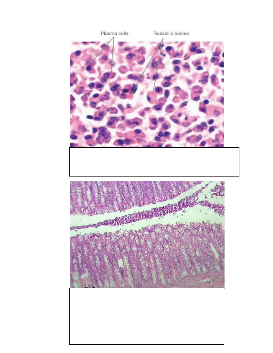
intracellular hyaline change
show excessive accumulation of immunoglobulines in plasma cell
which called Russell s bodies
.
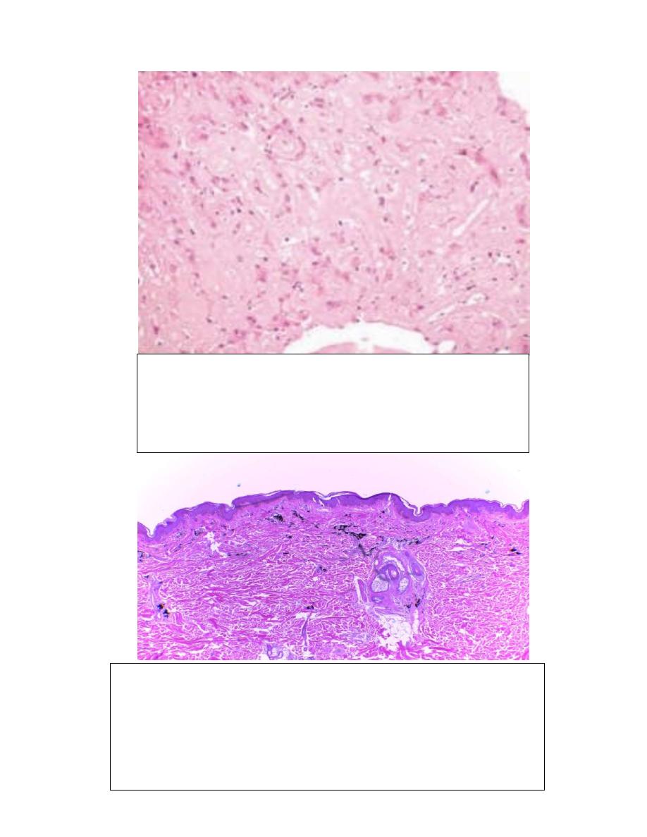
Organ :- small intestine
Lesion :- there is highly numerous goblet cells lining
epithelium of villi which enlarge due to accumulation
of mucine.
Diagnosis:-mucinous degeneration
.
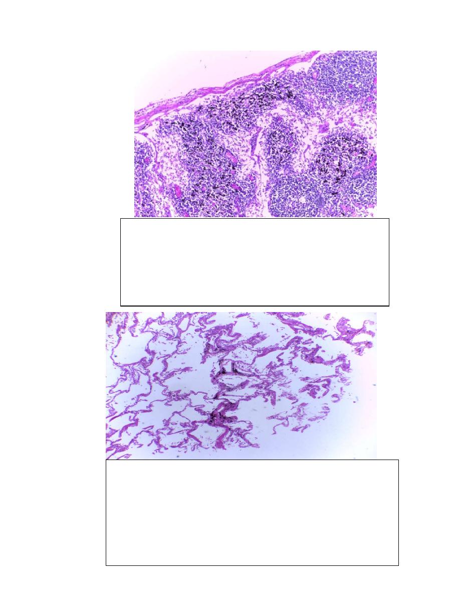
Organ :- liver
Lesion :- Accumulation of amyloid deposits in extracellular which
cause pressure atrophy of hepatocytes.
Diagnosis:- Amyloid degeneration
Organ :- skin
Lesion :- there is accumulation of exogenous pigments black in color
which injected in dermis layer of skin such as Indian ink or carbon
deposits .
Diagnosis:- Exogenous pigmentation(tattoo)
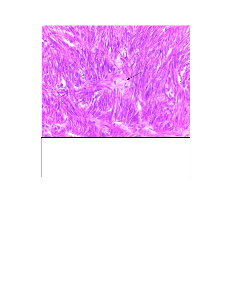
Organ :- Lymph node
Lesion :- there is macrophage laden with carbon particles(black
color) which came from the lung by lymphatic drainage to the
regional lymph node (tracheobronchial) lymph nodes
Diagnosis:- Anthracosis
Organ :- lung
Lesion :- there is accumulation of carbon particles (coal dust which
inhaled from pollutant air ) to lung parenchyma and then phagocytosed
by alveolar macrophages.
Diagnosis:- Anthracosis
Organ :- Uterus
Lesion :- there is glass like homogeneous materials between
myometrium.
Diagnosis:- Hyaline change