
THE Cardiovascular system
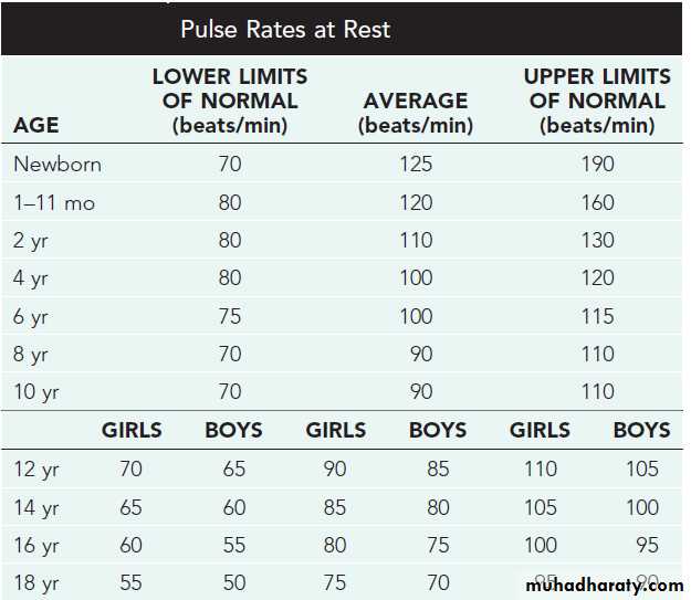
Objectives: The following lectures aim to teach the student about:Normal values of pulse rate in pediatrics
Congenital heart disease
Cyanotic: TOF,TGA,TA,EA.
Acyanotic: ASD,VSD, PDA,
Obstructive: Coarctation of Aorta.
Heart failure in infancy and childhood: Etiology, presentation, diagnosis,& treatment.
Rheumatic fever: Etiology, Diagnosis, Treatment, Prevention.
Infective endocarditis : Etiology, Diagnosis, Treatment, Prevention.
Cardiomyopathies : types with special focus on dilated CMP.
History
Children do not present with the typical features of congestive heart failure as seen in adults.Age is very important when assessing child.
Infants:
Feeding difficulties
Easily fatigued
Sweating while feeding
Rapid respirations
Older children:
Shortness of breath
Dyspnea on exertion
Physical examination:
• Tachycardia
• Rapid respiration• Tender hepatomegaly
• Pulmonary rales
REMEMBER THAT During physical examination:
− Need to refer to normal heart and respiratory rates for ages to determine tachycardia and tachypnea.− Height and weight should be assessed to determine proper growth.
− Always get upper and lower extremity blood pressures and pulses.
− Hepatosplenomegaly suggests right-sided heart failure.
− Always palpate for femoral pulses and compare with radials.
− Cyanosis and clubbing result from hypoxia.
Diagnostic tests
Chest radiograph for:° Heart size
° Lung fields
° Ribs for notching
° Position of great vessels
Electrocardiogram
Echocardiography definitive diagnosis
Other : MRI, cardiac catheterization, angiography, exercise testing
Congenital Heart Diseases
Classification of congenital heart diseases
• Group I : Left to right shunts (acyanotic): ASD, VSD, PDA• Group II: Right to lefts shunts (cyanotic): TOF, TGA,TA,EA
• Group III: Obstructive lesions: AS,PS,COA
Incidence : 8/1000 births
Etiology:Multifactorial
Some are associated with chromosomal disorders, or single gene defects.
Teratogens & maternal drugs.
Maternal metabolic disease.
Acyanotic Congenital Heart Disease
• Left-to-Right Shunt Lesions• Atrial Septal Defect (ASD)
• Ventricular Septal Defect (VSD)
• Atrioventricular Septal Defect (AV Canal)
• Patent Ductus Arteriosus (PDA)
Atrial Septal Defect
• ASD is an opening in the atrial septum permitting free communication of blood between the atria. Seen in 10% of all CHD.
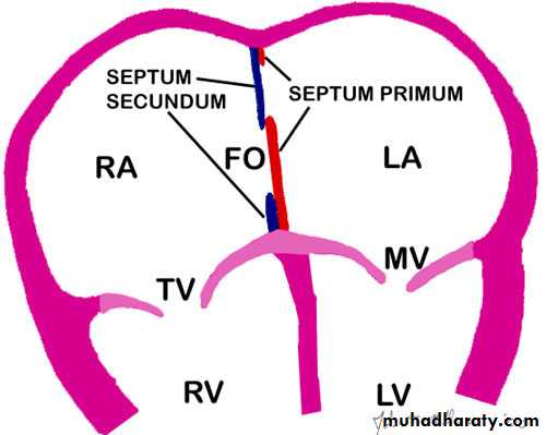
There are 3 major types:
• Secundum ASD – at the Fossa Ovalis, most common.• • Primum ASD – lower in position.
• • Sinus Venosus ASD – high in the atrial septum, associated with anomalous venous return & the least common.
• • Most are asymptomatic but may have easy fatigability or mild growth failure.
• • Cyanosis does not occur unless pulmonary HTN is present.• Examination:
• • Hyperactive precordium, RV heave, fixed widely split S2.
• • II-III/VI systolic ejection murmur at Left SB(2nd intercostal).
• • Mid-diastolic murmur heard over LLSB indicates large defect.
Diagnosis
• Chest x-ray—varying heart enlargement (right ventricular and right atrial); Increased pulmonary vessel markings, edema• ECG—right-axis deviation and RVH
• Echocardiogram definitive
• Treatment:
• Surgical or Catheterization laboratory closure is generally recommended for secundum ASD with a Qp:Qs ratio >2:1.
• • Closure is performed electively between ages 2 & 5 yrs to avoid late complications.
• • Surgical correction is done earlier in children with CHF or significant Pulmonary hypertension( HTN).
• • Once pulmonary HTN with shunt reversal occurs this is considered too late (Eisnmenger syndrome).
• • Mortality is < 1%.
• N.B. Infective Endocarditis is very rare in ASD.
Ventricular Septal Defect
VSD – is an abnormal opening in the ventricular septum, which allows free communication between the Rt & Lt ventricles. Accounts for 25% of CHD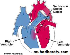
• Clinical Signs & Symptoms
• • Small - moderate VSD, 3-6mm, are usually asymptomatic and 50% will close spontaneously by age 2yrs.• • Moderate – large VSD, almost always have symptoms(dyspnea, feeding difficulties, poor growth, sweating, pulmonary infection, heart failure) and will require surgical repair
• Symptoms develop between 1 – 6 months.
• • CHF, FTT, Respiratory infections, exercise intolerance hyperactive precordium• • II-III/VI harsh pansystolic murmur heard along the Left SB (2nd intercostal) , more prominent with small VSD.
Imaging Studies
ECG and CX-ray findings depend on the size of the VSD.
• Small VSDs usually have normal studies.
• Larger VSDs, ECG findings of left atrial and ventricular enlargement and hypertrophy.
• A CXR may reveal cardiomegaly, enlargement of the left ventricle, an increase in thepulmonary artery silhouette, and increased pulmonary bloodflow.
• Pulmonary hypertension due to either increased flow or increased pulmonary vascular resistance may lead to right ventricular enlargement and hypertrophy.
• Treatment
• • Small VSD - no surgical intervention, nophysical restrictions, just reassurance and periodic follow-up and endocarditis prophylaxis.• • Symptomatic VSD - Medical treatment initially with afterload reducers & diuretics( ± digoxin).
• • Prophylaxis against infective endocarditis(after dental or GU procedures).
Indications for Surgical Closure:
• Large VSD with medically uncontrolled symptomatology & continued FTT.• Ages 6-12 mo with large VSD & Pulmonary HTN.
• Age > 24 mo w/ Qp:Qs ratio > 2:1.
• Supracristal VSD of any size.
Complications
Large defects lead to heart failure, failure to thriveEndocarditis
Pulmonary hypertension
Patent Ductus Arteriosus
Persistence of the normal fetal vessel that joins the PA to the Aorta.
• Normally closes in the 1st wk of life.
• Accounts for 10% of all CHD, seen in 10% of other congenital heart lesions and can often play a critical role in some lesions.• Female : Male ratio of 2:1
• Often associated with coarctation & VSD.
• Can be caused by congenital Rubella.
• Clinical Signs & Symptoms
• Small PDA’s are usually asymptomatic.• Large PDA’s can result in symptoms of CHF, growth restriction, FTT.
• Collapsing arterial pulses.
• Widened pulse pressure .
• Enlarged heart, prominent apical impulse.
• Classic continuous machinary systolic murmur.
• Mid-diastolic murmur at the apex.
Imaging Studies
• ECG and chest x-ray findings are normal with small PDAs• moderate to large shunts may result in a full pulmonary artery silhouette and increased pulmonary vascularity.
• ECG findings vary from normal to evidence of LVH. If pulmonary HTN is present, there is also RVH.
• Treatment:
• Indomethacin, inhibitor of prostaglandin synthesis can be used in premature infants.
• PDA requires surgical or catheter closure.
• Closure is required for heart failure & to prevent pulmonary vascular disease.
• Usually done by ligation & division or intra vascular coil.
• Prophylaxis against infective endocarditis(after dental or GU procedures).
Obstructive Heart Lesions
• Pulmonary Stenosis• Aortic Stenosis
• Coarctation of the Aorta
Coarctation of the Aorta
is narrowing of the aorta at varying points anywhere from the transverse arch to the iliac bifurcation. It is commonly associated with bicuspid valve.More common in Turner’s syndrome.
• 98% of coarctations are juxtaductal
• Male: Female ratio 3:1.
• Accounts for 7 % of all CHD
• The obstruction to blood flow will lead to LVH.

• Clinical Signs & Symptoms
• Classic signs of coarctation are diminution or absence of femoral pulses.• Higher BP in the upper extremities as compared to the lower extremities.
• 90% have systolic hypertension of the upper extremities.
• With severe coarctation, HF and shock.
• Differential cyanosis if ductus is still open.
• II/VI systolic ejection murmur at LSB.
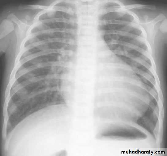
• Cardiomegaly, rib notching on X-ray.
notching of ribs in
coarctation.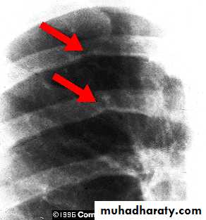
• Treatment
• With severe coarctation maintaining the ductus with prostaglandin E is essential.• Surgical intervention, to prevent LV dysfunction.
• Angioplasty is used by some centers.
• Re-coarctation can occur, balloon angioplasty is the procedure of choice.
• Prophylaxis against infective endocarditis(after dental or GU procedures).
Cyanotic congenital heart disease
Tetralogy of Fallot
It is the most common cyanotic congenital heart diseaseComponents:
• Pulmonary stenosis and infundibular stenosis (obstruction to right ventricular outflow)• VSD
• Overriding aorta (overrides the VSD)
• Right ventricular hypertrophy
Hemodynamics
Pulmonary stenosis plus hypertrophy of subpulmonic muscle (crista supraventricularis)→ Varying degrees of right ventricular outflow obstruction
→ Blood shunted right-to-left across the VSD with varying degrees of arterial desaturation and cyanosis.
Clinical Picture
• Symptomatic any time after birth• Paroxysmal attacks of dyspnea
• Anoxic spells
• Predominantly after waking up
• Child cry
• Dyspnea& cyanosis
• Loss of consciousness
• Convulsions
• Frequency varies from once a few days to many attack everyday
Examination
Varying degrees of cyanosis and a murmur, a single S2 and right ventricular impulse at the left sternal border are typical findings.
Clubbing of fingers & toes.
Cyanosis may not be apparent for the first few weeks of life until pulmonary stenosis reach a critical level that cause reduction in pulmonary blood flow.
Therefore; the only finding in the first few days may be the murmur of PS.
Hypoxic (Tet) spells
1. Restlessness and agitation2. Inconsolable cry
3. Squatting:
increases the peripheral vascular resistance, which diminishes the right-to-left shunt
increases pulmonary blood flow.
4. Predominantly after waking up
5. Dyspnea& cyanosis6. Loss of consciousness
7. Convulsions
8. Loss of the murmur
Frequency varies from once a few days to many attack everyday
Imaging studies
ECG : RAD &RVH
CXR : Boot-shaped heart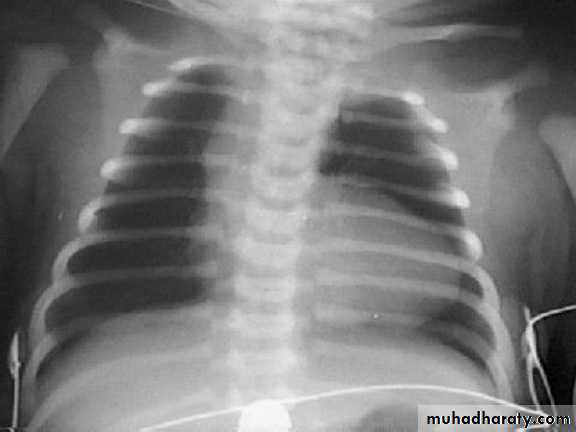
Complications:
• Each anoxic spell is potentially fatal• Polycythemia may lead to Cerebrovascular thrombosis
• Anoxic infaction of CNS
• Brain Abcess
• Infective endocarditis
Treatment
Management of anoxic spell:Knee chest position
Humified O2
Be careful not to provoke the child
Morphine 0.1 -0.2 mg/kg subcutaneously
Correct acidosis – sodium bicarb IV 1 mmol/kg slowly
Propanolol start (0.1mg/kg/IV) slowly during spells followed by (0.5 to 1.0) mg/kg/ 4-6hourly orally
Vasopressors: Methoxamine or Phenylephrine IM or IV drip
GA is the last resort
Surgery
Palliative procedure: Blalock Taussig shunt
Subclavian artery – Pulmonary artery anastomosis
Definitive operation: complete surgical repair with VSD closure and removal or patching of the pulmonary stenosis can be performed in
infancy
Transposition of Great Areries (TGA)
Aorta originating from the right ventricle, and pulmonary artery originating from the left ventricleAccounts for 5-7% of all congenital heart disease
Survival is dependent on the presence of mixing between the pulmonary and systemic circulation
Associated ASD, VSD, or PDA are essential for survival .
50% of patients have a VSD
Usually presents in the first day of life with profound cyanosis
More common in boys
Examination :
Cyanosis in an otherwise healthy looking babyLoud S2
Loud VSD murmur
CXR : Egg on side & narrow mediastinum
Acute management in newborn baby
Initial medical management includes prostaglandin E1 to maintain ductal patency. If significant hypoxia persists on prostaglandin therapy, a balloon atrial septostomy improves mixing between the two circulations.Surgical repair Aterial switch (old style).
Arterial switch (new)TRICUSPID ATRESIA:
The absence of the tricuspid valve results in a hypoplastic right ventricle. All systemic venous return must cross the atrial septum into the left atrium.A PDA or VSD is necessary for pulmonary blood flow and
survival.
Clinical Manifestations
Usually severely cyanosed.Single S2.
If a VSD is present, there may be a murmur.
ECG: LVH& LAD.
Treatment:PGE-1, and minimal O2 to maintain ductal patency
Palliative procedure: Blalock-Taussig procedure
Definitive: bidirectional cavopulmonary shunt (bidirectional Glenn) and Fontan procedure.
Ebstein anomaly
Downward displacement of abnormal tricuspid valve into right ventricle; the right ventricle gets divided into two parts: an atrialized portion, which is thin-walled, and smaller normal ventricular myocardium.
• Right atrium is huge; tricuspid valve regurgitant
• Right ventricular output is decreased because:
− Poorly functioning, small right ventricle
− Tricuspid regurgitation
− Variable right ventricular outflow obstruction—abnormal anterior tricuspid valve leaflet. Therefore, increased right atrial volume shunts blood through foramen Ovale or ASD → cyanosis.
Clinical presentation
− Severity and presentation depend upon degree of displacement of valve and degree of right ventricular outflow obstruction° May not present until adolescence or adulthood
° If severe in newborn → marked cyanosis, huge heart
− Pansystolic murmur of tricuspid insufficiency over most of anterior left chest (most characteristic finding)
− Chest x-ray—heart size varies from normal to massive (increased right atrium); if severe, decreased pulmonary blood flow.
− ECG—tall and broad P waves, RBBB, and a normal or prolonged PR interval
• Treatment:
− PGE1− Systemic-to-pulmonary shunt
− Then staged surgery
Congestive heart failure
Refers to a clinical state of systemic and pulmonary congestion resulting from inability of the heart to pump as much blood as required for the adequate metabolism of the body.
Etiology:
cardiaccongenital structural malformations
● excessive preload
● excessive afterload
no structural anomalies
● cardiomyopathy
● myocarditis
● myocardial infarction
● acquired valve disorders
● hypertension
● kawasaki syndrome
● arrhythmia (bradycardia or tachycardia)
NONCARDIAC
● Anemia● Sepsis
● Hypoglycemia
● Diabetic ketoacidosis
● Hypothyroidism
● Other endocrinopathies
● Arteriovenous fistula
● Renal failure
● Muscular dystrophies
History
Children do not present with the typical features of congestive heart failure as seen in adults.
Age is very important when assessing child.
Infants:
Feeding difficulties
Easily fatigued
Sweating while feeding
Rapid respirations
Older children:
Shortness of breath
Dyspnea on exertion
Physical examination:
• Tachycardia• Rapid respiration
• Tender hepatomegaly
• Pulmonary rales
IMAGING STUDIES
The absence of cardiomegaly on a chest x-ray usually rules out the diagnosis of heart failure.An Echocardiogram assesses the heart chamber sizes, measures myocardial function, and diagnoses congenital heart defects when present.
Treatment
The goals of medical therapy for congestive heart failure include the following:Reducing the preload
Enhancing cardiac contractilityReducing the afterload
Improving oxygen delivery
Enhancing nutrition
General measures:
Bed rest and limit activitiesNurse propped up or in sitting position
Expressed breast milk for small infants
Fluid restriction in volume overloaded
Correction of anemia ,acidosis, hypoglycemia and hypocalcaemia if present
Oxygen
Treatment: Phrmacological therapy
Preload reduction:
1. Diuretics: (po)or (IV) diuretics (furosemide, thiazide. Metolazone).
2. Venous dilators (eg, nitroglycerin).
Contractility support:
1. Dopamin, dobutamin
2. Digoxin
Afterload reduction
1.Oral (ACI) inhibitors
2. IV hydralazine, nitroprusside, or alprostadil
Doses:
Furosemide: 1 mg/kg/dose PO or IVHydrochlorothiazide: 2 mg/kg/d PO divided bid
Digoxin :TDD followed by maintenance.
IV Dopamine : 5-10 mcg/kg/min IV (usual dosage; maximal dosage May be up to
28 mcg/kg/min)
Dobutamine: 5-10 mcg/kg/min iv
Captopril: 0.1-0.5 mg/kg/d PO divided q8h
Enalapril: 0.1 mg/kg/d PO divided qd/bid, not to exceed 0.5 mg/kg/d
Carvidolol: 0.2-0.4 mg/kg/dose bid.
Spironolactone: 1-3 mg/kg/day.
Digoxin: Rapid digitalization can be achieved by administration of “total digitalizing dose (TDD) as follow:
Premature: 20 μg/kg
Full-term neonate (up to 1 mo): 20-30 μg/kg
Infant or child: 25-40 μg/kg
Adolescent or adult: 0.5-1 mg in divided doses
NOTE: these doses are PO; IV dose is 75% of PO dose.
½ TDD is given initially followed by 1/4 TDD in 2 doses 12 hrs apart.
Maintenance digoxin : 5-10 μg/kg/day, divided q12h
Managing Acute Congestive Heart Failure (Acute Pulmonary Edema) in Children:
Admit to the ICU.Head up position.
Oxygen.
IV furosemide: 1-2mg/kg.
???Digoxin (TDD).
Dopamine if ↓BP: (5-10 mcg/kg/min) .
Nitrates (nitroprusside, nitroglycerin) as venodilators if ↑ pulmonary capillary wedge pressure
Rheumatic fever:
Due to an immunologic reaction that is a delayed sequela of group A beta-hemolytic streptococcal infections of the pharynx.A family history of rheumatic fever and lower socioeconomic status are additional factors.
The infection often precedes the presentation of rheumatic fever by 2 to 6 weeks. Streptococcal antibody tests, such as the antistreptolysin O (ASOT) titer, are the most reliable laboratory evidence of prior infection.
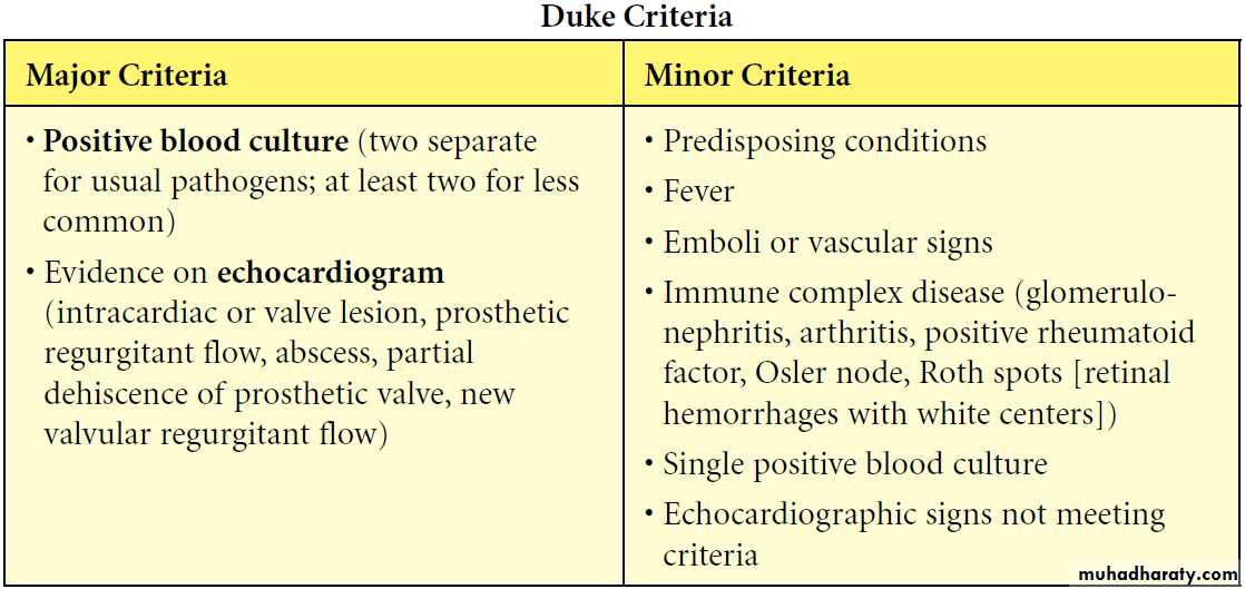
Diagnosis:
= {2 Major OR (1Major + 2Minor) Jones Criteria} + Evidence of antecedent Streptococcal infection.
Major : Migratory polyarthritis, Carditis, Erythema marginatum, Chorea,
Subcutaneous nodules.
Minor : Fever, arthralgias, previous rheumatic fever, leukocytosis, elevated ESR/C-reactive protein, and prolonged PR interval.
The presence of Sydenham’s chorea alone is sufficient for
diagnosis.
Evidence of recent group A Streptococcal disease (e.g. scarlet fever, positive throat culture, or elevated ASOT or other antistreptococcal antibodies) .
Treatment
Bed restBenzathine Penicillin 1.2 million unit im.
Salicylate: 50-70 mg/kg/day in 4divided doses PO for 3-5 days, followed by 50 mg/kg/day in 4 divideddoses PO for 3 wk and half that dose for another 2-4 wk
Prednisolone: 1-2mg/kg/day for 3 weeks for severe carditis or congestive HF.
Prevention:
Benzathine Penicillin 600,000 IU for children weighing ≤60 lb 1.2 million IU for children weighing >60 lb, every4 wk.Duration of prophylaxis for pt.
without carditis: 5years or until he is 21 years old.With carditis : 10 years or until age is 40.
Infective Endocarditis ;
Etiology/epidemiology
− Most are Streptococcus viridans (alpha hemolytic) and Staphylococcus aureus
− Organism associations
° S. viridans—after dental procedures
° Group D streptococci—large bowel or genitourinary manipulation
° Pseudomonas aeruginosa and Serratia marcescens— intravenous drug users
° Fungi—after open heart surgery
° Coagulase-negative Staphylococcus—indwelling intravenous catheters
− Highest risk with prosthetic valve and uncorrected cyanotic heart lesions
• Clinical presentation
− Prolonged intermittent fever, weight loss, fatigue, myalgia, arthralgia, headache,nausea, vomiting
− New or changing heart murmur
− Splenomegaly, petechiae, embolic stroke, CNS abscess, CNS hemorrhage, mycotic
aneurysm (all more with Staphylococcus)
− Skin findings—rare; late findings (uncommon in treated patients); represent vasculitis
from circulating Ag-Ab complexes; if present, are highly suggestive
° Osler nodes—tender, pea-sized, intradermal nodules on pads of fingers and toes
° Janeway lesions—painless, small erythematous or hemorrhagic lesions on
palms and soles
° Splinter hemorrhage—linear lesions beneath nail beds
º Roth spots —retinal exudates
Diagnosis
Three to 5 separate blood collections should be obtained after careful preparation of the phlebotomy site.
Complications
− Most common—heart failure from aortic or mitral lesions− Others—systemic or pulmonary emboli, myocardial abscess, myocarditis, valve
obstruction, heart block, meningitis, osteomyelitis, arthritis, renal abscess, immune
complex−mediated glomerulonephritis
TREATMENT
-Empirical antibiotic therapy may be started for acutely ill persons after blood cultures are obtained.-High doses of bactericidal antibiotics are required for an extended period of treatment (4 to 8 weeks).
Vancomycin or a β-lactam antibiotic, with or without gentamicin, for a 6-week course is the most common regimen.
CARDIOMYOPATHIES:
-CONGESTIVE(DILATED)-HYPERTROPHIC-RESTRICTIVEDilated Cardiomyopathy
Pathophysiology
- Extensive ventricular dilatation; mostly left ventricle.
- Vast majority is idiopathic (may be familial).
- Other causes-viral infection, endocrine (hypothyroidism), metabolic (storage disease), systemic disease (connective tissue), hereditary muscle or neurologic disease (muscular dystrophies), abnormality of coronary arteries.
Clinical presentation
- Initially nonspecific (respiratory symptoms, failure to thrive, abdominal complaints).- Then findings of failure:
- Tachycardia, decreased pulse pressure, cool and pale skin, decreased pulses, increased jugular venous pressure, hepatomegaly, edema, rales
- Cardiomegaly, mitral insufficiency, tricuspid insufficiency, gallop rhythm
Diagnosis
- ECG-atrial enlargement, left ventricular or right ventricular enlargement; nonspecific T-wave changes-Chest x-ray--cardiomegaly, pulmonary congestion.
-Echocardiogram-dilatation of left atrium and left ventricle ± right ventricle and decreased contractility; decreased flow velocity across aortic valve with mitral regurgitation.
Prognosis-downward progression; relapses; emboli; ventricular arrhythmias & sudden death.
Treatment
- Antifailure.- Antiarrhythmic agents
- May need an implantable cardioverter-defibrillator (ICD)
-Systemic anticoagulation
-Beta blocker (metoprolol, carvedilol)
- Trial of PO carnitine (for possibility of mitochondrial disorder)
-Referral to transplant center