department of Pathology
2
nd
class /2021-2022

Objectives
1. Define adaptation
2. What are the mechanisms and types of adaptation

Adaptation
: changes that occur in cells/tissues
in response to prolonged stimulation or chronic injury
It is reversible once the stress has been removed.
Adaptation includes:-
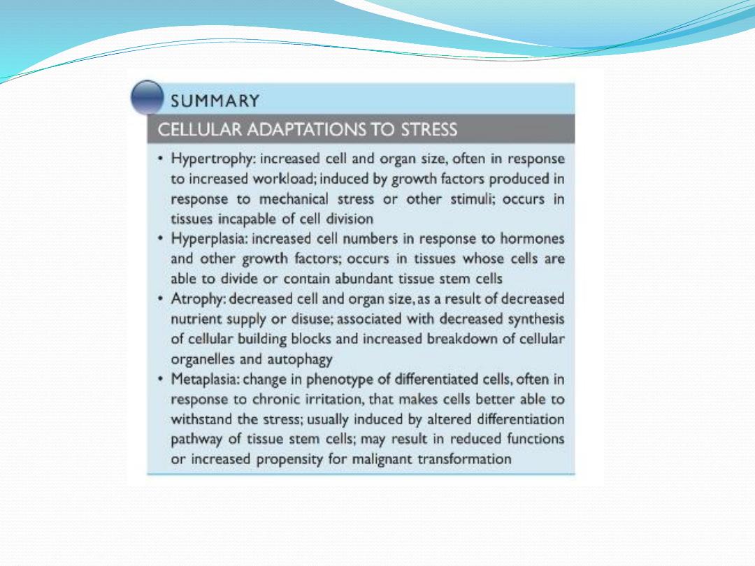
• Atrophy (decrease in cell size)
• Hypertrophy (increase in cell size)
• Hyperplasia (increase in cell number)
• Metaplasia (change in cell type)
• Intracellular accumulation of substances

Atrophy
definition
:
size of cell (then organ/tissue) due to
loss of cell substance (organelles). Physiologic or pathologic
Causes
:
1. Disuse: e.g.,
muscle bulk in bed ridden patients
2. Decrease stimulation: denervation (nerve damage
atrophy of muscle supplied by that nerve
or
hormonal stimulation (
ACTH atrophy of adrenal)
3. Decrease blood supply or nutrition: cerebral atrophy
in atheroma of internal carotid atrophy
4. Pressure atrophy: renal cortical atrophy in hydronephrosis
5. Aging (senile atrophy): e.g. testicular atrophy

Mechanism of atrophy
Degenerated organelles encircled by membrane from ER
Fuse with lysosomal vacoules degraded by hydrolytic
enzymes (autophagy) residual organelle membranes
accumulate in cytoplasm as a brown pigment called
lipofuscin

Hypertrophy
Definition
:
size of a tissue due to
size of individual cells.
Cause
:
1.
mechanical demand
* Physiologic (skeletal muscle hypertrophy in athletes)
* Pathologic (myocardial hypertrophy in hypertension)
2.
endocrine stimulation:
*
puberty (growth hormone, androgens/estrogens, etc.)
*Gravid uterus (estrogen)
*Lactating breast (prolactin and estrogen))
+
hypertrophy and hyperplasia are two distinct processes but
frequently both occur together
Mechanism of hypertrophy & hyperplasia
hypertrophy occurs in terminally differentiated cells
that are unable to divide.
(e.g., nerve, cardiac & skeletal muscle)
Mechanical or hormonal stimuli
stimulate synthesis of growth factors
expression of growth-promoting genes (proto=oncogenes)
protein synthesis &
organelle size
cell size
tissue/organ size (hypertrophy)

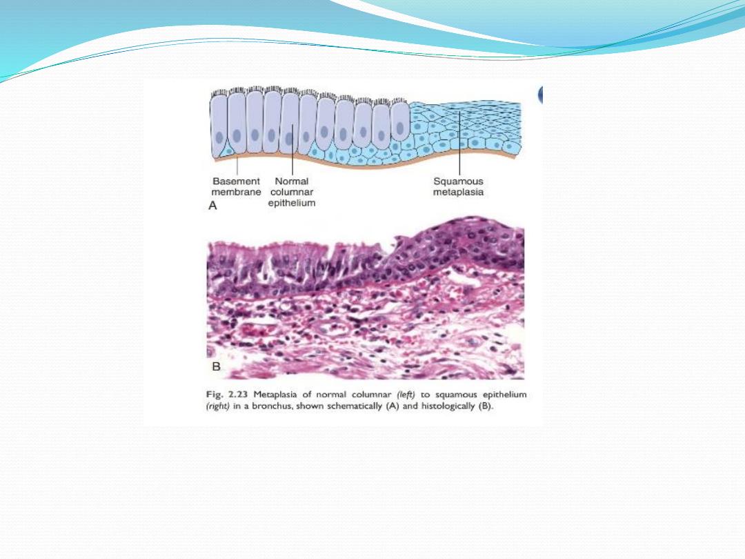
Metaplasia
Definition
: replacement of one adult cell type by another
adult cell type in response to chronic stimulation.
Metaplasia is reversible
Replacement cell is better tolerate environmental stress
e.g., bronchial epithelium undergoes squamous
Metaplasia in response to chronic irritation of tobacco smoking
Metaplasia could also involve mesenchymal cells
(e.g. osseous metaplasia i.e bone formation in area of
muscle trauma

Dysplasia
An abnormal proliferation of cells that is characterizes
by changes in cell size, shape, and loss of cellular
organization.
It is not cancer but may progress to cancer
(preneoplastic lesion)
Example: cervical dysplasia.

Accumulations
1. Exogenous material:
e.g., carbon dust in the lung of city dwellers, tattoos.
2. Endogenous material:
(Melanin
,
Bilirubin
,
Lipofuscin
,
hemosidrin
,
lipid)

Endogenous material:
1.Melanin
: brown black pigment normal in melanocytes.
Exposure to sunstimulate pituitary gland release
melanocyte stimulating hormone
melanin production
2.
Bilirubin
: in jaundice (prehepatic, hepatic, posthepatic)
3.
Lipofuscin
: yellow brown pigment “wear & tear pigment”
of lipid reminants of degenerated organelles.
4.
hemosidrin
: golden-brown iron-containing pigment.
*Localized hemosiderosis
hemorrageHb ingested/converted by macrophage to hemosidrin
5.
lipid
: alcoholismdisturbed lipid metabolismaccumulation
of lipid in hepatocytes fatty degeneration.

Calcification
Precipitation of calcium in tissues
1.
D
ystrophic calcification: precipitation of calcium in
previously
d
amaged tissue. Serum calcium is normal.
as in atherosclerosis plaques
2. Metastatic calcification: precipitation of calcium in
normal tissues due to hypercalcemia (
serum calcium)
(renal failure, hyperparathyroidism, parathyroid adenoma…)

Practical-adaptation
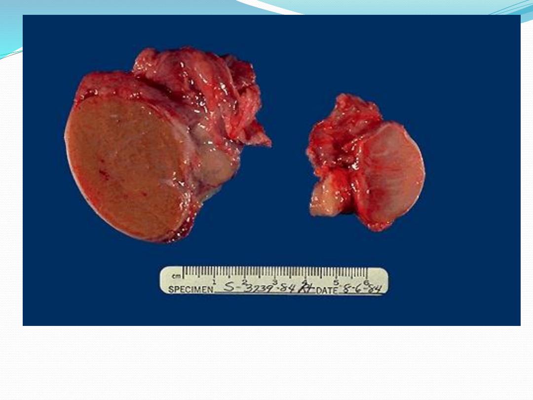
The testis at the right has undergone atrophy and is much
smaller than the normal testis at the left.
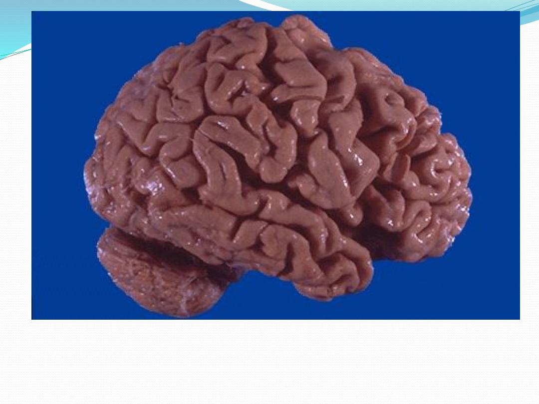
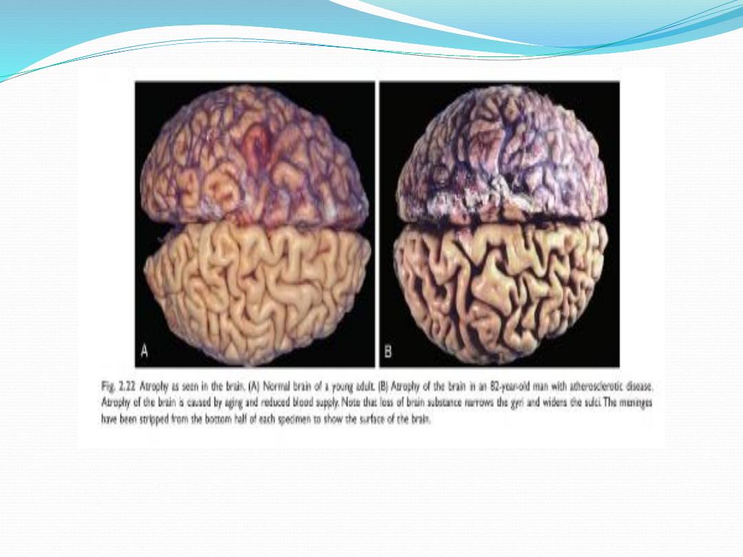
This is cerebral atrophy in a patient with Alzheimer's disease. The gyri are
narrowed and the sulci widened toward to frontal pole.
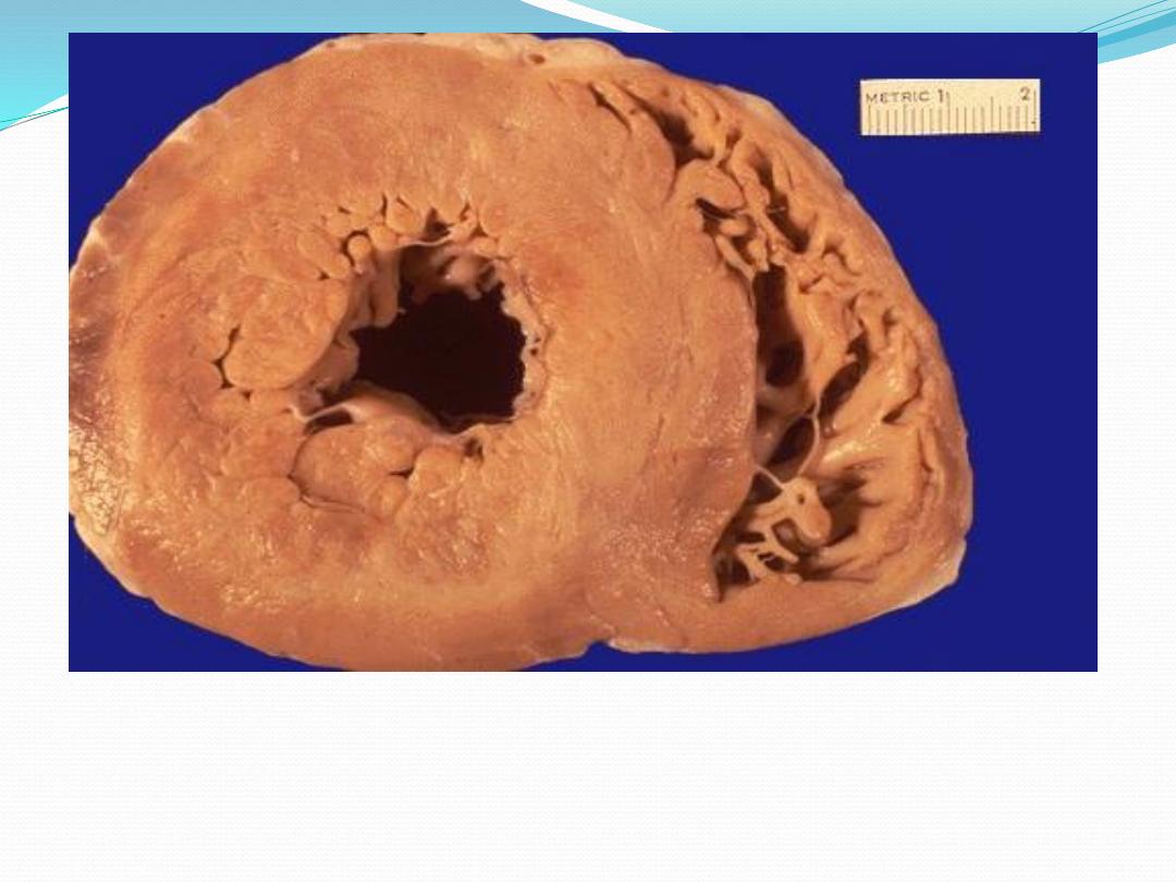
This is cardiac hypertrophy. The number of myocardial fibers never
increases, but their size can increase in response to an increased
workload, leading to the marked thickening of the left ventricle in this
patient with hypertension.

HYPERTROPHY (cont.)
In the heart two mechanisms leading to
hypertrophy:
mechanical stretch
activation of α-adrenergic receptors.
Eventually reaches a limit beyond which
enlargement of muscle mass is no longer able to
compensate.
At this stage a number of "degenerative" changes
occur.
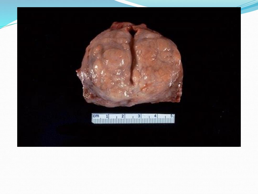
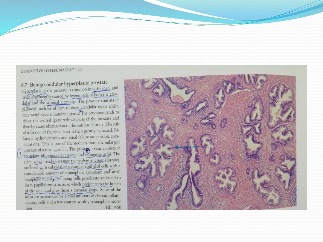
This is an example of prostatic hyperplasia. The normal prostate is about 3
to 4 cm in diameter. The number of prostatic glands, as well as the stroma,
has increased. The pattern of increase here is not uniform, but nodular. This
increase is in response to hormonal manipulation, but in this case is not a
normal physiologic process.
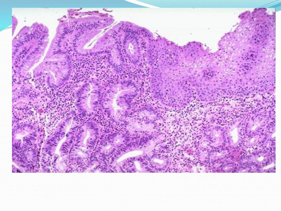
Metaplasia of esophageal squamous mucosa has occurred here, with
gastric type columnar mucosa at the left.