
{
Histology of the
lymph glands
By:-Dr .Elham majeed
TUCOM
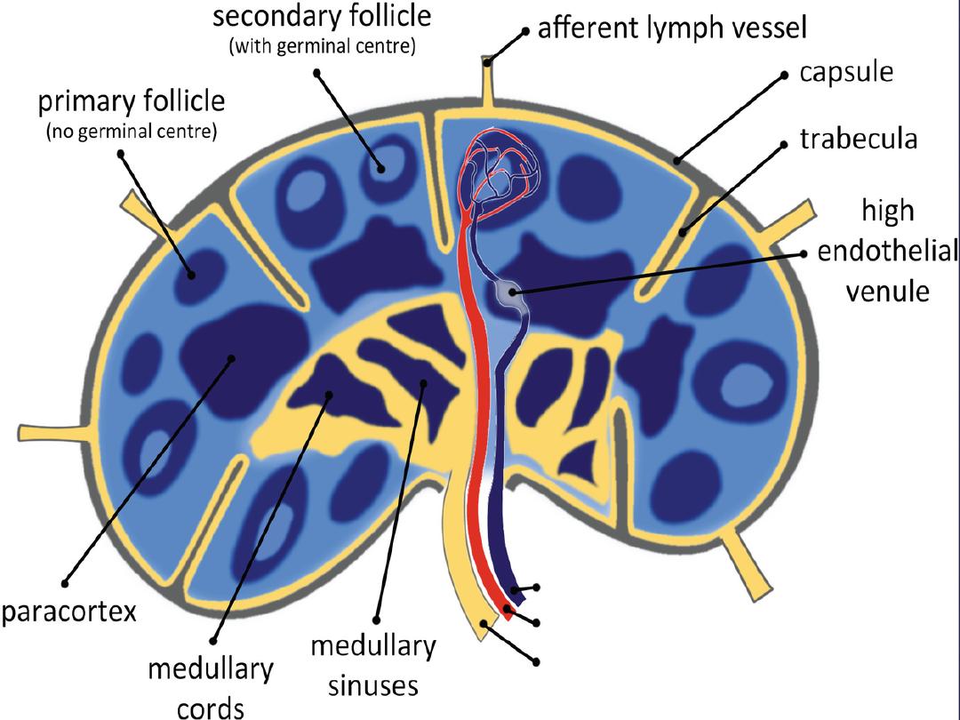

Lymph nodes
Lymph nodes are distributed through the body along the course of the lymphatic
vessels, they are found in the axilla and the groin ,along the great vessels of the neck ,
and in large number in the thorax and abdomen , especially in mesenteries, The convex
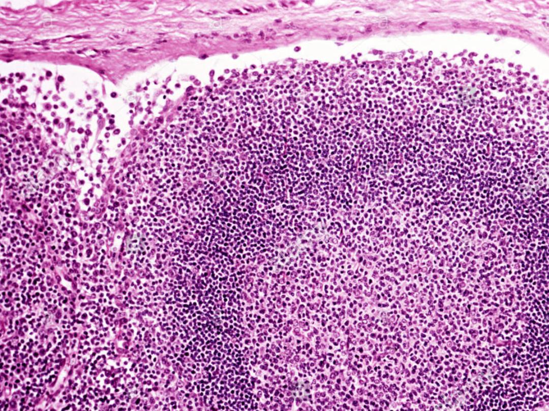
surface of the lymph node is pierced by numerous afferent lymph vessels. Lymph
nodes are bean shape, encapsulated by dense connective tissue comprised of elastin and
collagen fibers along with interspersed fibroblasts . They extend to the deeper areas of
the lymph node by way of the trabecular extensions of the cortex. As
the trabeculae penetrate the lymph node, they continue as reticulum fibrils (type III ,
collagen) that offer additional structural support to the gland, The convex surface of the
gland pierce by afferents lymphatic vessels that conduct the lymph to the lymph node
The most common cells of the lymph nods are lymphocytes, macrophages, APCs,
plasma cells and reticular cells and other , follicular dendritic cell are present within
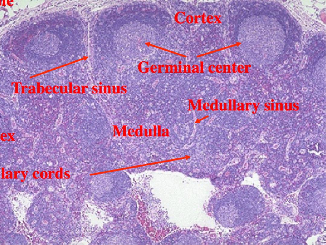
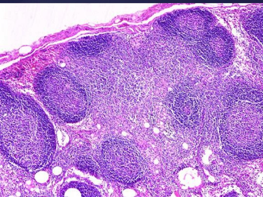
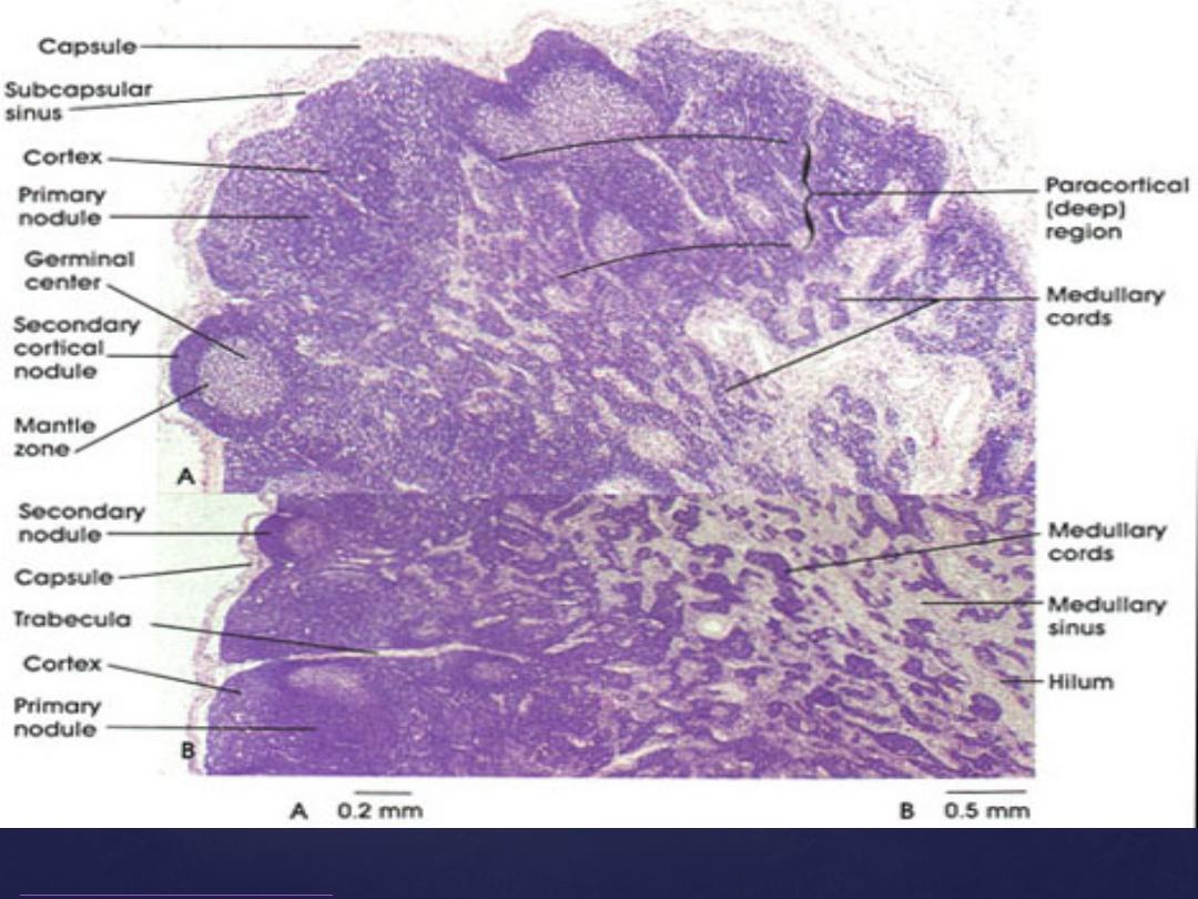
nodules The different arrangement of the cells within the nods creates two regions, a
cortex and a medulla the cortex can be subdivided into an outer cortex and an inner
cortex or paracortical region
Outer cortex
The outermost layer is the cortex. It is made up of a subcapsular sinus, cortical sinus
and lymphoid nodules. The subcapsular sinus is the first space that lymph fluid from
the afferent vessels enters within the node , they composed of loose network of reticular
cells and fibers . The fluid then travels from here to the cortical sinuses; which are
branches of the subcapsular sinus. The cortical sinuses are also known as the
intermediate or trabecular sinuses because they travel along the trabecular network
within the lymph node and share the same structure with the sub capsular sinus
.
Cross sectional analysis
of a lymph node reveals that it is subdivided into three regions
:
The endothelium of the trabecular sinuses are perforated by dendritic processes as well
as reticulin fibres. Antigen presenting cells (APCs), circulating antigen,
and lymphocytes flowing within the lymph can access the lymphatic tissue within the
nodes through the disrupted endothelium. The cortical layer also has relatively large
aggregates of helper T – lymphocytes and rapidly dividing B – lymphocytes in the
peripheral part of the lymph nodes. Although both T cells and B cells are present in the
cortex, B cells are more abundant than T cells are in this region. These lymphoid
nodules are situated around the branched, interlacing extensions of the follicular
dendritic cells (FDCs). The nodules may or may not have a germinal center depending
on if it is a primary or secondary follicle
.
The primary follicle is comprised of small dormant lymphocytes throughout, while
the secondary follicle has a heterogeneous collection of large B lymphocytes that have
already been activated by inciting antigens. Primary follicles absorb less histological
stains then secondary follicles. This is likely due to fewer cells in the primary follicle
when compared to the secondary follicle
.

The lymphatic nodules with or without germinal centers formed mainly by B
lymphocytes embedded in the irregular population of cortical cells. The germinal
center can be further subdivided into a dark zone, light zone and a mantle zone. Each
zone facilitates different aspects of B cell affinity maturation. In the peripherally aspect
of the germinal center, quiescent B cells are found in the mantle zone. These cells are
characterized by intense basophilic staining, small cytoplasmic volume and a
heterochromatic nucleus. Other cells in the mantle zone
include follicular dendritic cells as well as the occasional helper T
lymphocyte and macrophages. The fate of B cells in the mantle zone can go one of two
ways. These cells either remain in the lymph node and mature into antibody
secreting plasma cells and remain in the lymph node, or they transform into memory B
cells that re-enter the systemic circulation
.
The other two zones of the germinal center are the light zone and dark zone. The light
zone contains centrocytes that interact with follicular dendritic cells that express intact
antigen on their surface. Centrocytes with high affinity binding to the follicular
dendritic cell antigen will persist, while those with weak binding undergoes apoptosis.
While resident macrophages help to clean up apoptotic B cells, helper T cells support
the remaining B cells and foster the class switching phase of the cellular maturity
.
In the dark zone of the germinal centre, the centroblasts are highly mitotic and have a
strong likelihood of producing mutated antibodies. These are the source cells for the
light zone
.

Deep to the cortical layer is the
paracortex.
Its margins blend with the superficial
cortex and deep medulla. The principal distinguishing features are the absence of
lymphoid nodules and the large number of T lymphocytes within the stroma of the
paracortex
.
The paracortex also has unique venules known as high endothelial venules (HEVs).
Most of the lymphocytes that enter the lymph node do so via these channels. They are
made up of cuboidal endothelium . These specialized vessels are also present in the
mucosa associated lymphatic tissue distributed throughout the gastrointestinal tract.
However, they are at their highest level of development within the lymph nodes
.
Medulla
The deepest layer of the lymph node is the medulla. It is subdivided functionally and
histologically into two other regions; which are the medullary cords and sinuses. The
cords are populated by plasma cells, as well as B – cells and T – cells. The cells are
arranged in cord-like projections extending centrally from the paracortex
.
Interlacing between the cords are distended areas lined by discontinuous endothelium.
The luminal surface of the sinuses also contains a vast network of reticular cell
processes. They act as the final point of filtration of circulating lymph. The medullary
sinuses are the terminal continuations of the peripherally located cortical sinuses. They
eventually culminate at the hilum of the lymph node to form efferent lymphatic vessels
.
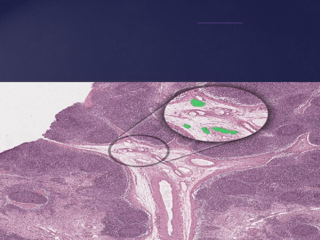
Lymph circulation inside the lymph node
:-
Lymph vessels are lined by a single layer of squamous
endothelium
. They are fi ed
with valves that promotes unidirectional flow of lymph from the afferent lymph vessels
to the lymph node and then to the efferent lymph vessels. The afferent lymph vessels
bring lymph into the subcapsular space. Of note, the subcapsular space extends around
the entire lymph node except at the hilum. The lymph then flows through the cortical
sinuses and to the medullary sinuses then leaves the lymph node through the afferent
vessels