
Lecture 1
النسائية
د
.
هبه
Pelvic Organ Prolapse
Page 1 of 9
Pelvic Organ Prolapse
Definition
A prolapse is a protrusion of an organ or structure beyond its normal
anatomical confines.
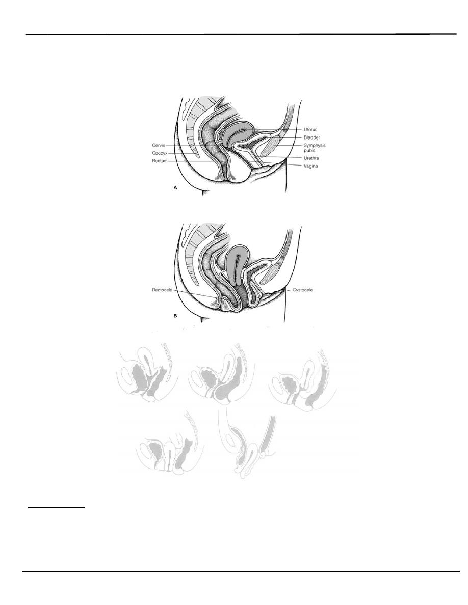
The pelvis is devided into three compartments
1. Anterior: contain urethra and bladder
2. Middle: contain utrerine or vault descent and enterocele
3. Posterior :contain rectum
.
Classification
Anterior vaginal wall prolapse
1. Urethrocele: urethral descent
2. Cystocele: bladder descent
3. Cystourethrocele: descent of bladder and urethra .
Posterior vaginal wall
1. Rectocele: rectal descent
2. Enterocele: small bowel descent
Apical vaginal prolapse
1- Uterovaginal: uterine descent with inversion of vaginal apex occur
when the lateral cervical ligaments become weakened .
2- Vault prolapse: post-hysterectomy inversion of vaginal apex , due to
inadequate support by lateral cervical ligaments
Prevalence
o
Uterovaginal prolapse is extremely common.
o
prolapse affects 12-30 per cent of multiparous
o
2 per cent of nulliparous women.
3
1
/
4
/
4114
العدد
:
(
5
)
Lecture 1
النسائية
د
.
هبه
Pelvic Organ Prolapse
Page 2 of 9
o
A woman has an 11 per cent lifetime risk of having an operation for
prolapse.
Grading
Three degrees of prolapse are described and the lowest or most
dependent portion of the prolapse is assessed whilst the patient is
straining (in the uterovaginal prolapse, the most dependent portion of the

Lecture 1
النسائية
د
.
هبه
Pelvic Organ Prolapse
Page 3 of 9
prolapse is the cervix)
1st: descent within the vagina
2nd: descent to the introits
3rd: descent outside the introits.
Aetiology
The vital structures for the maintenance of position of the pelvic organs
are:
1. The connective tissue lining the pelvic wall
2. Levator ani
3. Intact nerve of the levator ani
4. Intact cardinal and uterosacral ligaments
These are influenced by:
1. Pregnancy
2. Childbirth
3. Ageing (acquired) or
4. Congenital connective tissue defects
1- Congenital factor
Two per cent of symptomatic prolapse occurs in nulliparous
women, implying that there may be a congenital weakness of connective
tissue.
2- Racial variation
A decrease in prevalence of prolapse among black women may be due to
to better connective tissue or lumber lordosis that encourage
divertion of abdominal forces towards abdominal wall rather than pelvis
3- Childbirth and raised intra - abdominal pressure
o
The single major factor leading to the development of genital prolapse
appears to be vaginal delivery which lead to pelvic support damage (
nerve ,muscles and connective tissue ) .

Lecture 1
النسائية
د
.
هبه
Pelvic Organ Prolapse
Page 4 of 9
o
prolapse increases with the increasing parity which was up to seven
times more common in women who had more than seven children
compared to those who had one.
Prolapse occurring during pregnancy is rare but is mediated by:
1. the effects of progesterone and relaxin.
2. In the increase in intra-abdominal pressure will put an added strain on
the pelvic floor .
a raised intra-abdominal pressure outside of pregnancy ( e.g. chronic
cough or constipation ) is also a risk factor.
4- Ageing
o
The process of ageing can result in loss of collagen and weakness of
fascia and connective tissue.
o
These effects are noted particularly during the postmenopause as a
consequence of oestrogen deficiency.
5- Postoperative
o
Poor attention to vaginal vault support at the time of hysterectomy leads
to vault prolapse.
o
Mechanical displacement as a result of gynaecological surgery such as
colposuspension may lead to the development of a rectocele or
enterocele.
6- Others
a. Vitamine defeciency
b. Smoking
c. steroid therapy
Clinical features
A.
History
1. Women usually present with non-specific symptoms.

Lecture 1
النسائية
د
.
هبه
Pelvic Organ Prolapse
Page 5 of 9
2. Specific symptoms may help to determine the type of prolapse.
3. Risk factors should be looked for .
Symptoms of prolapse depends on the type &site of prolapse
Non - specific :
1- Feeling of a lump in the vagina which usually worse towards the end of
the day& relieved by lying down .
2- Local discomfort
3- Backache
4- Bleeding/infection if ulcerated
5- Dyspareunia.
Specific:
A-Uterine descent cause low backache,protrusion of cervix and blood
stained discharge.
B-Enterocele &vault prolapse may produce vague symptoms of discomfort
C- Rectocele: incomplete bowel emptying, digitation,
D-Cystocele may lead to
1- Discomfort & urinary symptoms:
2- Stress incontinence if there is descent of urethrovesical junction.
3- Voiding difficulty can occur if cystocele is present & bladder
neck is normal in position so the woman has to reduce the mass
digitally in order to pass urine .
4- Over flow incontinence with incomplete emptying of bladder
5- Alarge cystocele may lead to increased frequency due to persistant
residual urine or recurrent urinary tract infection because of stasis.
6- Urgency & frequency are found in association with cystocele
which may developed as self induced habit to keep the bladder empty
B.
Abdominal examination
Abdominal examination should be performed to exclude

Lecture 1
النسائية
د
.
هبه
Pelvic Organ Prolapse
Page 6 of 9
organomegaly or abdominopelvic mass that lead to increase intra-
abdominal pressure .
C.
Vaginal examination
o
Prolapse may be obvious when examining the patient in the dorsal
position if it protrudes beyond the introitus; ulceration and/or atrophy
may be apparent.
o
The anterior and posterior vaginal walls and cervical descent should be
assessed with the patient straining in the left lateral position , using
Sims' speculum.
o
Bimanual pelvic examination should be performed to exclude pelvic
mass .
o
Combined rectal and vaginal digital examination can be an aid to
differentiate rectocele from enterocele
Differential diagnosis
1. Congenital or inclusion vaginal dermoid cyst
2. Urethral diverticulum.
3. large uterine polyp.
4. Secondary from the uterine tumor
Investigations
1. If urinary symptoms are present, urine microscopy, cystometry and
cystoscopy is considered
2. If urination difficulty present and renal failure be suspected , serum
urea and creatinine should be evaluated
3. Ultrasound performed to diagnosed abdominopelvic mass
4. Pap smear
Prevention
1. Shortening the second stage of delivery
2. Reducing traumatic delivery may result in fewer women developing a
Lecture 1
النسائية
د
.
هبه
Pelvic Organ Prolapse
Page 7 of 9
prolapse.
3. Women should avoid smoking, constipation and heavy work.
4. The benefits of episiotomy and hormone replacement therapy at the
menopause have not been substantiated.
Treatment
The choice of treatment depends on
1. The patient's wishes.
2. level of fitness .
Prior to specific treatment
1- Attempts should be made to correct obesity, chronic cough or
constipation.
2- If the prolapse is ulcerated, a 7-day course of topical oestrogen should be
administered
3- If infection present a course of antibiotics.
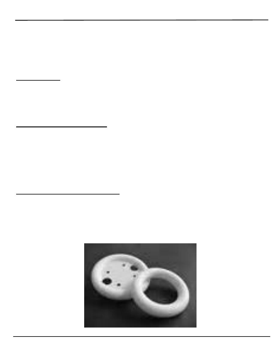
Medical (conservative )
o
Silicon-rubber-based ring pessaries , they are inserted into the vagina and
need replacement at annual intervals.
o
Shelf pessaries are rarely used but may be useful in women who
cannot retain a ring pessary.

Lecture 1
النسائية
د
.
هبه
Pelvic Organ Prolapse
Page 8 of 9
o
Indications for conservative treatment:
1. Patient's wish
2. As a therapeutic test
3. Childbearing not complete
4. Medically unfit
5. During and after pregnancy (awaiting involution)
6. While awaiting surgery.
Complications of conservative treatment:
1. vaginal ulceration and bleeding .
2. infection.
3. Incarceration .
4. Fistula formation
Surgery:
o
Is the main stay in the treatment of prolapse.
o
The aim of surgical repair is to restore anatomy and function.
o
Approach: the vaginal, abdominal and laparoscopic.
Anterior colporraphy(cystourethrocele )
o
Anterior repair (colporrhaphy) is the most commonly performed surgical
procedure but should be avoided if there is concurrent stress
incontinence.
o
An anterior vaginal wall incision is made and the fascial defect
allowing the bladder to herniate through is identified and closed .
With the bladder position restored, any redundant vaginal epithelium
o
Is excised and the incision closed.
Posterior colporraphy (rectocele)
Is common performed procedure. A posterior vaginal wall incision is
made and the fascial defect allowing the rectum to herniate through is
identified and close with the rectal position restored, any redundant vaginal
epithelium is excised and the incision closed.

Lecture 1
النسائية
د
.
هبه
Pelvic Organ Prolapse
Page 9 of 9
Enterocele
The surgical principles are similar to those of anterior and posterior
repair but the peritoneal sac containing the small bowel should be excised..
Uterovaginal prolapse
o
Vaginal hysterectomy with adequate support of the vault to the
uterosacral ligaments is sufficient If the woman does not wish to
conserve her uterus for fertility
o
If uterine conservation is required, the Manchester operation and
sacrohysteropexy
1- The Manchester operation involves partial amputation of the cervix.
2- Sacrohysteropexy is an abdominal procedure and involves attachment
of a synthetic mesh from the uterocervical junction to the sacrum.
Vault prolapse
o
Sacrocolpopexy is an abdominal procedure in which a mesh is used to
attached the vaginal vault to the sacrum .
o
Sacrospinous ligament fixation is a vaginal rocedure in which the vault
of the vagina is sutured to one or other sacrospinous ligament
By: Mu’taz Fathi