
Lecture 6
النسائية
د. أحمد جاسم
Malignant disease of the body of the uterus
Page 1 of 8
Malignant disease of the body of the uterus
uterine cancer.
o Endometrial cancer may sometimes be referred to as uterine cancer.
However, different cancers may develop not only from the
endometrium itself but also from other tissues of the uterus, including
cervical cancer, sarcoma of the myometrium, and trophoblastic
disease.
o Corpus cancer accounts for 3% of cancer in females.
1. Endometrial carcinoma which arise from the lining of the uterus.
0r
2. Sarcoma arise from the stroma of endometrium or from
myometrium.
o The incidence is at 60 years & more ,75% of cases usually occur in
the post menopausal period
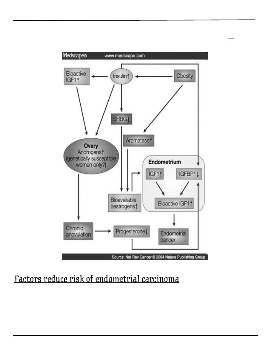
1. high levels of estrogen
2. endometrial hyperplasia
3. obesity
4. hypertension
5. polycystic ovary syndrome
[citation needed]
6. nulliparity (never having carried a pregnancy)
7. infertility (inability to become pregnant)
8. early menarche (onset of menstruation)
9. late menopause (cessation of menstruation)
10. endometrial polyps or other benign growths of the uterine lining
11. diabetes
12. Tamoxifen
13. high intake of animal fat
[citation needed]
14. pelvic radiation therapy
15. breast cancer
:العدد
4
9/3/2014
Lecture 6
النسائية
د. أحمد جاسم
Malignant disease of the body of the uterus
Page 2 of 8
16. ovarian cancer
17. heavy daily alcohol consumption (possibly a risk factor)
[3]
1. oral contraception.
2. progestogens.
3. Smoking.

Lecture 6
النسائية
د. أحمد جاسم
Malignant disease of the body of the uterus
Page 3 of 8
o The histopathology of endometrial cancers is highly diverse. The
most common finding is a well-differentiated endometrioid
adenocarcinoma, which is composed of numerous, small, crowded
glands with varying degrees of nuclear atypia, mitotic activity, and
stratification. This often appears on a background of endometrial
hyperplasia.
o Lymphatic spread occurs later and is less frequent than in cases of
cervical carcinoma.
o Remote metastases in lungs, bones or else-where are not common but
occur more often than with cervical carcinoma
Symptoms
1. The classic symptom is bleeding
A. Post menopausal bleeding in 75-80 %.
This symptom should be assumed to be caused by
carcinoma of endometrium until proved otherwise.
Women with post menopausal bleeding in women not
taking hormone replacement therapy has 10% risk of
having a genital tract cancer
B. In premenopausal women may present as:
intermenstrual bleeding
menorrhagia.
2. Watery or purulent vaginal discharge (blood stained).
3. Pain is a late symptom and denotes extensive spread of
disease.
4. Abnormal screening test.
Signs
o A full general and systematic examination is indicated.

Lecture 6
النسائية
د. أحمد جاسم
Malignant disease of the body of the uterus
Page 4 of 8
o Enlarged lymph nodes in the groin or supraclavicular fossa may be
found.
o Breast should be palpated.
o Uterine enlargement can be palpated.
o Pelvic examination:
Bleeding through cervix.
Secondary metastasis in vagina, urethra.
o A Pap smear may be either normal or show abnormal cellular
changes.
o Endometrial curettage is the traditional diagnostic method. Both
endometrial and endocervical material should be sampled.
o If endometrial curettage does not yield sufficient diagnostic material,
a dilation and curettage (D&C) is necessary for diagnosing the
cancer.
o Hysteroscopy allows the direct visualization of the uterine cavity and
can be used to detect the presence of lesions or tumours. It also
permits the doctor to obtain cell samples with minimal damage to the
endometrial lining (unlike blind D&C).
o Endometrial biopsy or aspiration may assist the diagnosis.
o Transvaginal ultrasound to evaluate the endometrial thickness in
women with postmenopausal bleeding is increasingly being used to
evaluate for endometrial cancer.
o An endometrial thickness exceeding 4 to 5 mm on ultrasound is
suggestive of endometrial pathology in such women.
o Sonohysterography: It may improve delineation of endometrial
polyps.
o both D&C and Pipelle biopsy curettage give 65-70% positive
predictive value. But most important of these is hysteroscopy which
gives 90-95% positive predictive value.

Lecture 6
النسائية
د. أحمد جاسم
Malignant disease of the body of the uterus
Page 5 of 8
o Recently, a new method of testing has been introduced called the
TruTest, offered through Gynecor. It uses the small flexible Tao
Brush to brush the entire lining of the uterus. This method is less
painful than a pipelle biopsy and has a larger likelihood of procuring
enough tissue for testing. Since it is simpler and less invasive, the
TruTest can be performed as often, and at the same time as, a routine
Pap smear, thus allowing for early detection and treatment
o Magnetic resonance imaging (MRI):
It is expensive and not practical to screen all women.
It used for evaluation of endometrial thickness and to predict
myometrial invasion.
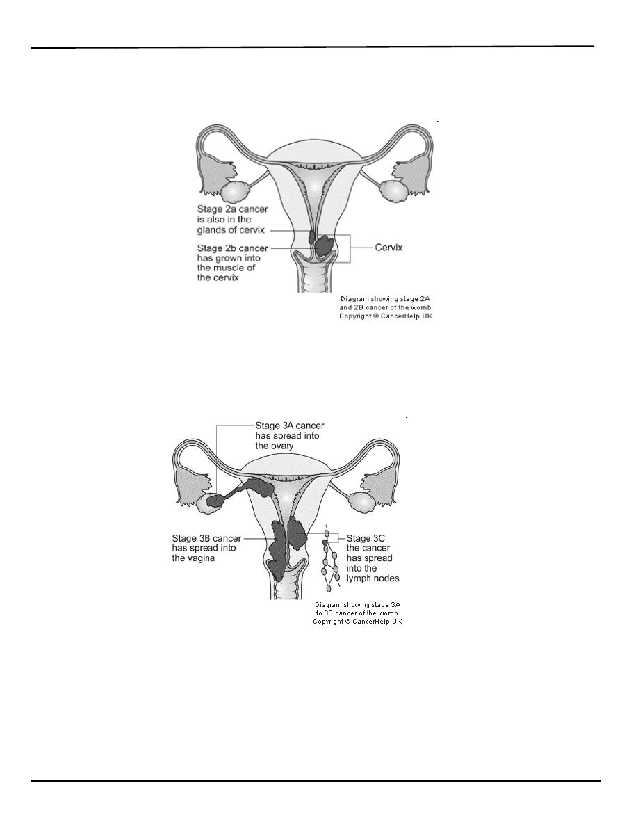
I: Confined to uterine corpus
IA: limited to endometrium
IB: invades less than ½ of myometrium
IC: invades more than ½ of myometrium
Lecture 6
النسائية
د. أحمد جاسم
Malignant disease of the body of the uterus
Page 6 of 8
II: invades cervix but not beyond uterus
IIA: endocervical gland involvement only
IIB: cervical stroma involvement
III: local and/or regional spread
IIIA: invades serosa/adnexa, or positive cytology
IIIB: vaginal metastasis
IIIC: metastasis to pelvic or para-aortic lymph nodes
IVA: invades bladder/bowel mucosa
IVB: distant metastasis

Lecture 6
النسائية
د. أحمد جاسم
Malignant disease of the body of the uterus
Page 7 of 8
1. Stage I: 81-91%
72% diagnosed at this stage
2. Stage II: 71-78%
3. Stage III: 52-60%
4. Stage IV: 14-17%
3% diagnosed at this stage
1. Direct extension (most common)
2. Transtubal
3. Lymphatic (Pelvic usually first, then para-aortic)
4. Hematogenous
a. Lung most common
b. Liver, brain, bone
1. Stage IB or less: total hyst/BSO/PPALND, cytology
2. Stage IC to IIB: total hyst/BSO/PPALND, cytology, adjuvant pelvic
XRT
3. Stage III: total hyst/BSO/PPALND, cytology, adjuvant chemotherapy
4. Stage IV: palliative XRT and chemotherapy

Lecture 6
النسائية
د. أحمد جاسم
Malignant disease of the body of the uterus
Page 8 of 8
The treatment of endometrial carcinoma is usually:
1. surgical.
2. Radiotherapy
3. Hormone therapy: Progestogens inhibit the rate of growth and
spread of endometrial carcinoma.
Other Types of Uterine Cancer
1. Leiomyosarcoma
Rapidly growing fibroid should be evaluated
2. Stromal sarcoma
3. Carcinosarcoma (MMMT) *
(Malignant Mixed Mullerian Tumor)
Uterine Sarcomas
o Account for fewer than 10% of all corpus cancers.
o Types:
( Carcinosarcoma, leiomyosarcoma, Endometrial stromal
sarcoma, adenosarcoma)
o Exposure to radiation may enhance the development of pelvic
sarcomas
o Abnormal vaginal bleeding most frequent presenting symptom for all
histologic types.
o No specific staging system (commonly use staging of endometrial
carcinoma)
Surgery is the hallmark of treatment with total abdominal
hysterectomy and bilateral salpingo-oopherectomy (TAH/BSO)
being the standard procedure.
By: Mu’taz Fathi