
Musculoskeletal
system
بسم هللا الرحمن الرحيم
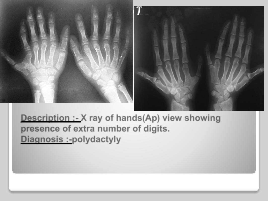
Description :- X ray of hands(Ap) view showing
presence of extra number of digits.
Diagnosis :-polydactyly
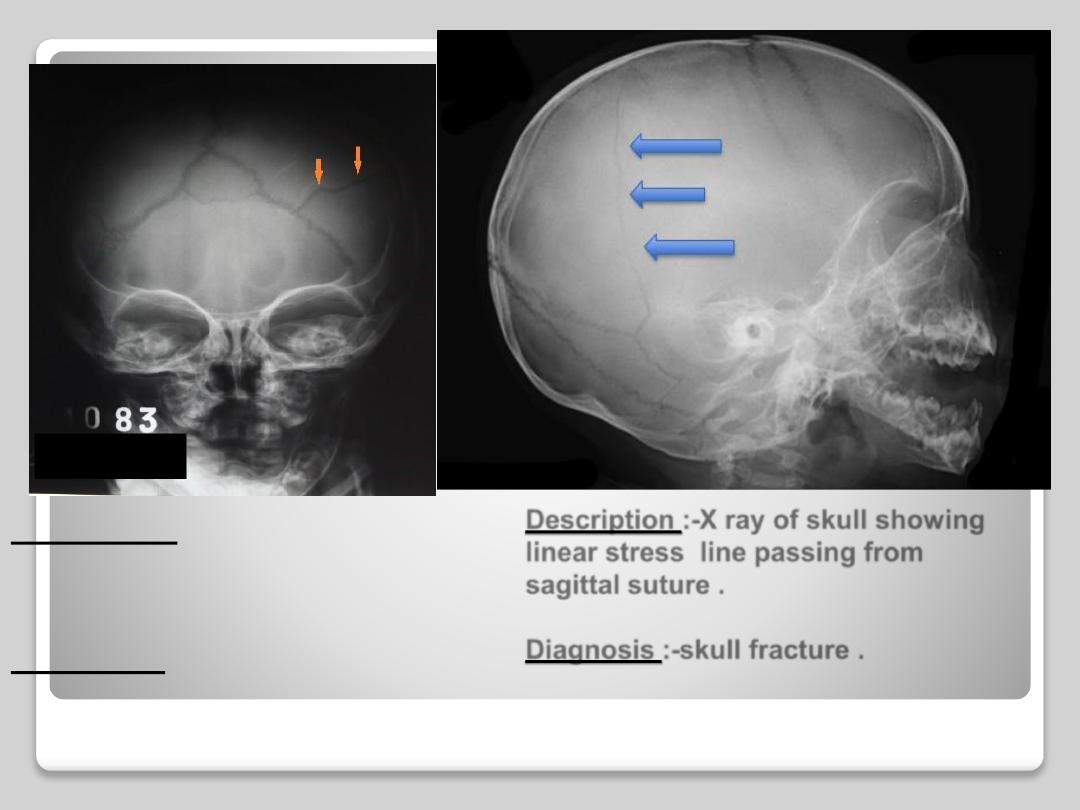
Description :-X ray of skull showing
linear stress line passing from
sagittal suture .
Diagnosis :-skull fracture .
Description:-X ray of skull
showing linear stress line
passing from coronal suture.
Diagnosis :-skull fructure
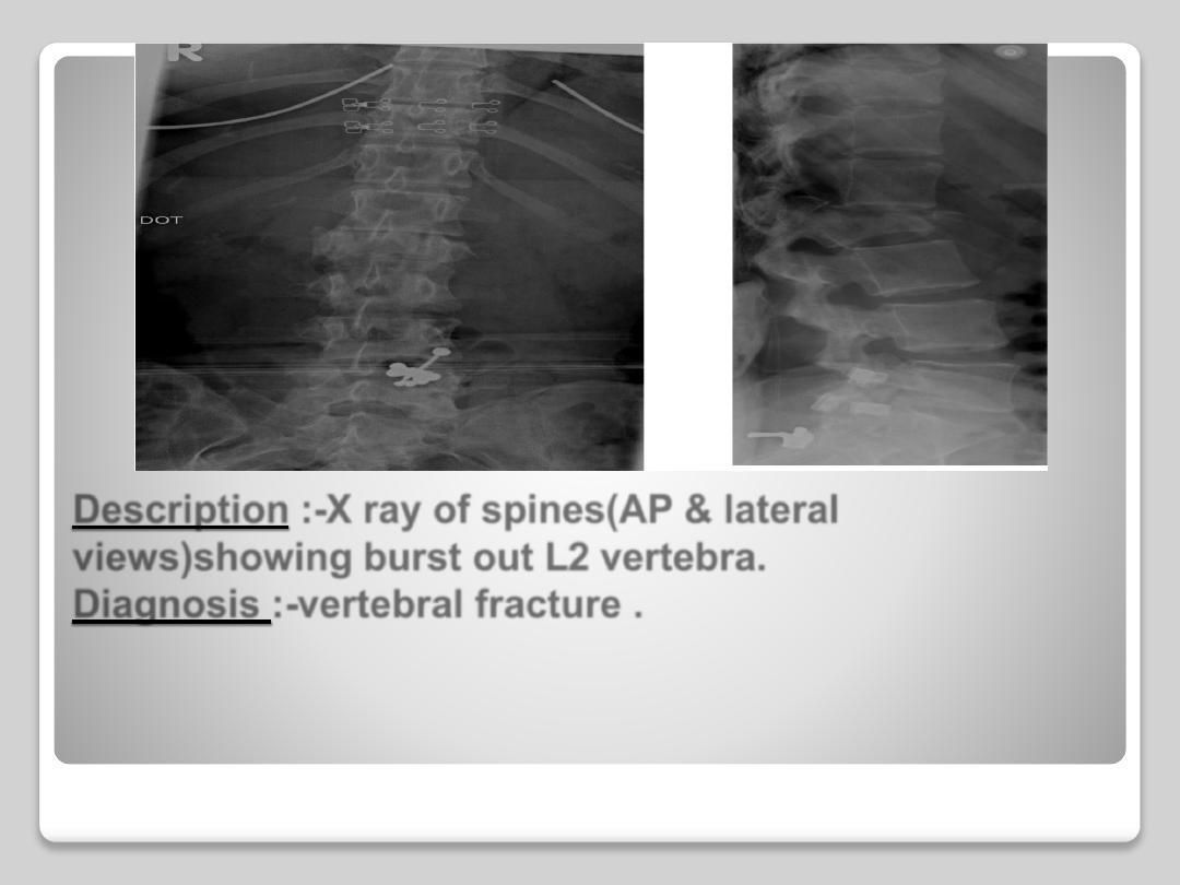
Description :-X ray of spines(AP & lateral
views)showing burst out L2 vertebra.
Diagnosis :-vertebral fracture .
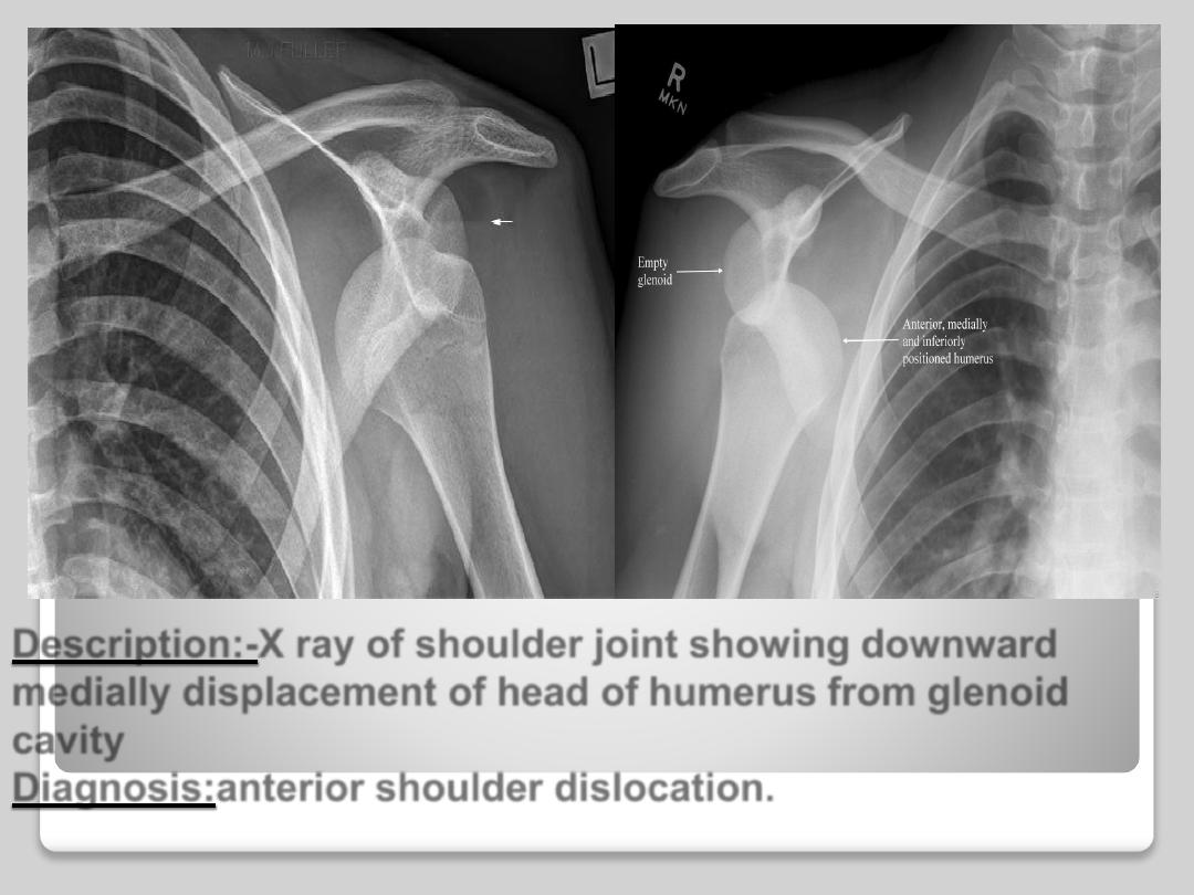
Description:-X ray of shoulder joint showing downward
medially displacement of head of humerus from glenoid
cavity
Diagnosis:anterior shoulder dislocation.
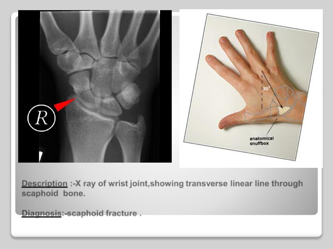
Description :-X ray of wrist joint,showing transverse linear line through
scaphoid bone.
Diagnosis:-scaphoid fracture .
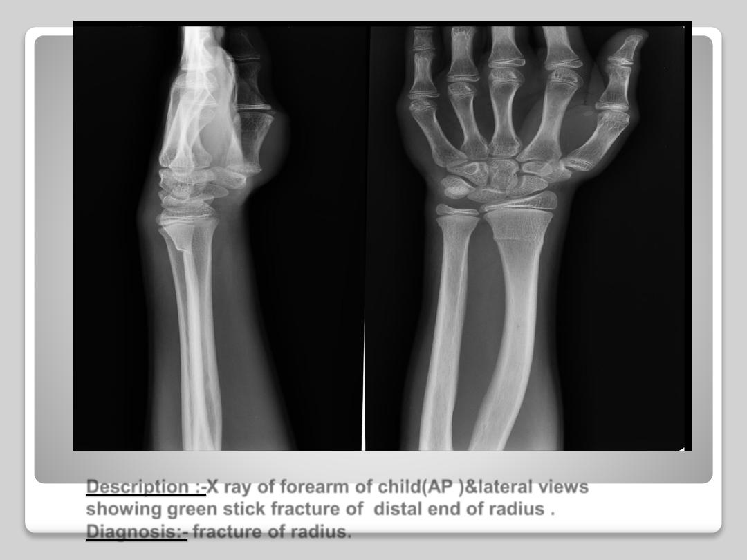
Description :-X ray of forearm of child(AP )&lateral views
showing green stick fracture of distal end of radius .
Diagnosis:- fracture of radius.
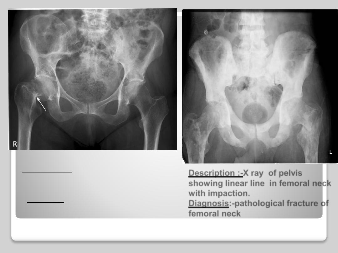
Description :-X ray of pelvis
showing linear line in femoral neck
with impaction.
Diagnosis:-pathological fracture of
femoral neck
Description :-X ray of pelvis showing
linear line in femoral neck with
impaction.
Diagnosis:- fracture of femoral neck
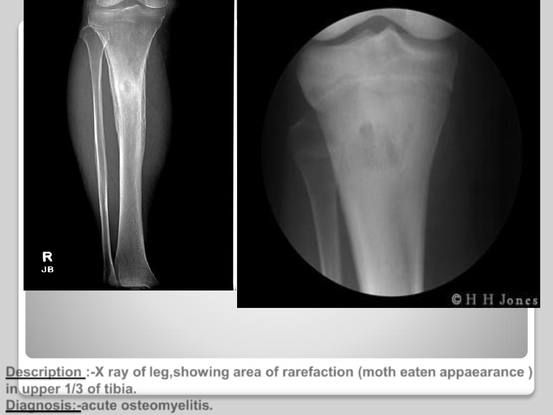
Description :-X ray of leg,showing area of rarefaction (moth eaten appaearance )
in upper 1/3 of tibia.
Diagnosis:-acute osteomyelitis.
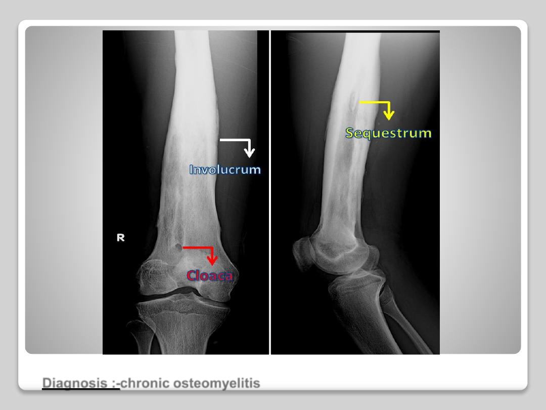
Diagnosis :-chronic osteomyelitis
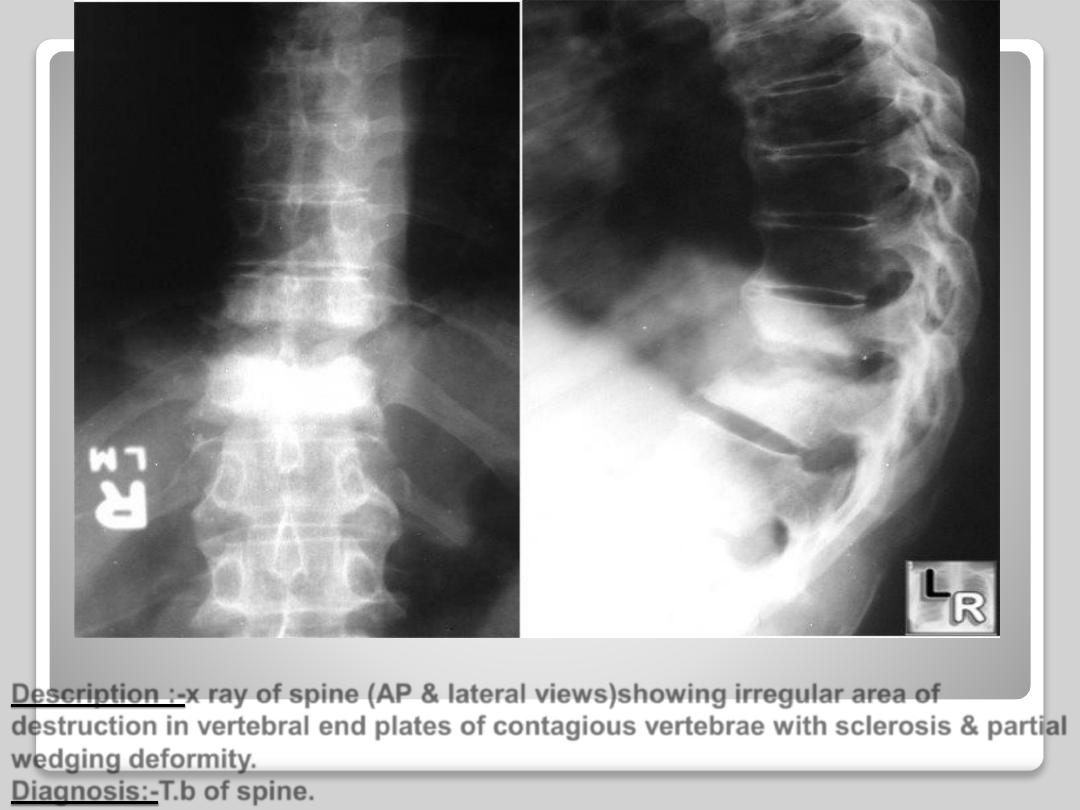
Description :-x ray of spine (AP & lateral views)showing irregular area of
destruction in vertebral end plates of contagious vertebrae with sclerosis & partial
wedging deformity.
Diagnosis:-T.b of spine.
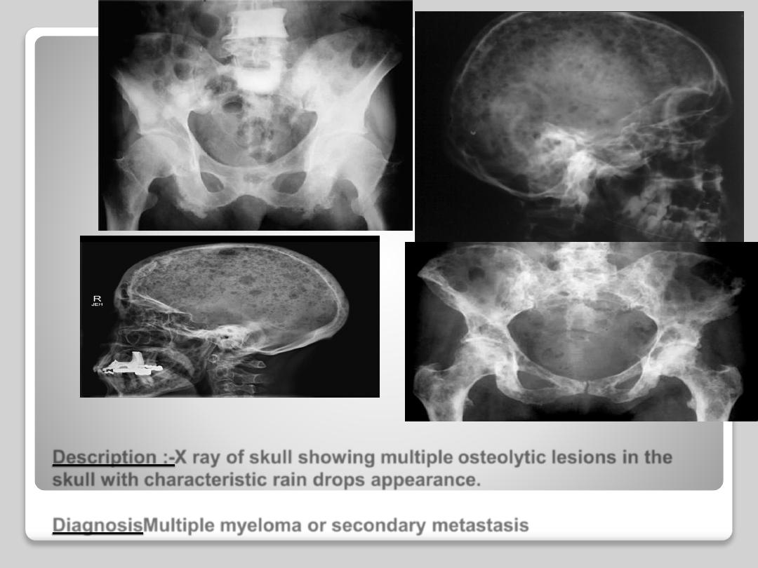
Description :-X ray of skull showing multiple osteolytic lesions in the
skull with characteristic rain drops appearance.
DiagnosisMultiple myeloma or secondary metastasis
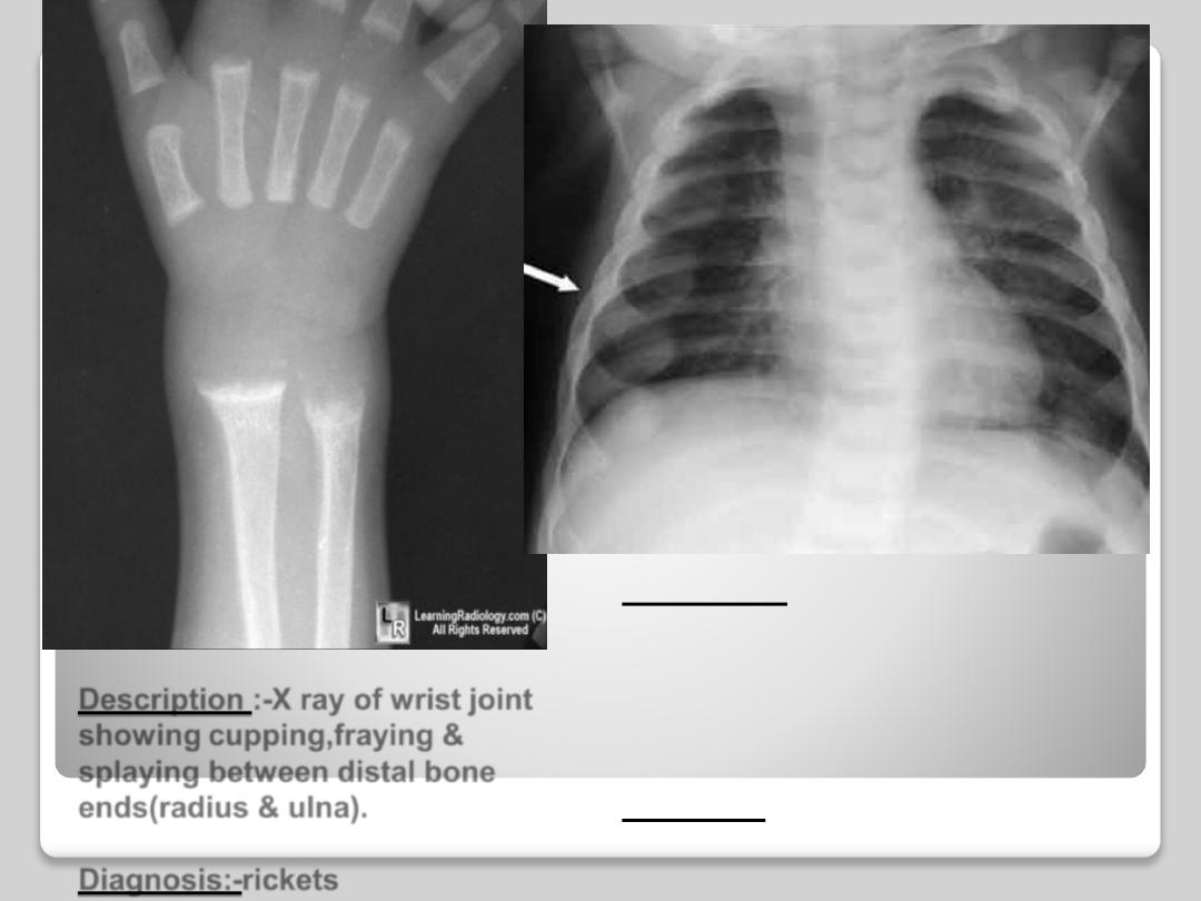
Description :-X ray of wrist joint
showing cupping,fraying &
splaying between distal bone
ends(radius & ulna).
Diagnosis:-rickets
Description :-X ray of chest
showing widening of anterior end
of ribs in the point of their meeting
with costal cartilages with
characteristic cupping,fraying &
splaying between distal bone ends
Diagnosis :- rachitic rosary
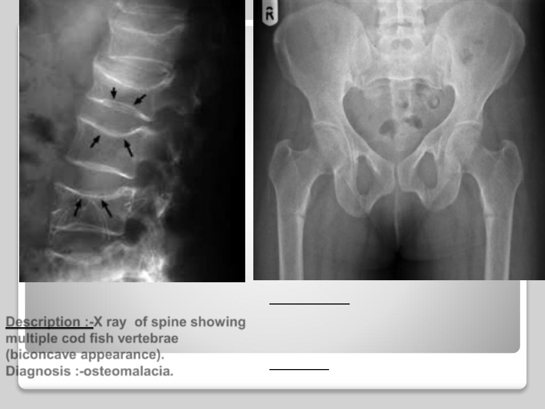
Description :-X ray of spine showing
multiple cod fish vertebrae
(biconcave appearance).
Diagnosis :-osteomalacia.
Description :-X ray of pelvis showing
triradiate pelvis deformity(inward
invagination of pelvic walls) with
invagination of both femoral heads.
Diagnosis :- osteomalacia.
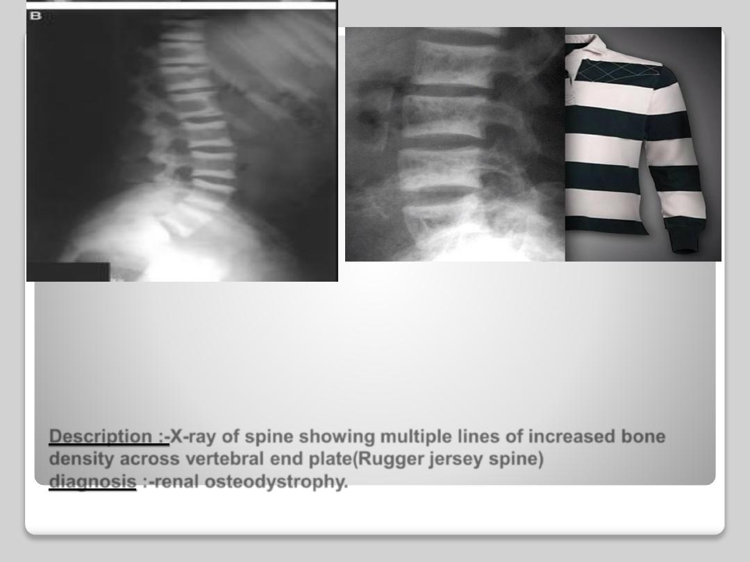
Description :-X-ray of spine showing multiple lines of increased bone
density across vertebral end plate(Rugger jersey spine)
diagnosis :-renal osteodystrophy.
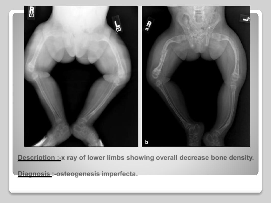
Description :-x ray of lower limbs showing overall decrease bone density.
Diagnosis :-osteogenesis imperfecta.
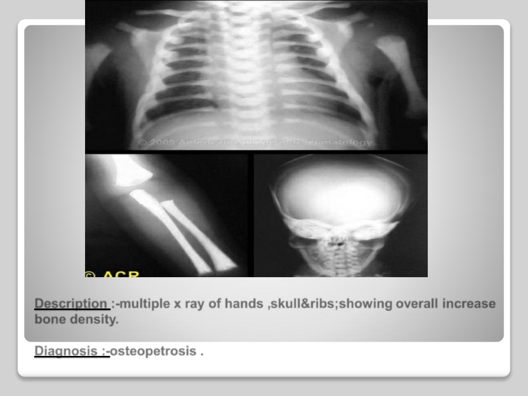
Description :-multiple x ray of hands ,skull&ribs;showing overall increase
bone density.
Diagnosis :-osteopetrosis .
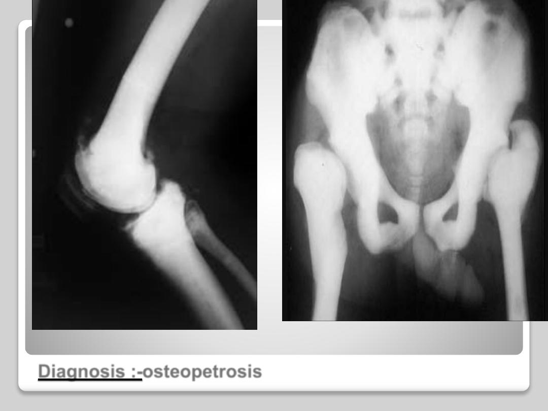
Diagnosis :-osteopetrosis
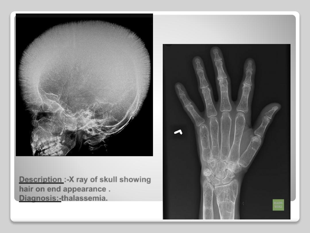
Description :-X ray of skull showing
hair on end appearance .
Diagnosis:-thalassemia.
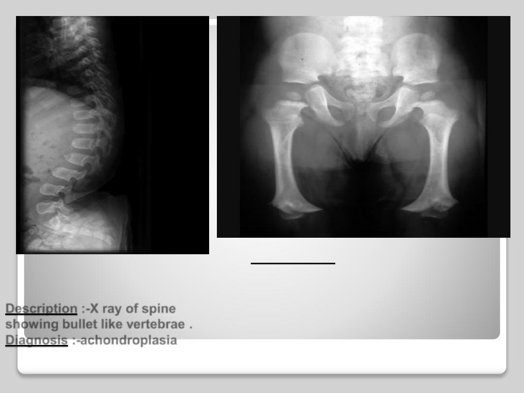
Description :-X ray of spine
showing bullet like vertebrae .
Diagnosis :-achondroplasia
Description :-X ray of pelvis showing
shortening of femur with flaring & peaking
of distal metaphysis.
Diagnosis:-achondroplasia
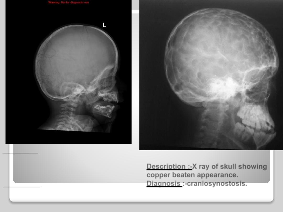
Description :-X ray of skull showing
copper beaten appearance.
Diagnosis :-craniosynostosis.
Description :-x ray of skull showing
multiple intrasutural bones in the
lambdoid suture .
Diagnosis :-wormian bone
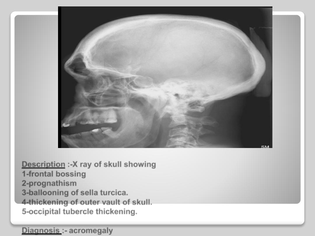
Description :-X ray of skull showing
1-frontal bossing
2-prognathism
3-ballooning of sella turcica.
4-thickening of outer vault of skull.
5-occipital tubercle thickening.
Diagnosis :- acromegaly
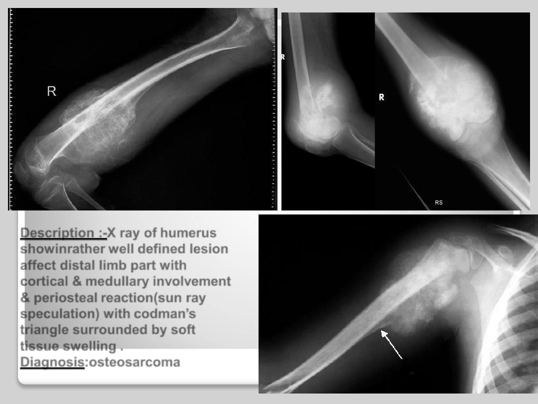
Description :-X ray of humerus
showinrather well defined lesion
affect distal limb part with
cortical & medullary involvement
& periosteal reaction(sun ray
speculation) with codman
’s
triangle surrounded by soft
tissue swelling .
Diagnosis:osteosarcoma
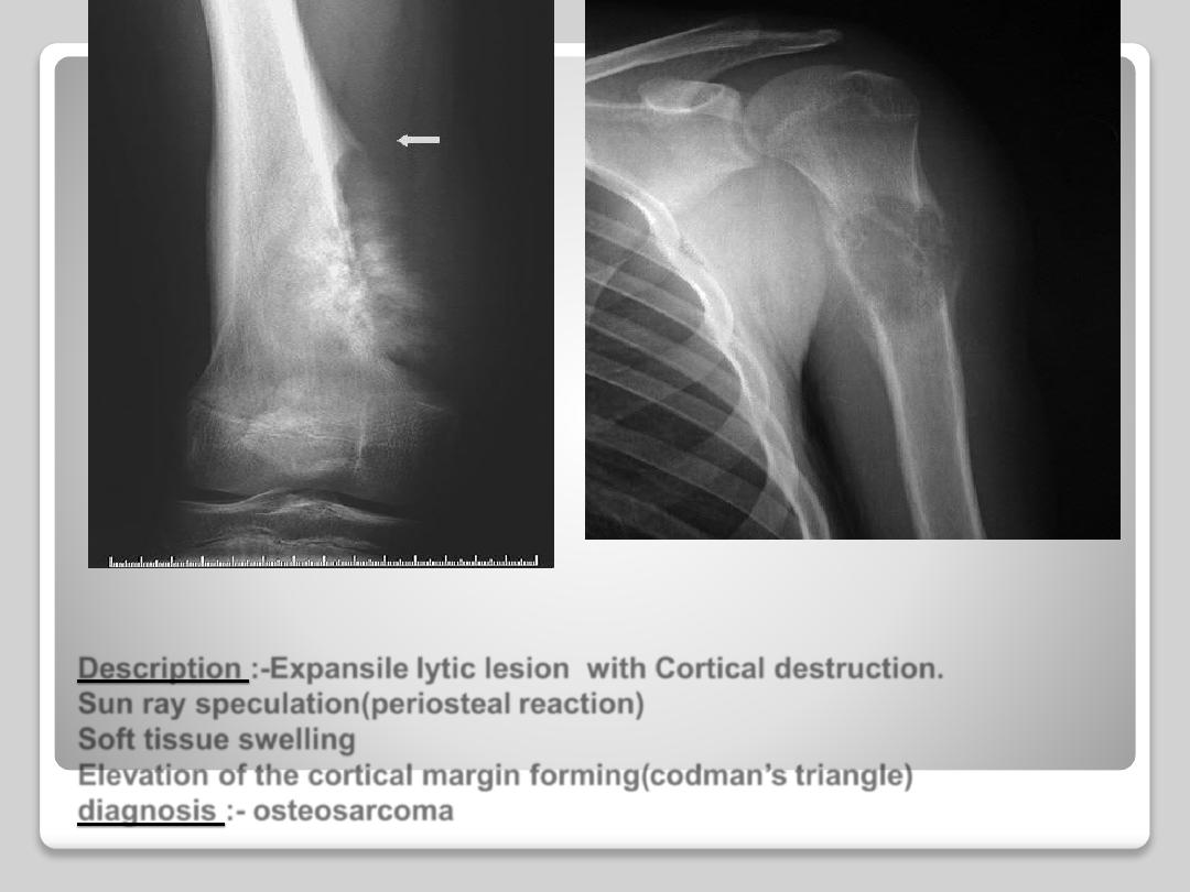
Description :-Expansile lytic lesion with Cortical destruction.
Sun ray speculation(periosteal reaction)
Soft tissue swelling
Elevation of the cortical margin forming(codman
’s triangle)
diagnosis :- osteosarcoma
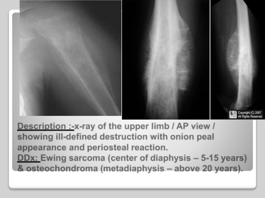
Description :-x-ray of the upper limb / AP view /
showing ill-defined destruction with onion peal
appearance and periosteal reaction.
DDx: Ewing sarcoma (center of diaphysis
– 5-15 years)
& osteochondroma (metadiaphysis
– above 20 years).
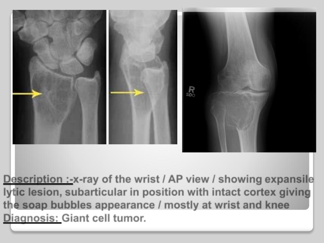
Description :-x-ray of the wrist / AP view / showing expansile
lytic lesion, subarticular in position with intact cortex giving
the soap bubbles appearance / mostly at wrist and knee
Diagnosis: Giant cell tumor.
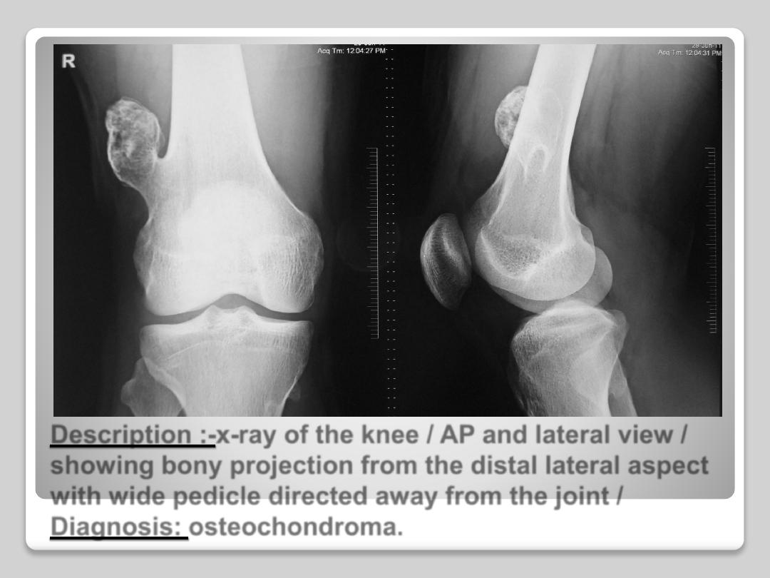
Description :-x-ray of the knee / AP and lateral view /
showing bony projection from the distal lateral aspect
with wide pedicle directed away from the joint /
Diagnosis: osteochondroma.
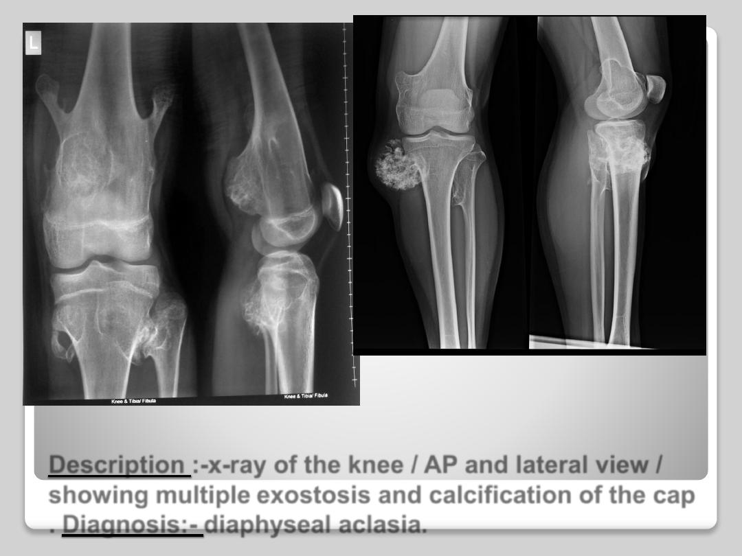
Description :-x-ray of the knee / AP and lateral view /
showing multiple exostosis and calcification of the cap
. Diagnosis:- diaphyseal aclasia.
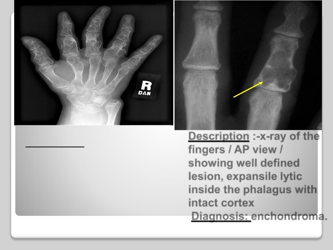
Description :-x-ray of the
fingers / AP view /
showing well defined
lesion, expansile lytic
inside the phalagus with
intact cortex
Diagnosis: enchondroma.
Description :-x-ray of
hand / AP view /
showing multiple
Enchondromas
Diagnosis :-ollier
’s
disease
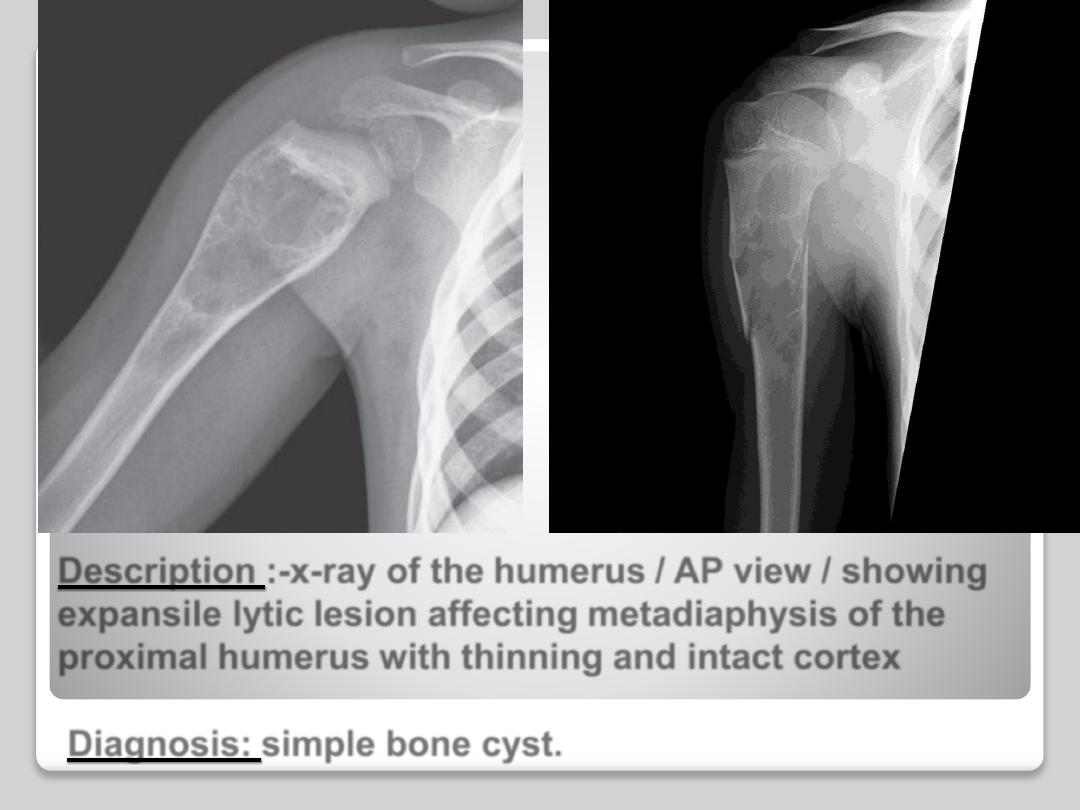
Description :-x-ray of the humerus / AP view / showing
expansile lytic lesion affecting metadiaphysis of the
proximal humerus with thinning and intact cortex
Diagnosis: simple bone cyst.
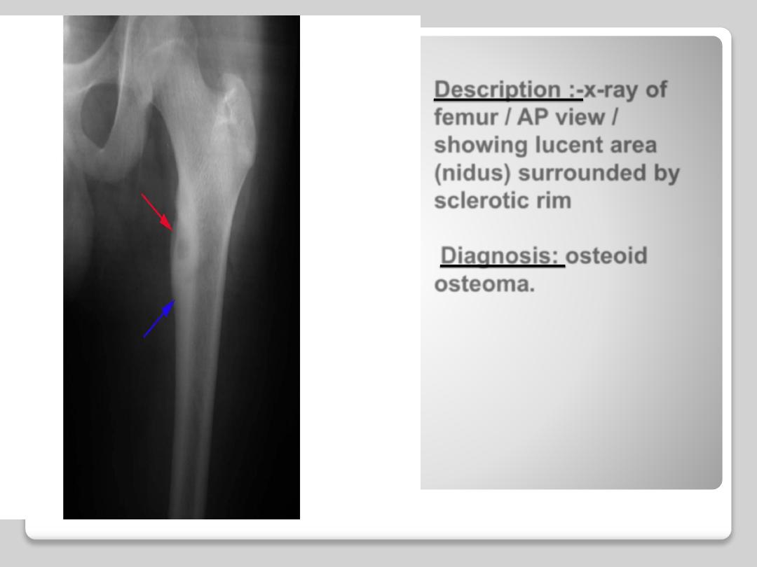
Description :-x-ray of
femur / AP view /
showing lucent area
(nidus) surrounded by
sclerotic rim
Diagnosis: osteoid
osteoma.
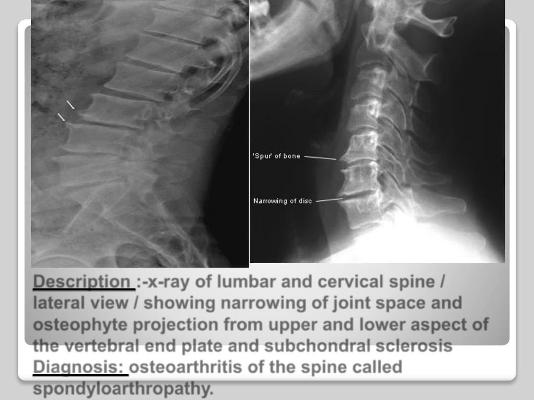
Description :-x-ray of lumbar and cervical spine /
lateral view / showing narrowing of joint space and
osteophyte projection from upper and lower aspect of
the vertebral end plate and subchondral sclerosis
Diagnosis: osteoarthritis of the spine called
spondyloarthropathy.
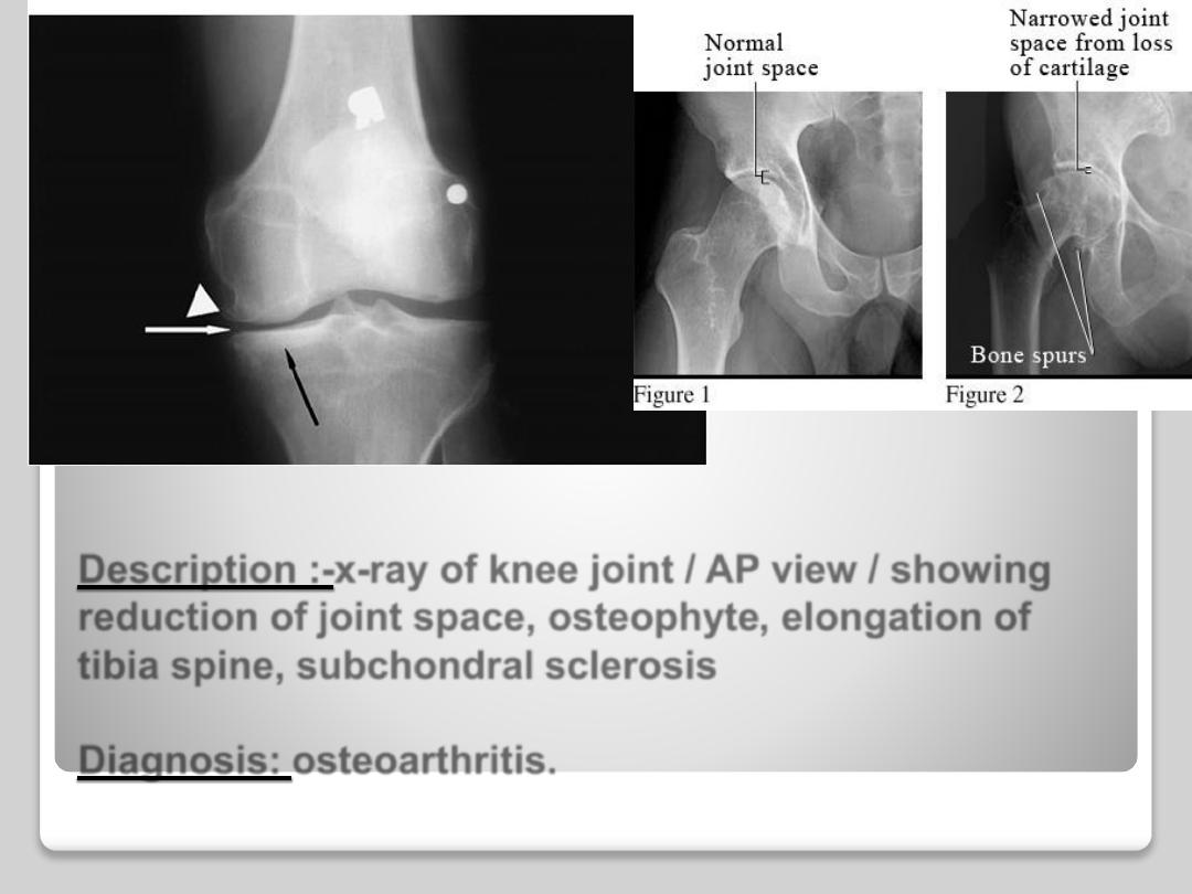
Description :-x-ray of knee joint / AP view / showing
reduction of joint space, osteophyte, elongation of
tibia spine, subchondral sclerosis
Diagnosis: osteoarthritis.
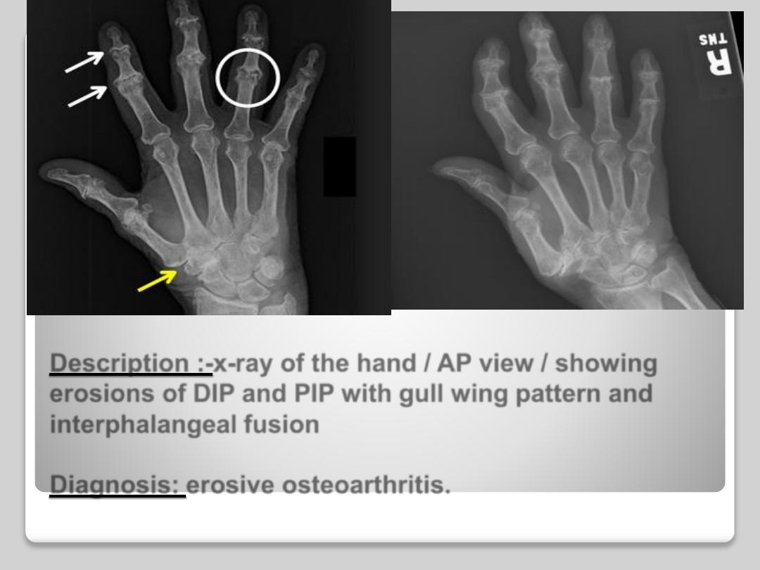
Description :-x-ray of the hand / AP view / showing
erosions of DIP and PIP with gull wing pattern and
interphalangeal fusion
Diagnosis: erosive osteoarthritis.
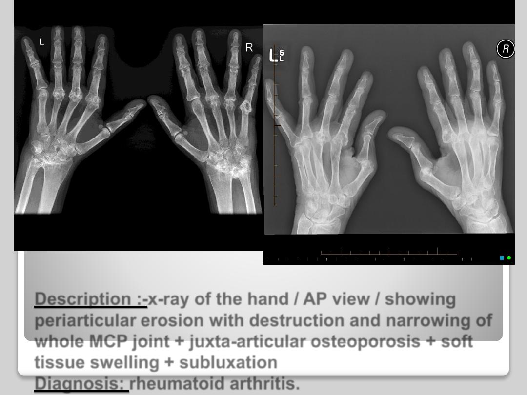
Description :-x-ray of the hand / AP view / showing
periarticular erosion with destruction and narrowing of
whole MCP joint + juxta-articular osteoporosis + soft
tissue swelling + subluxation
Diagnosis: rheumatoid arthritis.
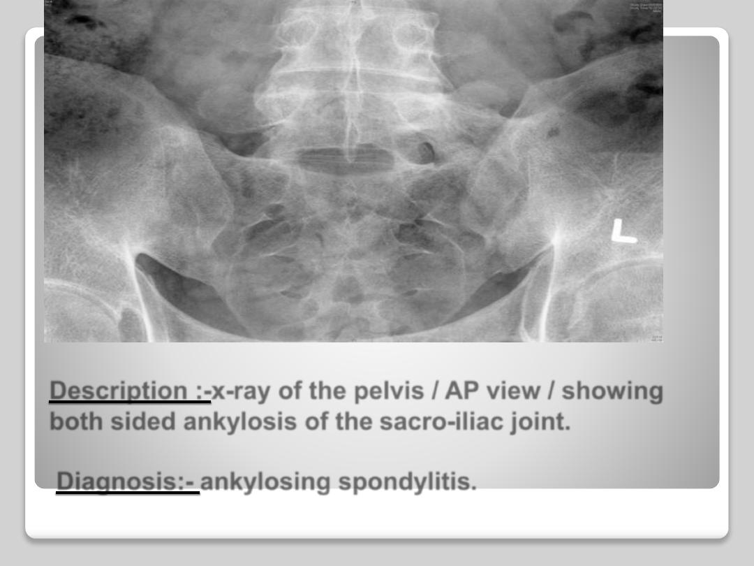
Description :-x-ray of the pelvis / AP view / showing
both sided ankylosis of the sacro-iliac joint.
Diagnosis:- ankylosing spondylitis.
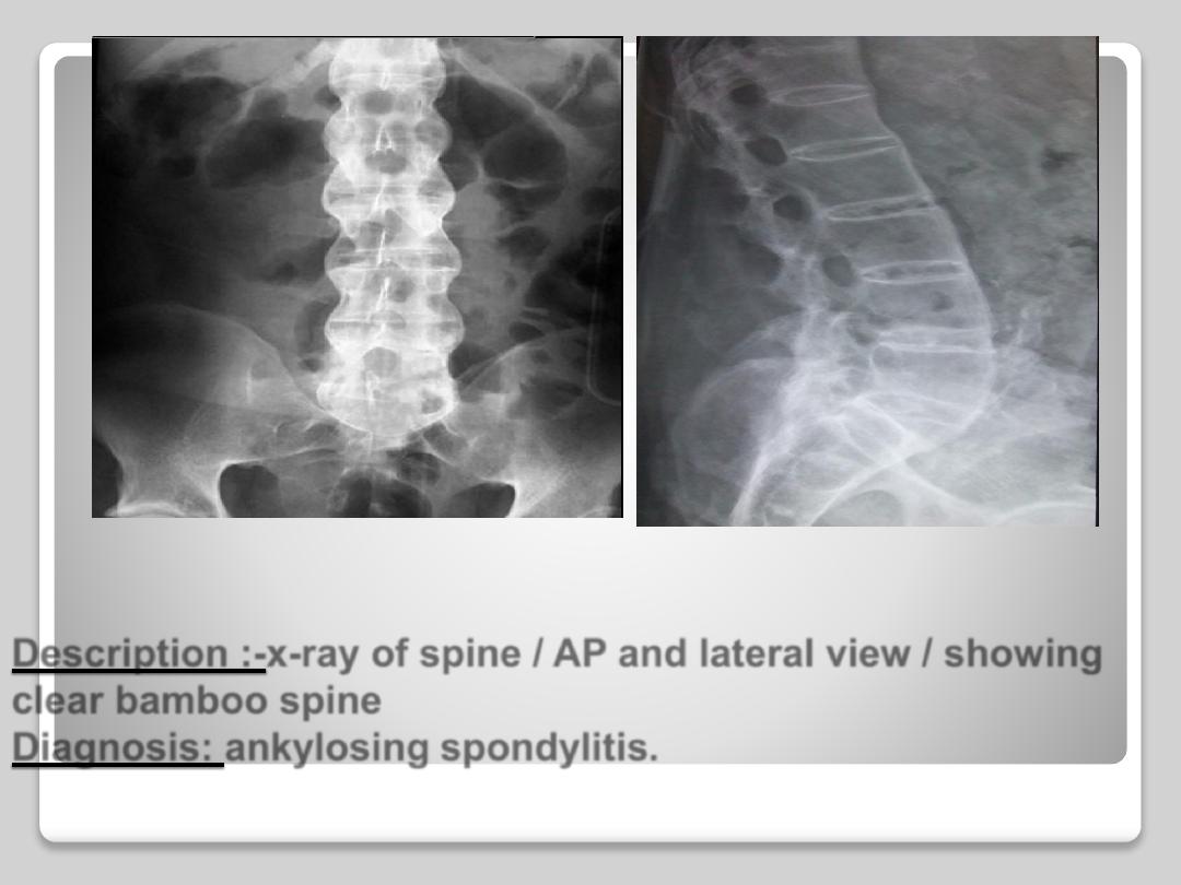
Description :-x-ray of spine / AP and lateral view / showing
clear bamboo spine
Diagnosis: ankylosing spondylitis.
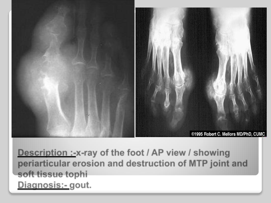
Description :-x-ray of the foot / AP view / showing
periarticular erosion and destruction of MTP joint and
soft tissue tophi
Diagnosis:- gout.

Nothing
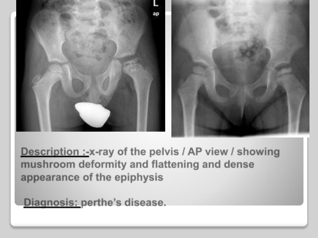
Description :-x-ray of the pelvis / AP view / showing
mushroom deformity and flattening and dense
appearance of the epiphysis
Diagnosis: perthe
’s disease.
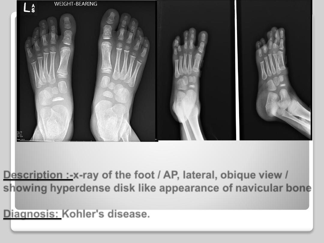
Description :-x-ray of the foot / AP, lateral, obique view /
showing hyperdense disk like appearance of navicular bone
Diagnosis: Kohler's disease.
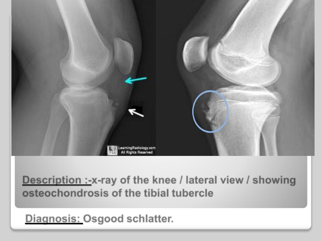
Description :-x-ray of the knee / lateral view / showing
osteochondrosis of the tibial tubercle
Diagnosis: Osgood schlatter.
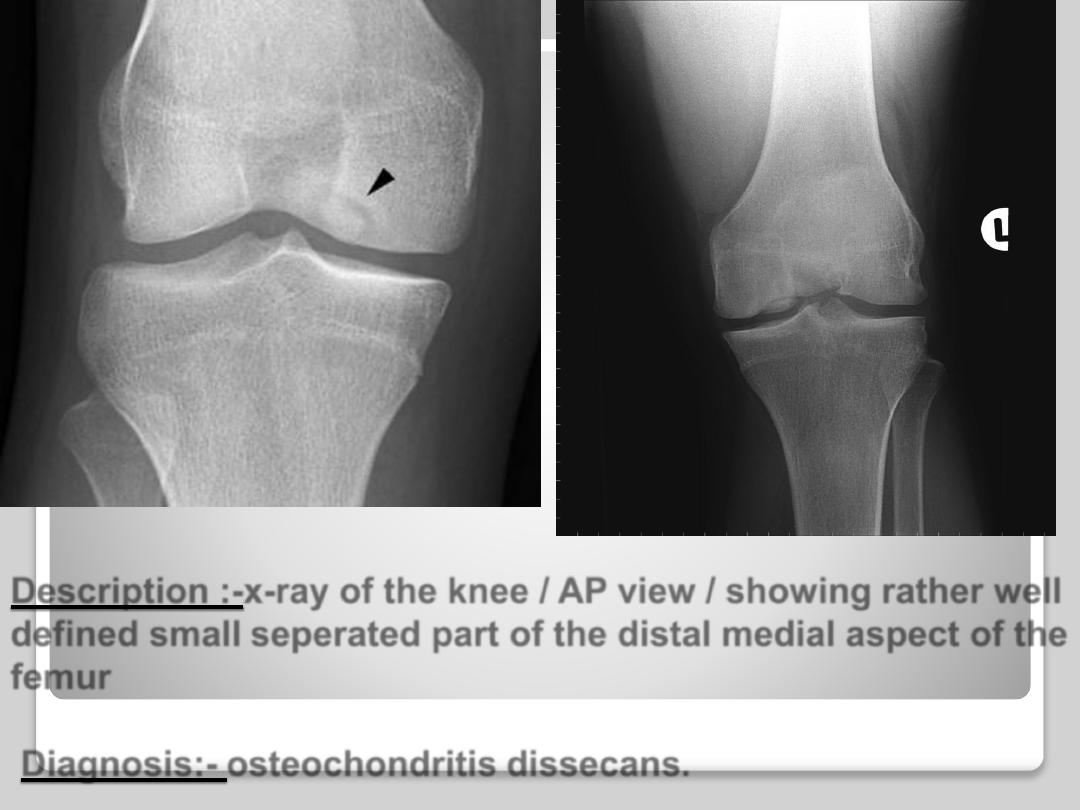
Description :-x-ray of the knee / AP view / showing rather well
defined small seperated part of the distal medial aspect of the
femur
Diagnosis:- osteochondritis dissecans.
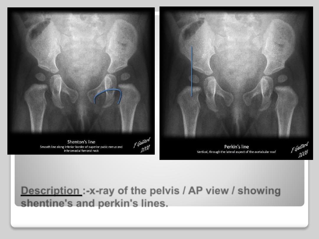
Description :-x-ray of the pelvis / AP view / showing
shentine's and perkin's lines.
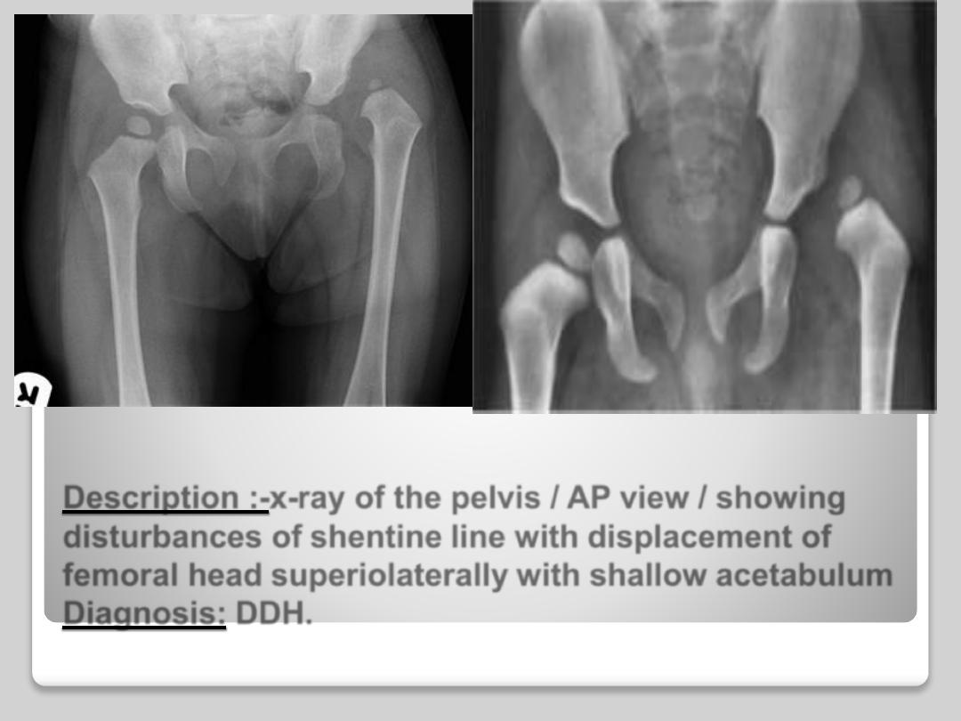
Description :-x-ray of the pelvis / AP view / showing
disturbances of shentine line with displacement of
femoral head superiolaterally with shallow acetabulum
Diagnosis: DDH.
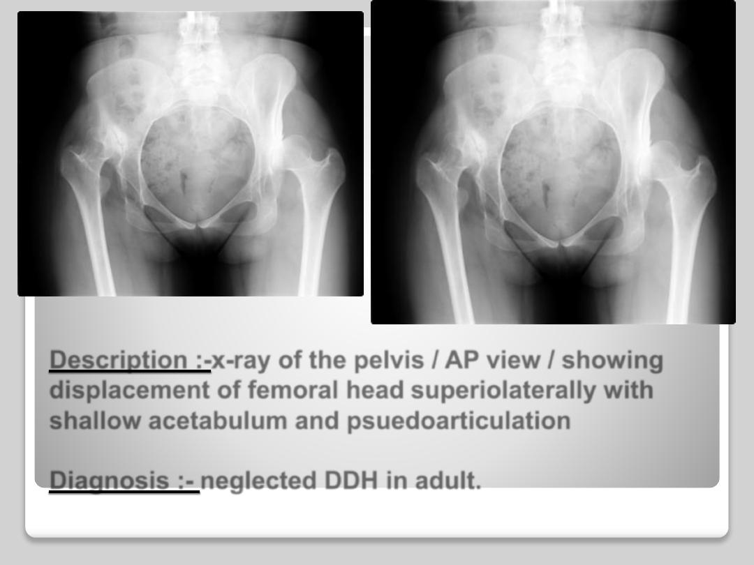
Description :-x-ray of the pelvis / AP view / showing
displacement of femoral head superiolaterally with
shallow acetabulum and psuedoarticulation
Diagnosis :- neglected DDH in adult.