1
Fifth stage
ENT
Lec-10
د.سعد
7/12/2015
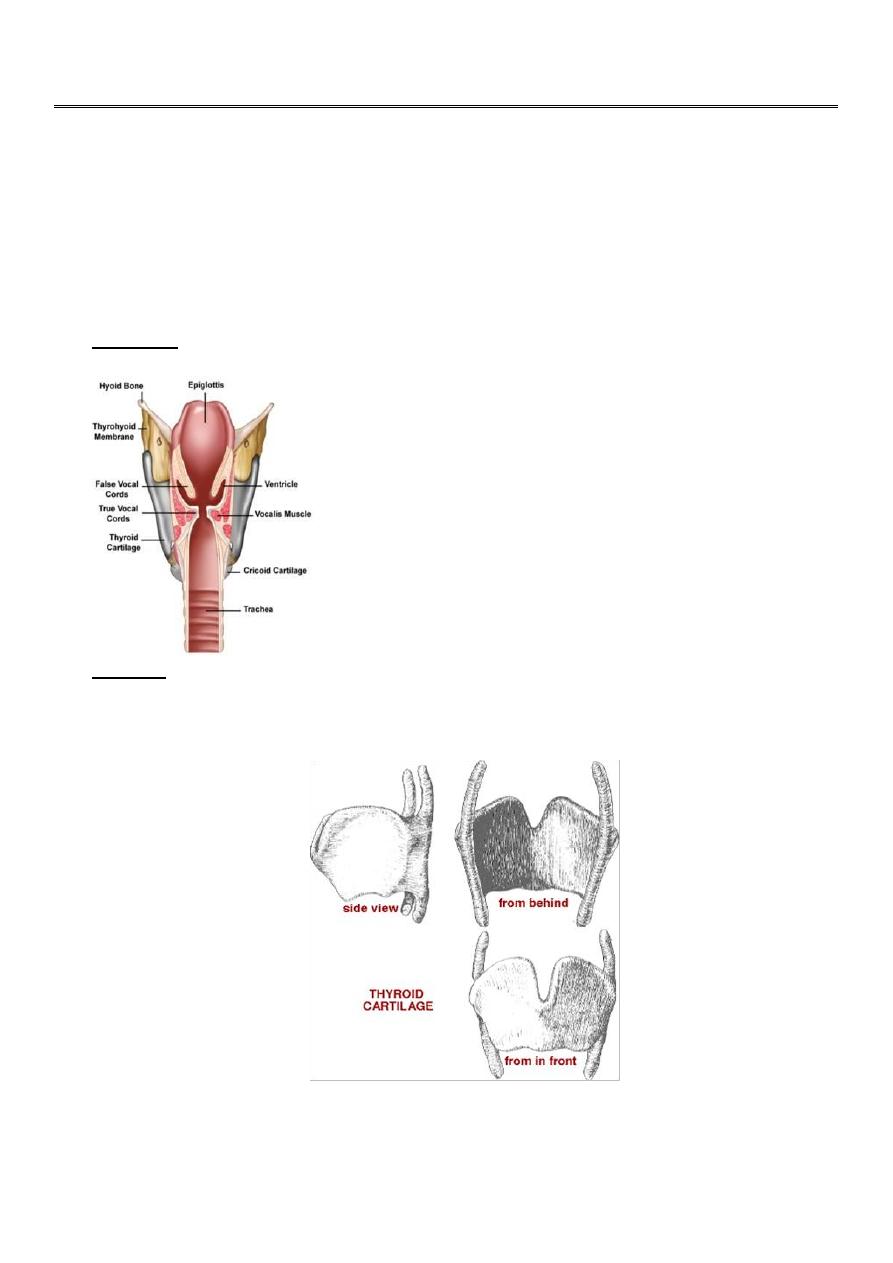
THE LARYNX
The larynx lies in front of the hypopharynx opposite the third to sixth cervical vertebrae. In adult,
the larynx ends at the lower border of C6 vertebra.
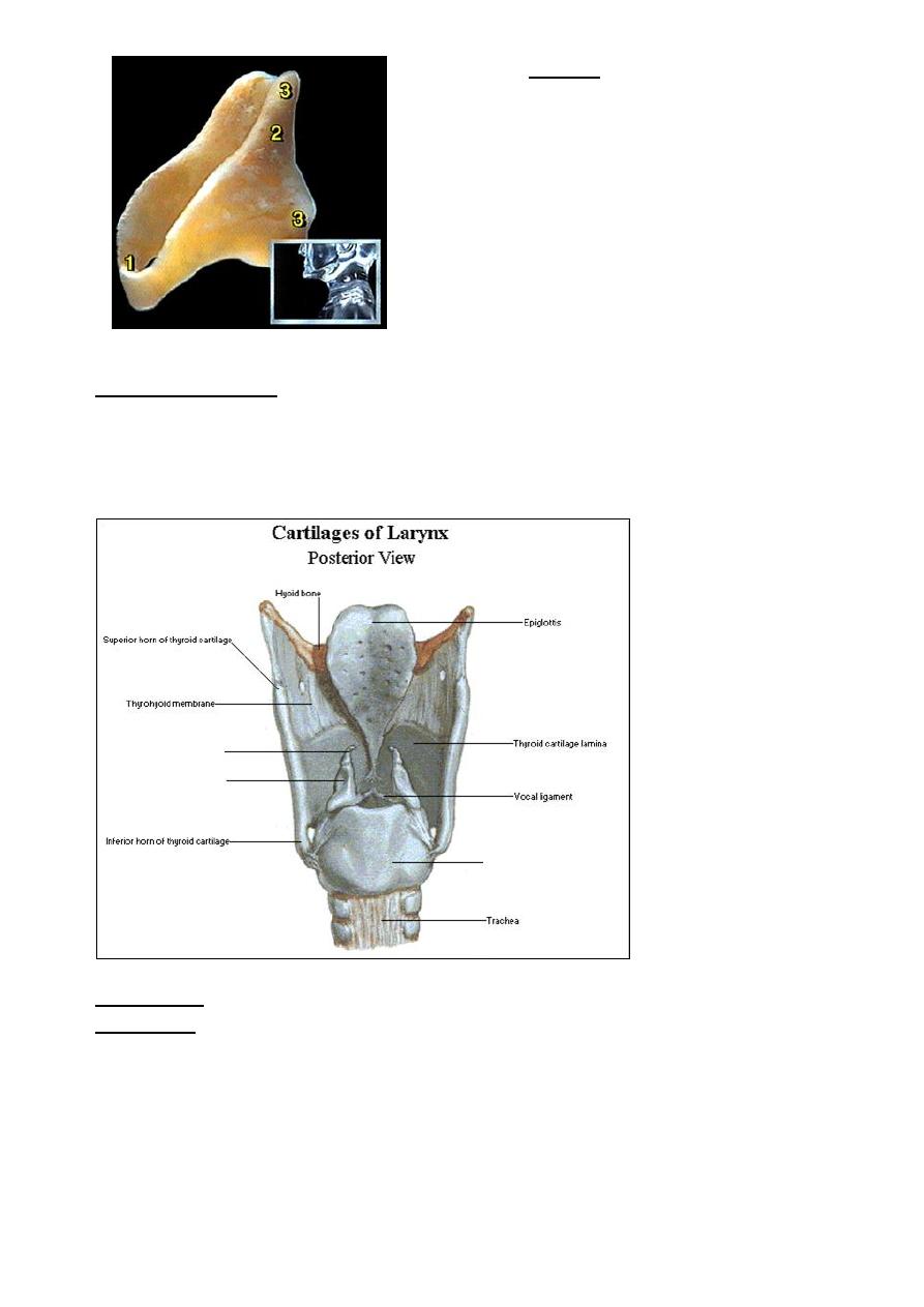
Laryngeal cartilages
1. Epiglottis: Leaf-like yellow elastic cartilage that never ossify.
2. Thyroid: Is the largest of all and consist of two alae meeting anteriorly at an angle, called
"Adam's Apple". It's a hyaline cartilage, ossifies at 20-30 years of age, and begins in the
inferior margin and progress cranially.
2
3. Cricoid: It is the only cartilage forming a
complete ring. Its posterior part is
expanded to form a lamina while
anteriorly it is narrow forming an arch.
It
is also a hyaline cartilage and it ossifies
after the thyroid cartilage, first part to be
calcified being the superior portion
(which can be mistaken for a foreign
body) Calcification progresses caudally.
cricoid cartilage
4. Arytenoid cartilages: they are paired. Each is pyramidal in shape. It has:
Base---articulate with cricoid cartilage.
Muscular process---laterally and give attachment to intrinsic muscle.
Vocal process--- directed anteriorly, giving attachment to vocal cord.
Apex---support the corniculate cartilage.
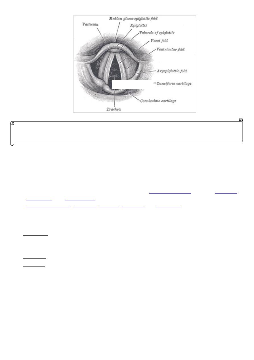
5. Corniculate: small cartilages that lie over the arytenoids.
6. Cuneiform: Small cartilages that lie in the aryepiglottic fold.
Aytenoid
Corniculate
Cricoid cartilage
3
Laryngeal Joints
Cricoarytenoid joint and cricothyroid joint. Both are synovial joints.
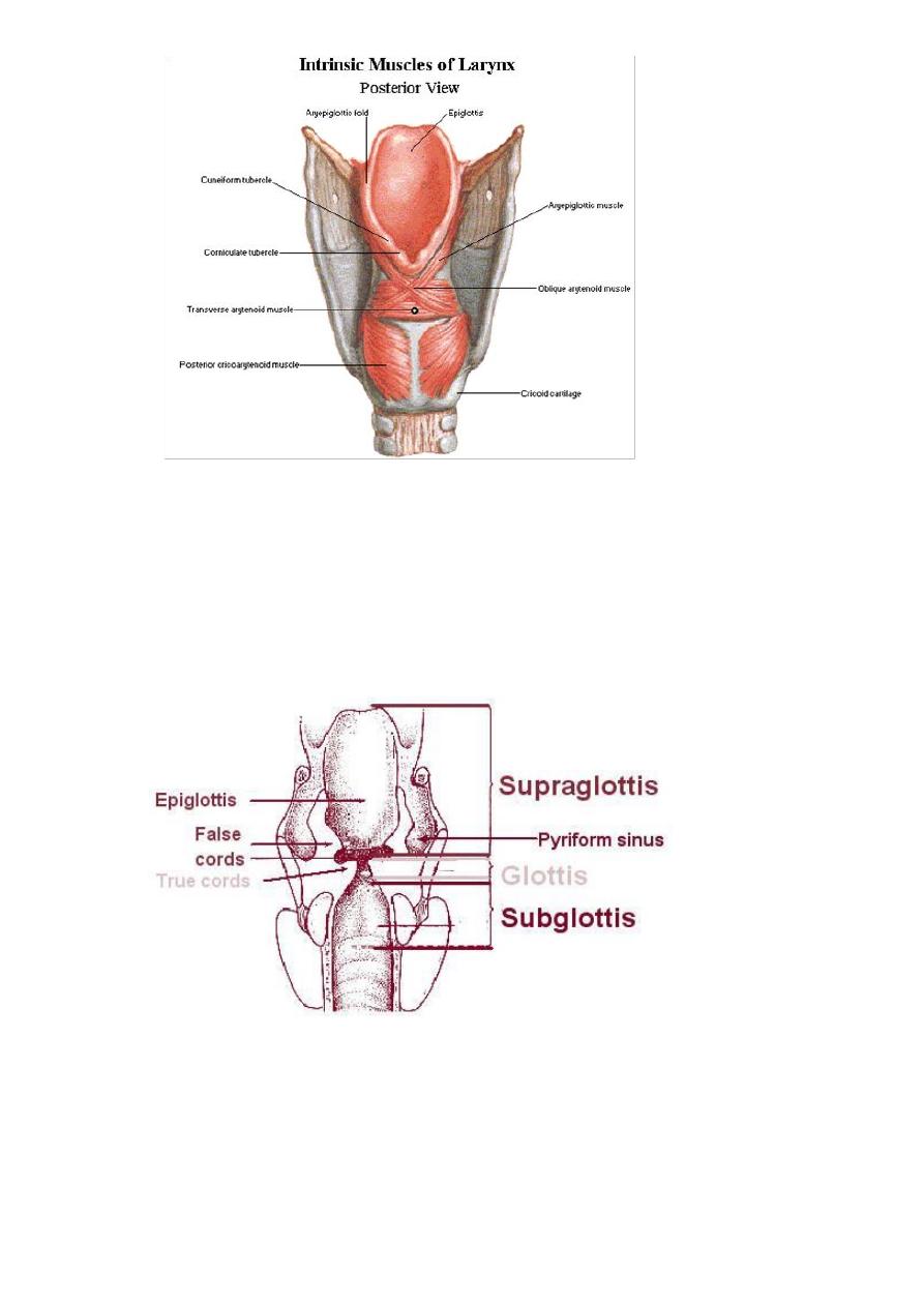
Laryngeal Muscles:
1. Extrinsic muscle:
The extrinsic muscles move the larynx as a whole. The
) are depressors of the hyoid bone and the larynx, whereas the
and
) and the stylopharyngeus
are elevator of the hyoid bone and larynx.
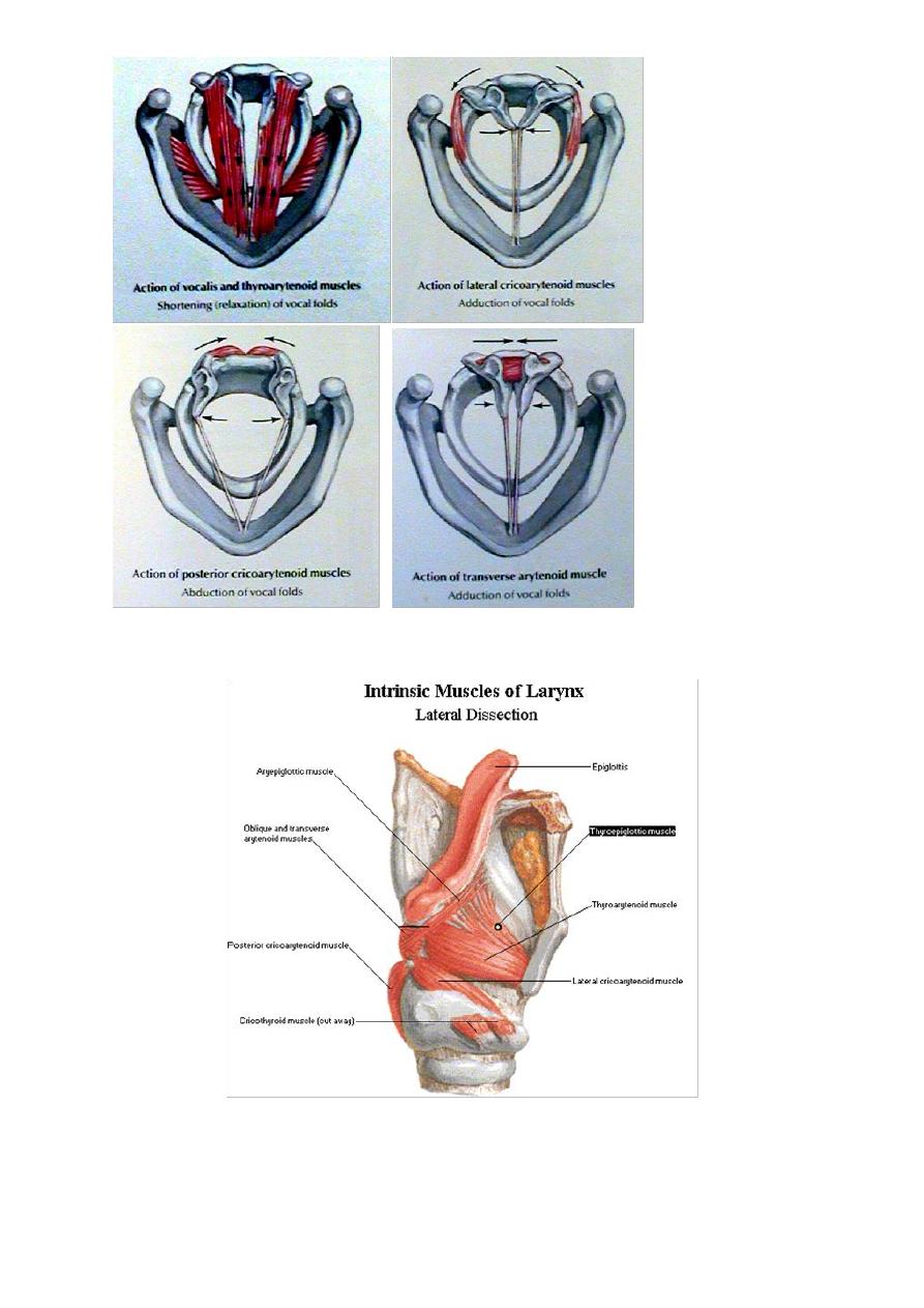
2. Intrinsic muscles: All are paired except interarytenoid muscle. They open and close the glottis;
and they are of three groups:
A. Abductors: consist of posterior cricoarytenoid muscle. It opens the glottis and it is the most
important muscle of the body. Paralysis of this muscle leads to adduction of the vocal cords and
suffocation.
B.
Adductors: Lateral cricoarytenoid, interarytenoid, thyroarytenoid (external part)
C. Tensors: cricothyroid and vocalis(internal part of
thyroarytenoid
)
Cuneiform
NOTE
:
(The first three laryngeal cartilages are single while the rest are paired )
4
5
Laryngeal compartments
1. The glottis: composed of the vocal cords from the extreme lateral edge of the cord to the
inferior border of the medial surface.
2. The subglottis: from the lower border of the glottis to the inferior border of the cricoid. Below
this is the trachea.
3. The supraglottis: extend from the upper border of the glottis inferiorly to the hyoid bone
superiorly.
Histology
Lining epithelium: squamous over the vocal cord and the upper 1/4 of the posterior surface of
the epiglottis and low columnar ciliated over the rest of the larynx. The latter type commonly
undergoes squamous metaplasia in response to atmospheric pollution and smoking.
Mucous glands and lymphatics: rich in supraglottis, nil in glottis and very few in subglottis.
6
The mucosa of the glottis and supraglottis is firmly bound down to the underlying tissue, but not
so in the subglottic region. Here, the laxity of tissue allows a dangerous degree of oedema,
especially in children, where the diameter of the area is relatively smaller than in adult.
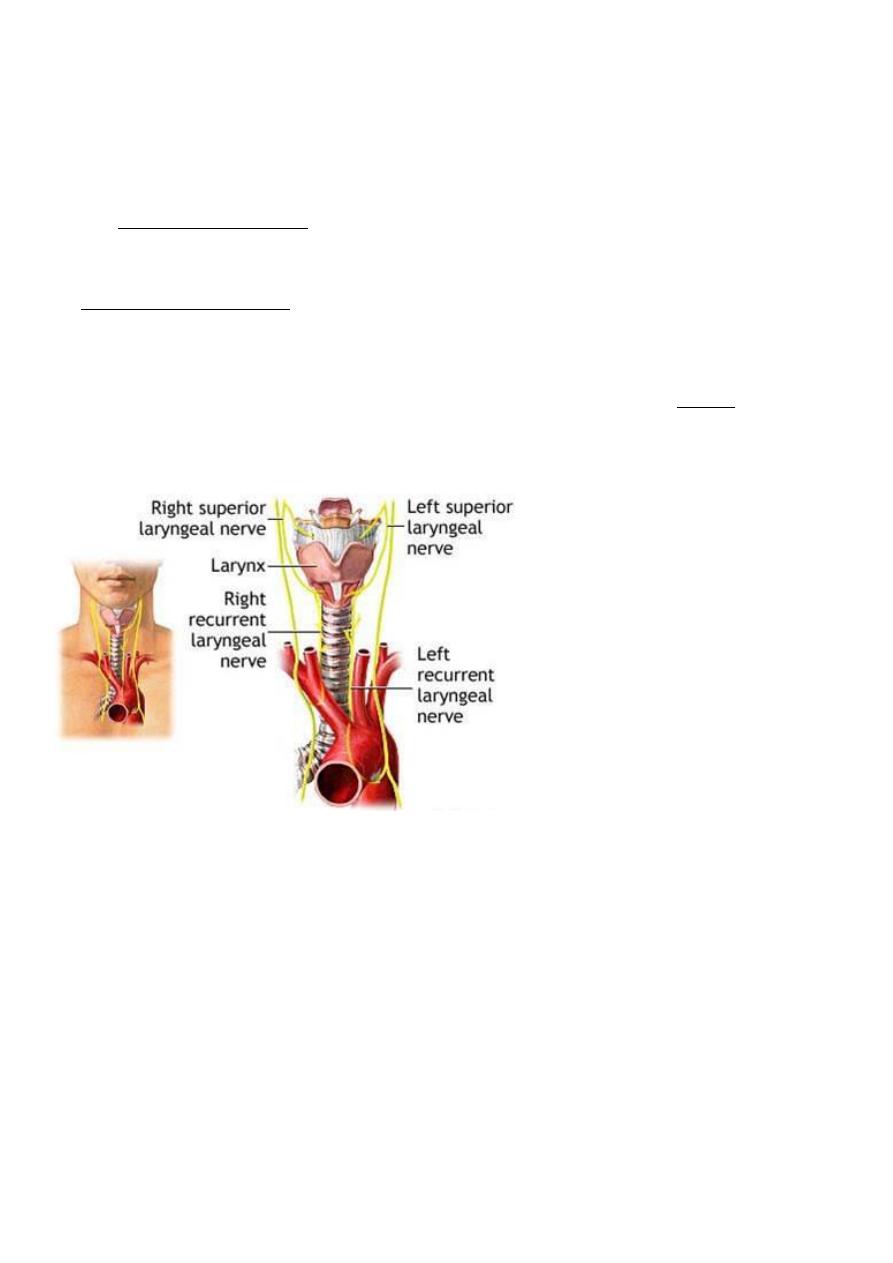
N
erve Supply:
1.
T
he superior laryngeal nerve (from the vagus) divides into external and internal branches:
The external is motor to cricothyroid muscle only.
The internal pass through the thyrohyoid membrane and is sensory to supraglottic area.
2. Recurrent laryngeal nerve: supply all the intrinsic muscles (abductors and adductors), and also it
is sensory to the subglottic area.
All muscles of the larynx are innervated by the recurrent laryngeal nerve, except cricothyroid
muscle which is innervated by the external branch of the superior laryngeal nerve.
Blood supply and venous drainage:
The supraglottis is supplied and drained by the superior laryngeal artery and vein, which enter the
larynx through thyrohyoid membrane. The glottis and subglottis are supplied and drained by the
inferior laryngeal artery and vein( branches of the inferior thyroid artery).
Lymphatic Drainage:
The main lymphatic drainage is to the deep cervical lymph nodes. The glottic area has NO
lymphatic network; that's why cancer of vocal cords does not give rise to lymph node enlargement.
7
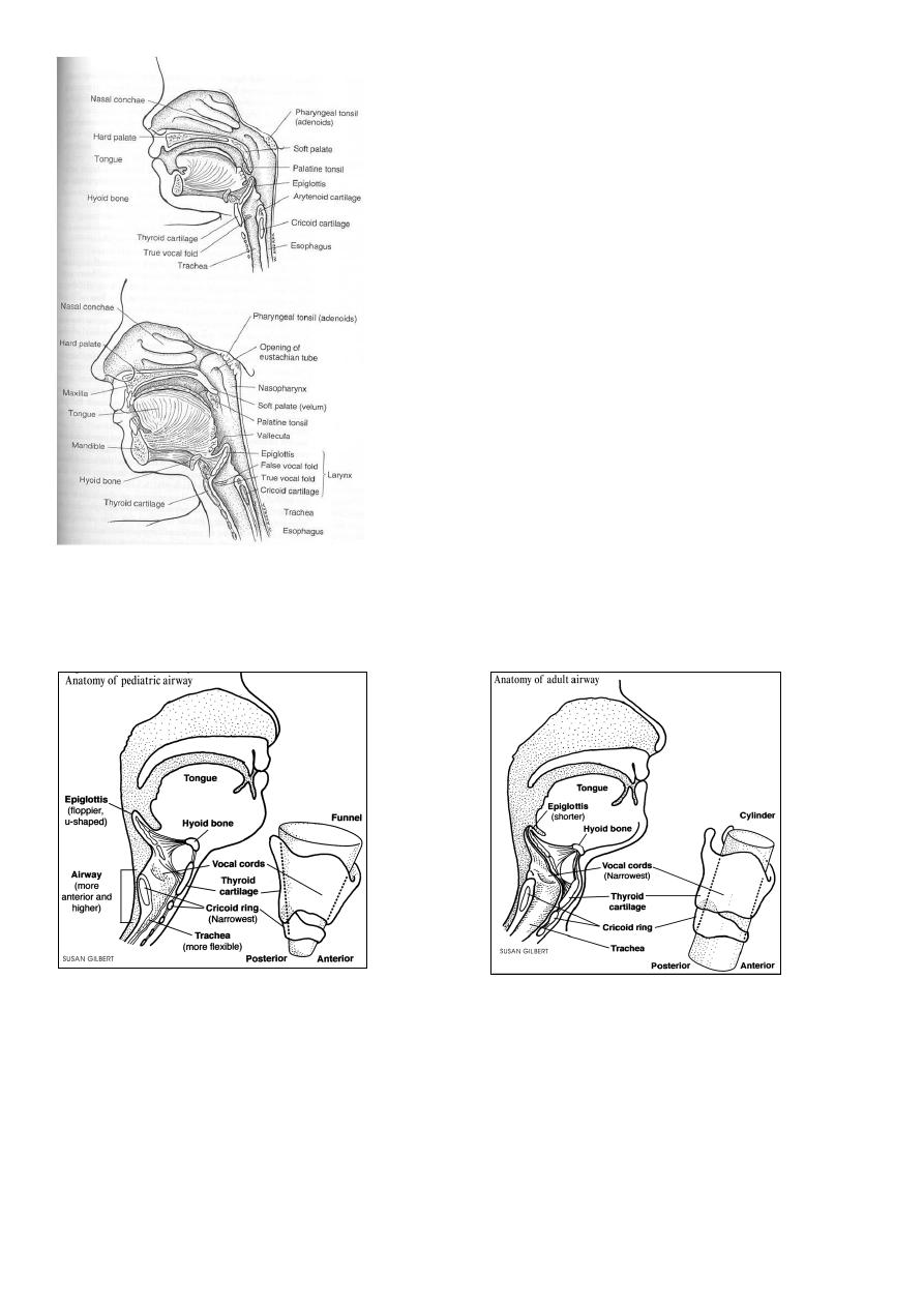
Paediatric larynx
The larynx of an infant differs considerably
from that of an adult and has great clinical
significans:
1. It is positioned high in the neck opposite C3
or C4 (level of vocal cord) at rest and
reaches C1 or c2 during swallowing.
2. The laryngeal cartilage are soft and collapse
easily.
3. The thyroid cartilage in an infant is flat and
the cricothyroid and thyrohyoid spaces are
narrow.
4. It is small and conical in shape (while it is
cylindrical in adult).
5. Submucosal tissues of infant's larynx are
loose and easily undergo oedematous
changes with trauma or inflammation
leading to obstruction.
Adult larynx
Infant larynx

8
Adult Infant
Physiology of the larynx
1. Protection: This is a vital function; it prevents entry of any substance into the air passages
(e.g. food, F.B…) , by sphincteric closure of laryngeal inlet and cough reflex.
2. Respiration: larynx regulates flow of air into the lungs. Vocal cords abduct during
expiration and adduct during expiration.
3. Phonation (sound production): air column from the chest causes vibration of the adducted
vocal cords thereby producing a sound.
4. Fixation of chest: closure of the glottis fixes the chest to give support to the powerful
voluntary muscular use of the arms. Closure of the glottis fixes the diaphragm to assist in
the act of straining.