
1
Fifth stage
Surgery
Lec-2
.د
اسامة
13/12/2015
Congenital abdominal wall defects
Congenital abdominal wall defects:
1-Omphalocele
2-Umbilical cord hernia
2-Gastroschiasis
3-Prune belly Syndrome
Omphalocele:
Abdominal wall defect at umbilicus with covering (sac may rupture)
Frequently associated with other anomalies
Giant omphaloceles: It contain liver,small bowel, large bowel, and stomach .
Omphalocele Associated Anomalies
Chromosomal abnormalities (50%)
o Trisomies 13, 18, 21
Congenital heart disease (50%)
Neural tube defects (40%)
Beckwith-Wiedemann syndrome
o hyperinsulinism, visceromegaly, macroglossia, hepatorenal tumors, cloacal
extrophy
o Pentalogy of Cantre
o omphalocele, ectopia cordis, anterior diaphragmatic hernia, intracardiac defect,
sternal cleft
2
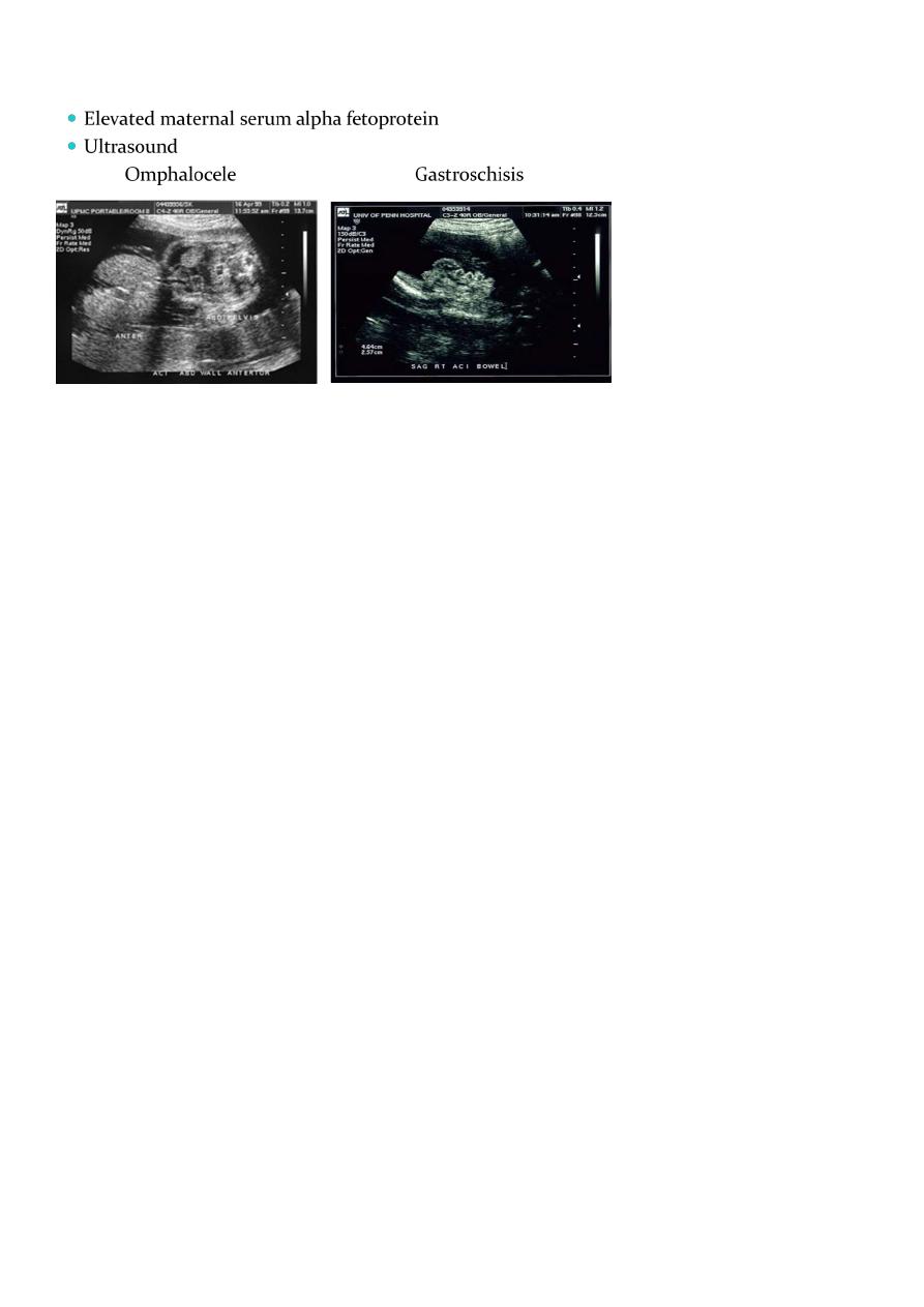
Prenatal Diagnosis
What is the mode of delivery in case of omphalocele?
If the Omphalocele is larger than 5 cm delivary should be through CS ,while if less than
5 cm could be with normal vaginal delivary
Initial Management
Early management aimed at maintaining circulation to bowel and preventing infection
while stabilizing infant (temperature/fluids) :
o Cover the defect with sterile dressing soaked in warm saline to prevent fluid loss
o Incubator
o Nasogastric decompression
o Urinary catheter for drainge
o IV fluids with glucose(150cc /kg/24hr)
o Invistigation(U/S of renal system,Echocardiograghy,Blood glucose level,Blood
group)
o Vitamine K
Surgical Treatment
The operation is not an emergency unlike gastroschiasis.
There are three approuch to manage omphalocele:-
1-Primary surgical Closure.
Under general anasthesia with endotreacheal tube,we excise the sac and return the viscera
to the abdominal cavity and close the defect.we have to avoid high increase in
3
intraabdominal pressure.and some times we have to close the skin only or to do release
inscion or to use special type of mesh.
Our aim is to do Primary Surgical Closure: Success dependent on size of the defect and
size of the abdomen.
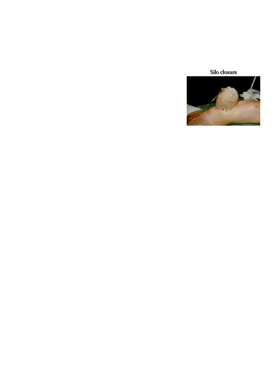
2-Staged Closure: When the Abdominal cavity is small,we
start with Gradual reduction of the contents into the
abdominal cavity using an extra-abdominal extension of the
peritoneal cavity (termed a silo) and using gentle pressure.
Usually requires 1-3 weeks, after which the defect is then
primarily closed.
3-Non-surgical treatment of omphalocele:
“paint” membrane with betadine to Thicken the memberane and later surgery.
After 2-3 years surgery done .
Gastroschisis
Abdominal wall defect to right of umbilicus with no covering over intestines
Gastroschisis Rarely associated with other anomalies
But, it may associated with gastrointestinal problems (25%)
o Including atresia, volvulus, stenosis
o Loss of bowel secondary to ischemia
Compromised bowel function
Rarely will have significant infarction of most of small bowel (i.e. lethal)
Initial Management
Early management aimed at maintaining circulation to bowel and preventing infection
while stabilizing infant (temperature/fluids) :
o Cover the defect with sterile dressing soaked in warm saline to prevent fluid loss
o Incubator
o Nasogastric decompression

4
o Urinary catheter for drainge
o IV fluids with glucose(200 cc /kg/24hr)
o Invistigation(U/S of renal system,Echocardiograghy and Blood group)
o Vitamine K
Gastroschisis
Surgery is an emergency operation due to exposure of intestine to the environment
1-Primary closure is attempted with the same principles of omphalocele closure.
2-May require silo with slow return of intestine into small abdominal cavity to Maintain
good perfusion and prevent ischemia.
No role for non-surgical management.
o Feeding difficulties are main post-op problem
o The patient is at risk for adhesions throughout life
Prune Belly Syndrome
Characterized by:
o deficiency of abdominal wall musculature
o a dilated, non-obstructed urinary tract
o bilateral cryptorchidism
o talipes equinovarus and hip dislocation
Incidence is 1/35-50,000
>95% occur in males