Radiology
For
5
th
stage
http://goo.gl/rjRf4F
I
LOKA
©
http://www.muhadharaty.com/radiology
I

Content
Topics:
Page:
Radiology of GIT system
6
Normal esophagus film
6
Tertiary contraction
7
Diffuse esophageal spasm
7
Congenital anomalies
7
Benign strictures of esophagus
7
Malignant structures of esophagus
8
Candida esophagitis
8
Esophageal web
8
Esophageal varieces
8
Achalasia cardia
9
Esophageal diverticulum
9
Hiatus hernia
9
Normal stomach film
10
Peptic ulcer
10
Gastric cancer
11
Hypertrophic pyloric stenosis
12
Gastric outlet obstruction
12
Normal duodenum film
12
Duodenal ulcer
13
Duodenal diverticulum
13
Atresia or stenosis
13
Pneumo-peritoneum
13
Sub-phrenic abscess
14
Congenital diaphragmatic hernia
14
Normal small intestine film
15
Crohn's disease
15
Ulcerative colitis
16
Toxic megacolon
16
Lymphoma of small bowel
16
Malabsorption syndrome
17
Bowel obstruction
17
Normal large intestine film
18
Colorectal carcinoma (CRC)
18

Irritable bowel syndrome (IBS)
18
Intussusception
19
Colonic diverticulosis
19
Familial adenomatous polyposis syndrome (FAPS)
19
Hirschprung disease
19
Anal atresia (imperforated anus)
20
Radiology of Renal system
21
Stone diseases
28
Urinary tract neoplasm
30
Urinary tract infections
31
Congenital anomalies
32
Other conditions
34
Radiology of musculoskeletal system
35
Solitary Bone Lesion
38
Malignant bone tumors
39
Benign bone tumors and tumor like conditions
40
Multiple focal bone lesions
44
Bone infections
45
Generalized decrease in the bone density
47
Generalized increase in the bone density
49
Arthritis
50
Other conditions
54
Radiology of chest
58
Cardiothoracic ratio (CTR)
62
Mitral valve disease
62
Congestive cardiac failure
63
Pulmonary edema
63
Pericardial effusion
64
Tetralogy of Fallot
65
Transposition of the great arteries
65
Ventricular septal defects
65
Pulmonary arterial hypertension
66
Pulmonary venous hypertension
66
The mediastinum
67
Normal thymus gland
67
Retro sternal goiter
67
Lymphadenopathy
68

The lung
69
Consolidation
69
Collapse
72
Bronchiectasis
74
Pulmonary emphysema
74
Subcutaneous emphysema
75
Pneumomediastinum
75
Bronchogenic carcinoma
76
Pancoast tumor
76
Secondary lung tumor
77
TB of the lung
77
Lung abscess
78
Hydatid cysts
79
Pleural effusion
79
Empyema
80
Pneumothorax
81
Tension pneumothorax
81
Hydropneumothorax
82
CXR signs
83
Radiology in women
84
Obstetric Ultrasound
84
Age of the pregnancy
85
Placenta
86
Liquor
86
Fetal sex
87
Doppler ultrasonography
87
Blighted ovum
87
Molar pregnancy
87
Ectopic pregnancy
88
Anencephaly
88
Hydropis fetalis
88
Polyhydramnios
89
Gynecological Ultrasound
90
Fibroid
90
Ovarian cyst
90
Polycystic ovarian syndrome
91
Ovarian tumors
91

Mammography
92
Breast cancer
94
Invasive ductal carcinoma
95
Infiltrating or invasive lobular carcinoma
95
Microcalcifications
95
Vascular calcifications
95
Popcorn calcifications
96
Cyst
96
Fibroadenoma
96
Computerized tomography (CT scan)
97
Encephalitis
98
Brain abscess
99
Infarction
99
Hemorrhage
100
Meningioma
101
Glioma
102
Posterior fosse tumor
102
Supra seller tumor (cranio-phyrengioma)
103
Hydrocephalous
104
Hydatid cyst of the brain
104
Hydatid cyst of the abdomen
105
Secondary metastasis in the liver
105
Pancreatitis
105
Magnetic resonance imaging (MRI)
106
Congenital hydrocephalus
107
Arnold Chiari malformation
107
Arachnoid cyst
107
White matter disease
108
Neuroradiology of the spine
109
Spondylosis
110
Disc prolapse
110
For more photos:
Part1
: Radiology of GIT system
Esophagus
Type of study:
Single contrast (SC): use barium swallow or gastrograffin.
Double contrast (DC): use sodium bicarbonate.
Indications of study:
Odynophagia.
Dysphagia.
Hematemesis.
Abdominal pain.
Unexpected weight loss.
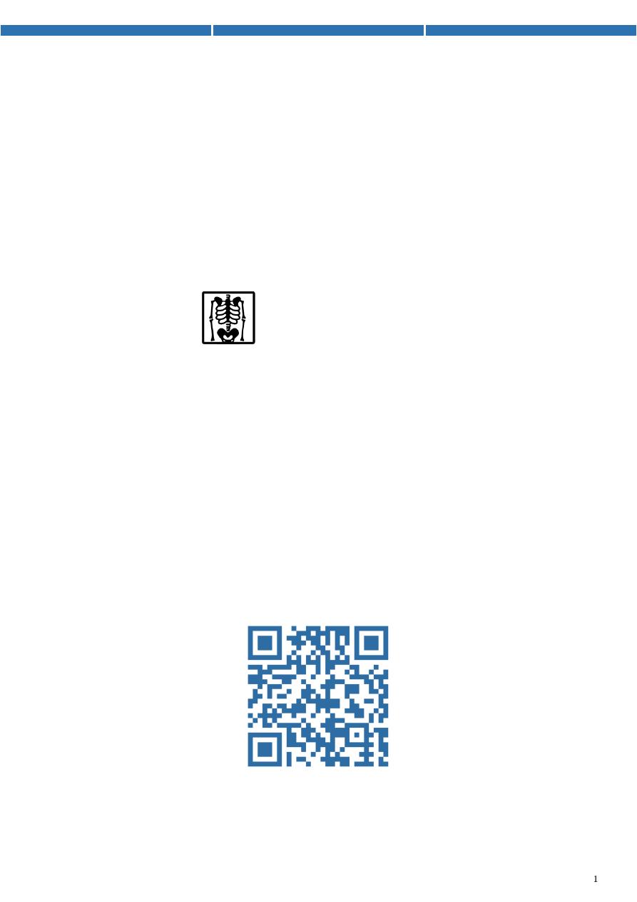
1- Normal esophagus film:
Start at C5 and end in the cardia of stomach.
25 cm length.
Mucosa thin, regular, longitudinal, parallel.
Number of lines of mucosa = 4-5 lines.
Indentations at aortic arch, left atrium, diaphragmatic
hiatus, body of cervical vertebra, right bronchus.
On the Hypo pharyngeal part common structures that we
can visualize are:
Epiglottis
(red arrow in the left)
Post cricoid impression
(yellow arrow in the left)
lateral pharyngeal pouches
(white arrow in the left)
Crico pharyngeal muscle impression
(white arrow in the right)
This is lateral oblique view
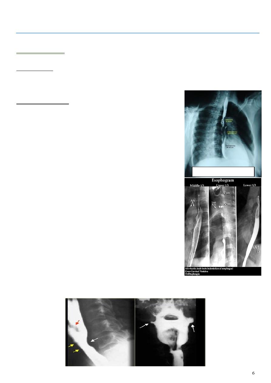
2- Tertiary contraction:
The film show that the esophagus is
contracted and dilated as the peristaltic wave
move.
Occur in:
o Diffuse esophageal spasm.
o Nutcracker esophagus (crock screw
esophagus).
o Decreased peristalsis (resulting from achalasia, scleroderma, dermatomyositis,
polymyositis, esophagitis, and secondary to many other diseases)
3- Diffuse esophageal spasm:
Intermittent contractions of the mid and distal esophageal smooth
muscle.
Tertiary peristalsis (
crock screw appearance
)
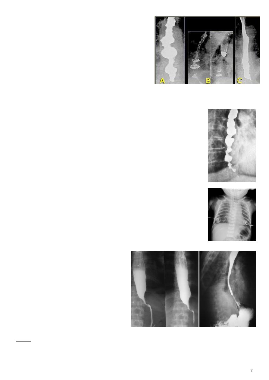
4- Congenital anomalies:
Atresia with TEF most common one is esophageal atresia with
distal TEF).
Congenital short esophagus.
Congenital duplication.
5- Benign strictures of esophagus:
Due to peptic esophagitis,
corrosive, traumatic.
Ba swallow show:
o
Funnel
(tapering shape).
o No shouldering sign.
o Smooth and regular.
o Long length (lower third).
o Constant narrowing.
o Mid proximal dilatation.
Note:
If the stricture is due to GERD short part of esophagus is narrow.
If the stricture is due to corrosive long part of esophagus is narrow.
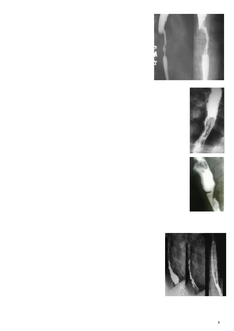
6- Malignant strictures of esophagus:
Due to SCC and adenocarcinoma.
Types infiltrative, as filing defect, ulcerative.
Ba swallow show:
o
Shouldering sing = apple core sign = minscus sign
.
o Constant narrowing.
o Irregular and variable length.
o Soft tissue shadow of the mass.
o Fistula (double tract).
7- Candida esophagitis:
Type of study: Ba. swallow.
Showing: numerous fine erosions and plaques causing
shaggy outline
of the esophagus.
8- Esophageal web:
Occur in the cervical part of esophagus.
Ba swallow show:
o Thin mucosal fold arise from the anterior wall of esophagus and
extend posteriorly.
o
Shelf like filling defect
with proximal dilatation.
o Single or multiple.
9- Esophageal varieces:
Dilated veins of the wall of esophagus.
Due to portal hypertension, lead to hematemesis.
Ba swallow (DC) show:
o Early changes loss of parallelism with thick tortious
folds.
o Later multiple filling defects (
fine cobble stone
).
o Advanced larger filling defects (
coarse cobble stone
).
o More advanced elongated (
worm like filling defect
).
These changes could be seen at the lower third of
esophagus and at the cardia.
10- Achalasia cardia:
Ba swallow show:
Tapering (
Rat tail
,
Tip of pencil
,
cigar shape
)
under left dome of diaphragm.
Proximal part dilitation (sac like).
Regular and smooth.
No shouldering sign.
The narrowing is constant short length (confined to cardia).
Sluggish peristalsis (undulating or spiky out line).
Food particles (non- homogeneity of Barium).
Air Barium level.
Absence of fundal gas shadow.
CXR shows widening of mediastinum, basal fibrosis (due to repeated aspiration
pneumonia).
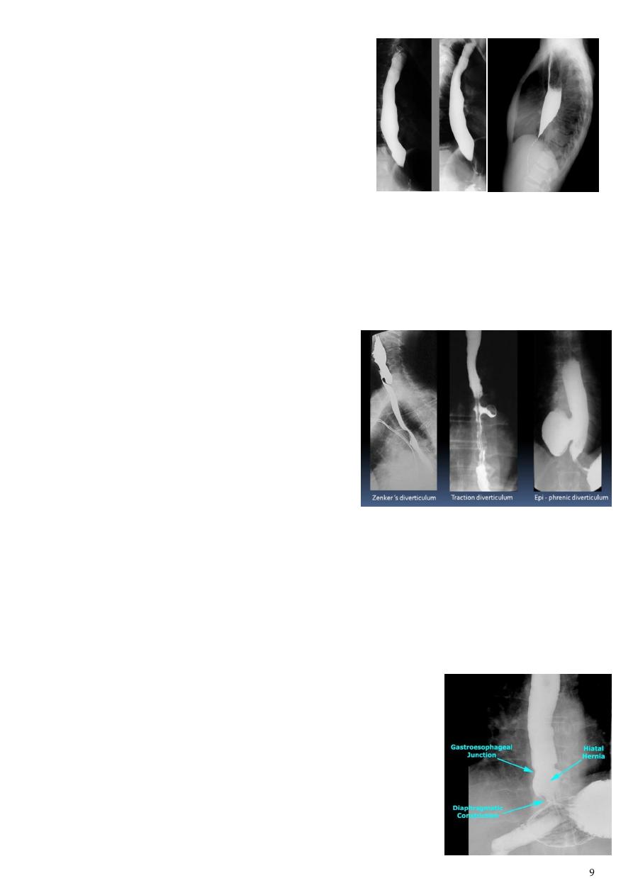
11- Esophageal diverticulum:
Pulsion diverticulum:
o In the upper third of esophagus.
o Causes: chocking after meal, increased
intraluminal pressure.
o Ba show: abnormal dilatation and
pouching due to increased pre-vertebral
space, A/F level (
killence dehiscent
) posteriorly.
Traction diverticulum:
o In the middle third of esophagus.
o Causes: post-TB.
o Ba show: upward direction of diverticulum and irregular base.
Epi-phrenic diverticulum:
o In the lower third of esophagus.
o Ba show: arise from the esophagus, above the diaphragm.
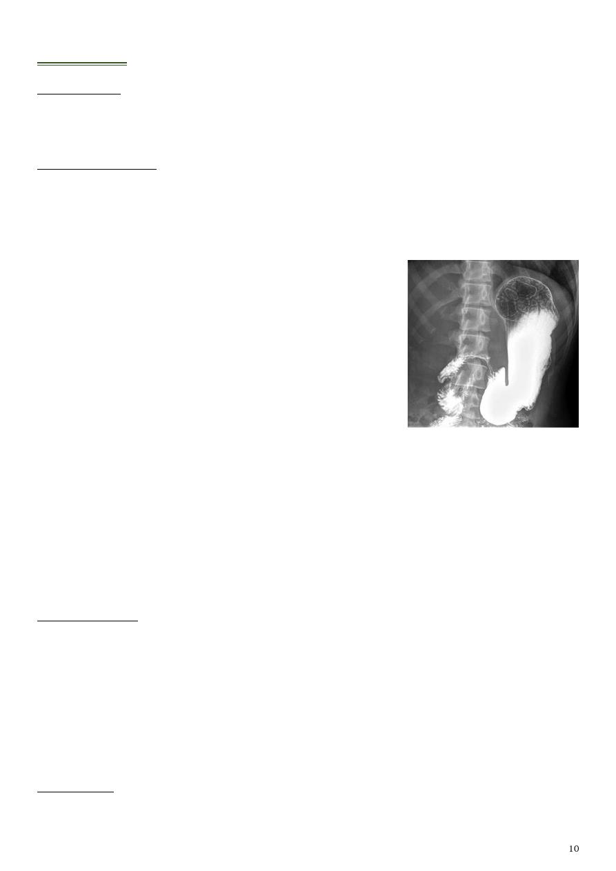
12- Hiatus hernia:
Sliding (95%) gastro-esophageal junction above the
hiatus, stomach protrude through the hiatus.
Para-esophageal (5%) gastro-esophageal junction below
the diaphragm, gastric fundus protrude through hiatus.
Mixed gastro-esophageal junction above diaphragm,
gastric fundus herniate beside distal esophagus.
Stomach
Type of study:
Barium meal (use barium sulphate).
Two views AP or PA view (enface), lateral view (profile).
Indications of study:
Gastric or duodenal obstruction.
GIT hemorrhage.
Malignancy.
Upper abdominal mass.
Motility disorders.
Systemic diseases (T.B).
1- Normal stomach film:
Anatomy:
o Note the shape of the stomach.
o Not the size of the stomach.
o Anatomical parts cardia, fundus, body, pylorus.
o Mucosal pattern regular, longitudinal lines, more than 5 lines.
Supine position of the patient lead to white color (SC) of the body, pyloric antrum
and black color (DC) of the fundus.
Prone position of the patient lead to black color (DC) of the body, pyloric antrum
and white color (SC) of the fundus.
2- Peptic ulcer:
Ba meal findings:
Direct signs:
o Ulcer crater (nitch) in enface or profile views.
o Associated signs spasm, radiated mucosal folds, edema (Hampton's line).
Indirect signs:
o Hyper-peristaltic waves.
o Companion B sign.
o Thick mucosal folds and hyper-peristaltic stomach (angry mucosa).
Benign ulcer:
Profile view:
o
Ulcer crater
(collection of Ba projects from stomach
wall).
o
Hampton's line
(1mm thin straight line at the neck of
ulcer).
o
Ulcer mound
(smooth sharply delineated soft tissue mass
surrounding a benign ulcer).
Enface view:
o Ulcer crater.
o Zone of edema with mucosal lines.
Malignant ulcer:
Profile view large circle with large zone of edema.
No enface view no projection.
Complicated benign ulcer:
Filling defect with central ulcer crater.
No mucosal radiation due to destruction.
Meniscus sign the ulcer crater is not projected outside the
lumen.
Hour glass deformity (X) of stomach.
3- Gastric cancer:
95% of benign ulcer arising form the lesser curvature, but
malignant ulcer arise from the greater curvature.
Site of predilection of gastric cancer is pylorus.
Presentation polypoidal, ulcerative, infiltrative (localized
or generalized).
Ba meal show:
o Single or multiple irregular filling defect.
o Alternation of nearby mucosal pattern.
o Localized or generalized narrowing of the stomach.
o Stomach rigid in appearance.
o
Shouldering sign
: in either sided aspect of the narrowing.
Ulcerative cancer:
o Polypoidal tumor with ulceration.
o Complicated benign gastric ulcer.
4- Hypertrophic pyloric stenosis:
Causes congenital type, adult type.
Information M:F = 4:1 / Caucasian / 4-8 weeks
of life / +ve family history.
Normally:
o Pyloric muscle thickness = less than 3 mm.
o Pyloric length = less than 15 mm.
Ba meal show:
o Thickening of pyloric muscle (better by US).
o Narrowing of pyloric region.
o Dilatation of other regions.
o Contraction not go to the duodenum.
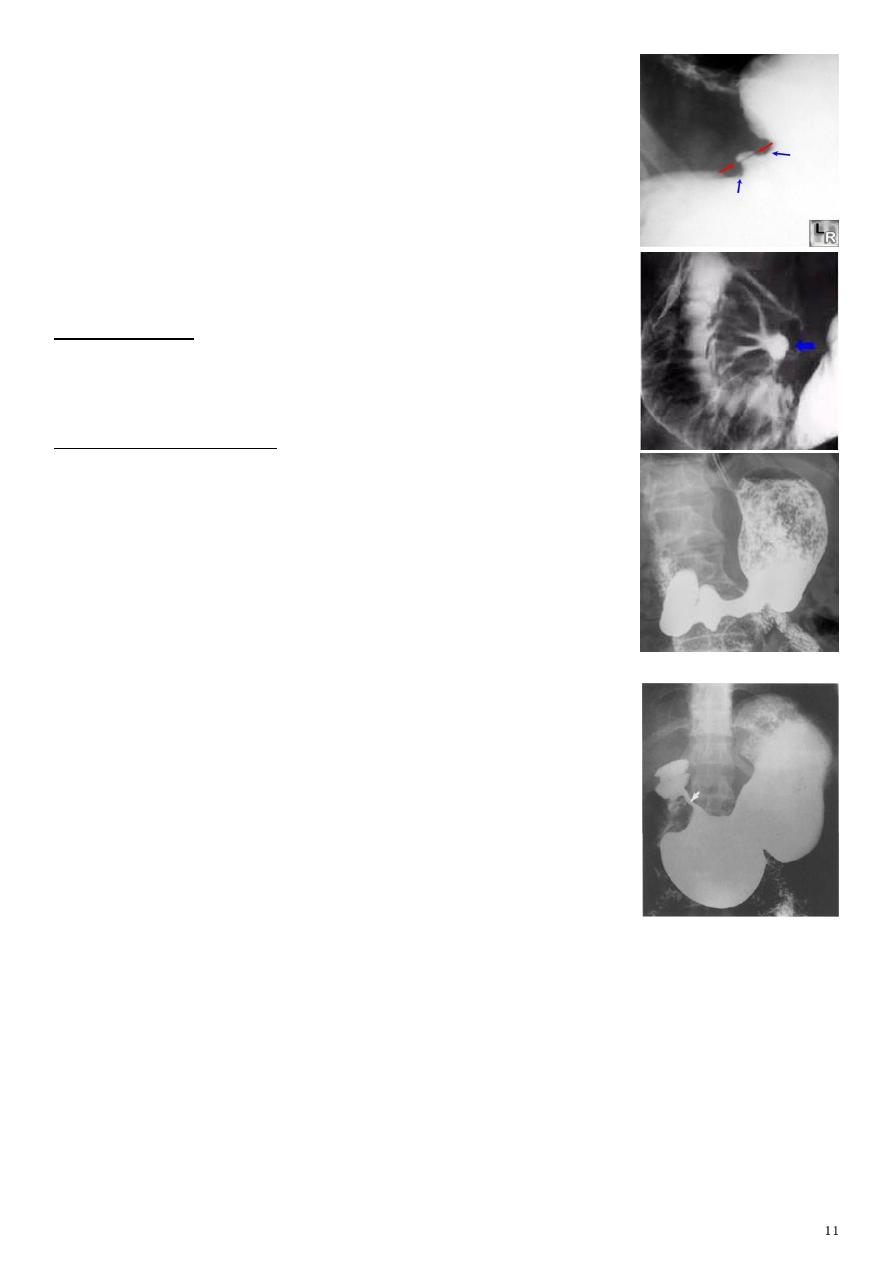
5- Gastric outlet obstruction:
Information:
o Lead to recurrent vomiting of food.
o Stomach dilate to accommodate food intake and
secretions.
o Causes peptic ulcer (benign), gastric carcinoma
(malignant).
o Other benign causes of narrowing is corrosive obstruction (like the photo )
Ba meal findings in pyloric stenosis:
o Marked dilatation of the stomach.
o Failure of passage of content in to the duodenum.
o Multiple filling defect within the stomach due to retained food materials.
Duodenum
Study by barium meal
1- Normal duodenum film:
C shape, start at pylorus, end at jejunum.
Consist from 4 parts.
Normal Anatomy of Duodenum:
o Duodenal cap.
o Duodenal loop.
2- Duodenal ulcer:
Information:
o 95% of DU occur in the duodenal bulb (2 cm above the ampulla of vator).
o 5% of DU occur in the post bulbar duodenum.
o Half of DU occur in the anterior wall of the bulb.
o Most of DU round or ovoid pools of barium.
o 5% of DU may be linear.
o Most DU smaller than 1 cm in diameter.
o Giant DU above 2 cm in diameter.
o Multiple DU (15% of patient) lead to Zollinger
Ellison syndrome.
Complicated chronic DU:
o
Trifolate
duodenum (tri foil tree shape).
o Pseudo diverticulum (
Akerland
diverticulum).
o Gastric outlet obstruction (Pyloric obstruction).
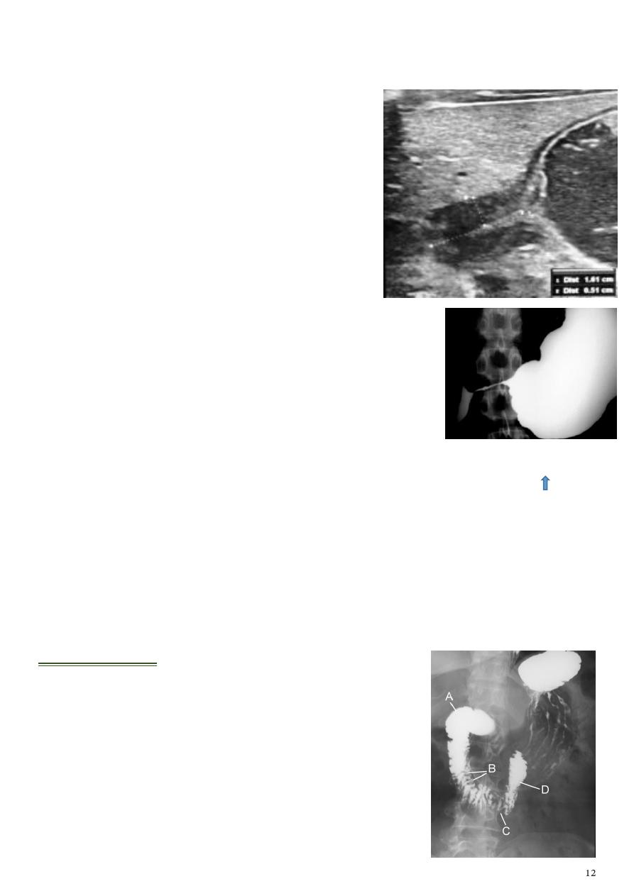
3- Duodenal diverticulum:
95% in the second part of the duodenum.
Well defined oval or round shape smooth wall out
pouching from the 2nd part of the duodenum with or
without
A/F level
.
4- Atresia or stenosis:
Pyloric stenosis
single bubble sign
.
Duodenal stenosis
double bubble sign
.
Jejunal atresia
triple bubble sign
.
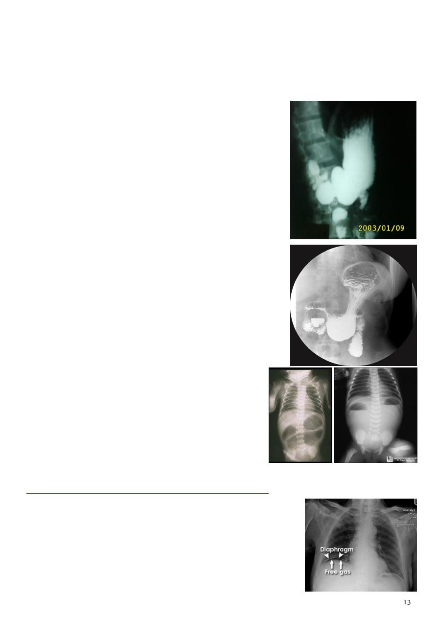
Abdominal problems in the chest
1- Pneumo-peritoneum:
Use erect CXR.
Free intra-peritoneal gas.
Due to critical illness perforated viscus or ulcer.
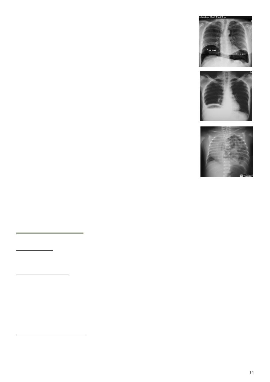
Finding:
Crescent shape of lucency
below the right diaphragm
described as sub-diaphragmatic free gas.
Could lead to huge collection of gas below both sides of
diaphragm.
2- Sub-phrenic abscess:
Lesion like abscess (not free air).
Use erect CXR.
Findings air below the diaphragm, A/F level, thick wall.
3- Congenital diaphragmatic hernia:
Mostly is
Bockdolik
type.
Use plain x-ray of chest and abdomen.
Findings:
o Multiple air filled loops of bowel in hemithorax (mostly left
side).
o Indistinct left dome of the diaphragm.
o Shifting of mediastinum (cardiac shadow) to the right side.
Small intestine
Type of study:
Barium follow through (SBFT).
Indications of study:
Inflammatory bowel disease.
Mal absorption syndrome.
Tumors.
Ulcer.
Swelling or inflammation of small intestine walls.
Contraindications of study:
Suspected bowel perforation.
Bowel obstruction.
Conditions when aspiration of barium is likely (cough, choking).
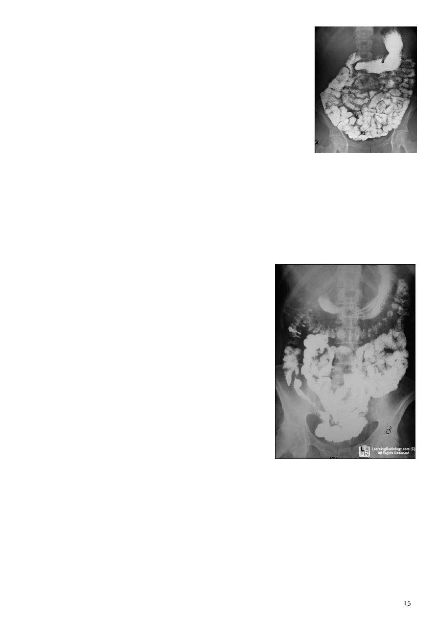
1- Normal small intestine film:
Information:
o Jejunum in the upper left abdomen, ileum in the lower
right abdomen.
o Valvule conniventis give normal
feathery appearance
in Ba. study.
o Small bowel diameter = 3-3.5 cm (above 4 is abnormal).
o Ileum loop diameter = 2-2.5 cm (above 3 is abnormal).
o Distance between loop and loop should be less than 1-2
mm.
Normal SBFT:
o Complete filling of the lumen of small intestine.
o Normal distance between small bowel loops.
o No any flocculation or segmentation of Ba.
o No loss of feathery appearance.
2- Crohn's disease:
Information:
o Transmural lesion.
o Skip area.
o Rectal sparing.
o Any region of GIT.
o Terminal ileitis.
o Stricture in chronic Crohn's.
o No gender preference.
o Small bowel involvement 70-80%, colonic
involvement 15-20%.
SBFT findings:
o Multiple mucosal ulcers (aphthous ulcers).
o Multiple skip lesions.
o Longitudinal fissures.
o Widely separated loops of bowel (due to fibro-fatty proliferation).
o Thickened folds (due to edema).
o May lead to sinus tracts and fistulae.
o Severe CD lead to
cobblestone appearance
: apthae enlarge and merge with
edematous mucosa and deep ulcers.
o
Pseudo diverticula
formation: due to contraction at the site of ulcer with
ballooning of the opposite site.
o
Stricturing
:
Active disease (string sign, edema, spasm).
Fibrotic diseases (irreversible stricture, lead to obstruction and fistula).
o
Pseudo polyps
:
Inflammatory cobble stoning, nodular filling defects, edematous mucosa
surrounded by ulcerations.
Post-inflammatory filiform, mucosal overgrowth during healing process.
3- Ulcerative colitis:
Information:
o Affect the mucosa.
o Continuous lesion.
o Involves the rectum.
o Usually limited to the colon.
o Back-wash ileitis.
o Lead pipe appearance.
o
Thumb-printing
= lead pipe + granulation in acute UC.
o Male predilection.
o Rectal involvement 95%, terminal ileum only involved in
pan colitis (back wash ileitis).
Palin film: mural thickening + Thumb printing.
Ba enema:
o Bowel wall and hastura thicken.
o Button shaped ulcers.
o
Pseudo polyp appearance
mucosa lost + mucosa
remain.
o
Lead pipe sign
featureless bowel + loss of hastura +
luminal narrowing + bowel shortening.
Note: colorectal carcinoma in UC sessile or simple stricture.
4- Toxic megacolon:
Seen in UC more than in CD.
The colon (
transverse colon
) is dilated more than 6 cm.
Additional loss of hastural markings.
Risk of perforation.
Splenic and hepatic flexure are high.
It is dangerous condition so do plain x-ray before Ba study.
5- Lymphoma of small bowel:
Splaying
تفلطحand
separation
(>1-2 mm) of the bowel loops
due to enlarged L.N.
Saw tooth appearance
thickening of mucosa (lead to
nodular appearance), irregular outline.
Loss of normal feathery appearance.
Later stage signs of malabsorption syndrome
(flocculation and segmentation of Ba.).
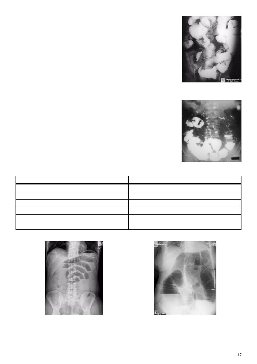
6- Malabsorption syndrome:
Loss of feathery appearance.
Dilated small bowel loops > 3.5 cm.
Flocculation and segmentation of Ba. (
Mosaic appearance
).
Splaying and increase distance between small bowel loops.
7- Bowel obstruction:
Small bowel obstruction 80%
Large bowel obstruction 20%
Central location (cross transversely).
Peripheral location.
Maximum diameter is 5 cm.
Maximum diameter is 8 cm.
Valvule connictivae are visible.
Hasturation is present.
In erect position A/F level small and many.
In erect position A/F level large and few.
Colon is filled with feces which has bubbly
appearance.
Type of study plain x-ray, KUB, Ba (more than 3 cm is small bowel obstruction).
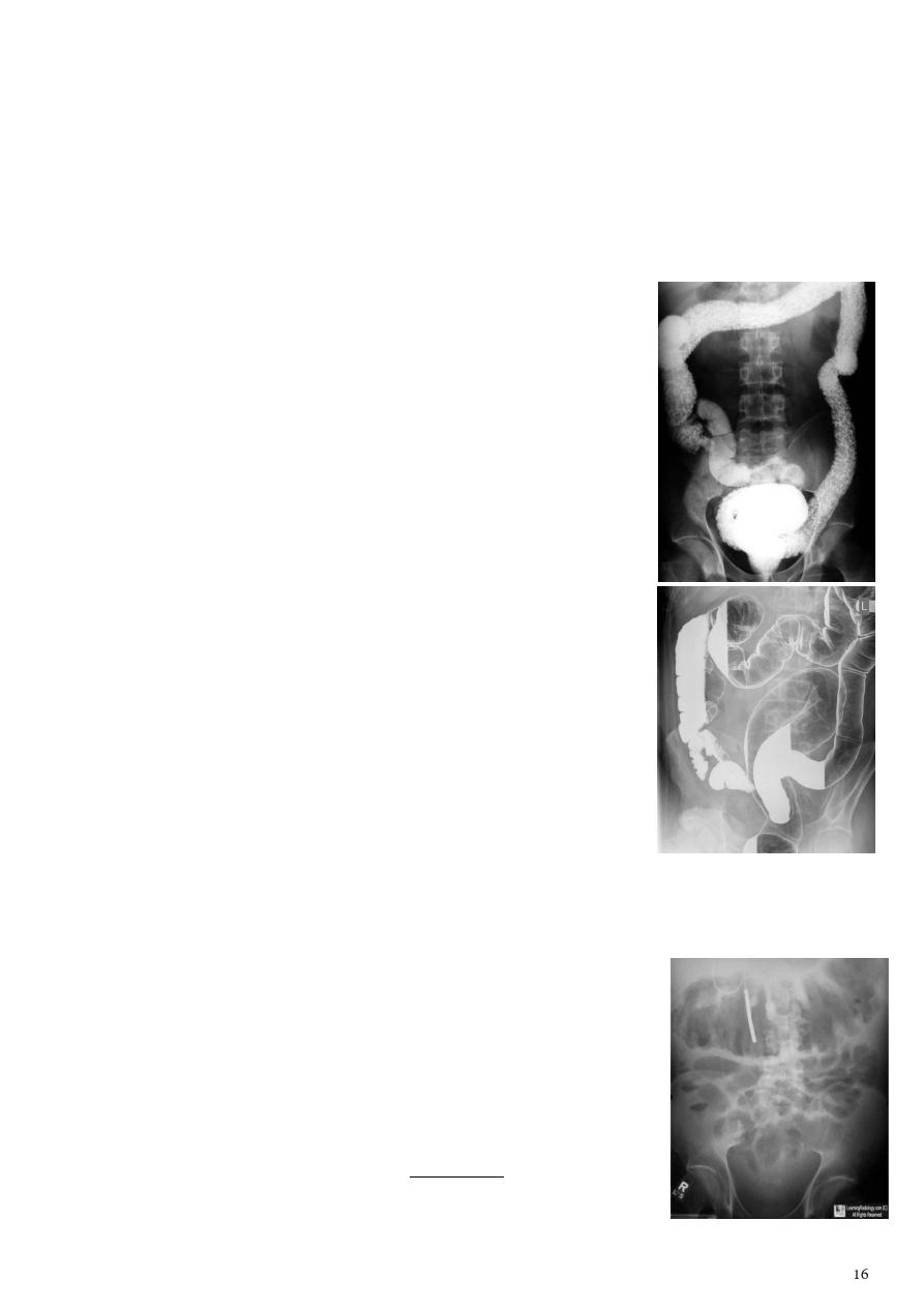
Large intestine
Type of study:
Barium enema.
Before examination empty the colon by restricted diet, laxative, enema.
After examination get rid of any barium still in the body by laxative.
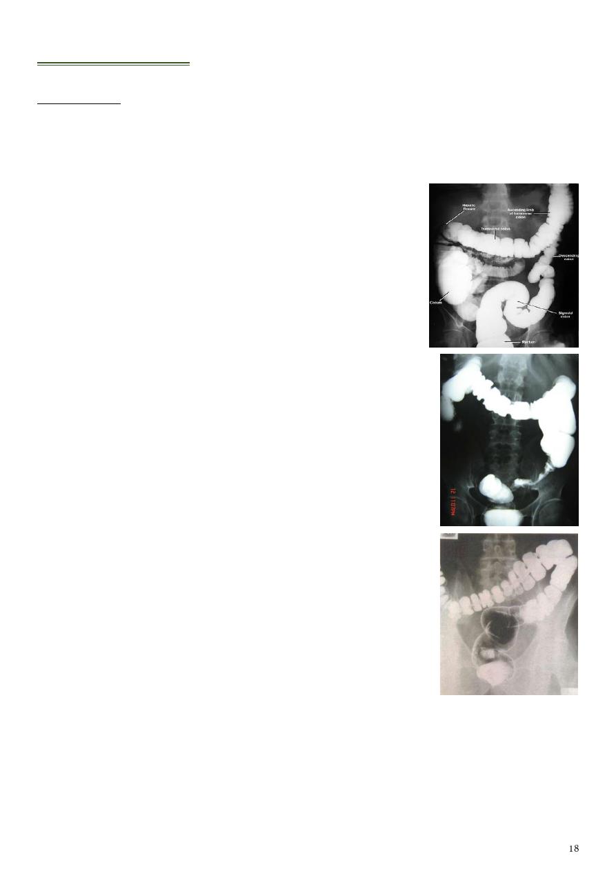
1- Normal large intestine film:
Diameter of large bowel normally 5-6 cm.
Above 8 cm means large bowel obstruction.
Normally we see hastura.
2- Colorectal carcinoma (CRC):
Most common cancer in GIT.
Most commonly occur in the recto-sigmoid region.
Ba enema:
o Infiltrative
apple core sign
(the lesion infiltrate the
bowel wall from outside).
o Ill-defined filling defect within the lumen of bowel.
o Could be ulcerative nodule or ulcerative lesion.
Area of stoppage, narrowing, missing, filling defect,
shouldering means that there is problem here.
3- Irritable bowel syndrome (IBS):
see a lot of hastura in the
descending colon.
4- Intussusception:
Information:
o Causes inflammation of L.N (90% child), CA colon (90% adult).
o In pediatric most common cause of intestinal obstruction is intussusception
and lead to acute abdomen.
o 90% of intussusception occur in the ileo-colic region.
Abdominal plain film:
o Elongated soft tissue mass (typically in the right upper quadrant in children).
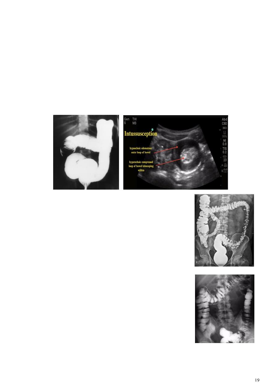
o With a bowel obstruction proximal to it.
Ultrasound sigs:
o
Target of doughnut sign
(ileum is hypo-echoic, Colon is hyper-echoic).
o Pseudo kidney shape sign.
o Area of devoid of gases shadow called spring sign.
o If there is no vascularity it means ischemic segment (in the ileum).
Contrast enema "gold standard":
o
Coiled spring appearance
demonstrating the intussusception as occluding
mass prolapsing into the lumen.
o Contraindicated in perforation.
o Diagnostic and therapeutic.
5- Colonic diverticulosis:
SC or DC Ba enema.
Diverticula few mm to few cm.
Description
barium filling out pouching
.
Here the pouching of mucosa from outline of the bowel
(outside).
6- Familial adenomatous polyposis syndrome (FAPS):
Innumerable adenomatous polyps.
Size < 5 mm.
Pouching mucosa
inside
the lumen of colon.
Predisposition to CRC.
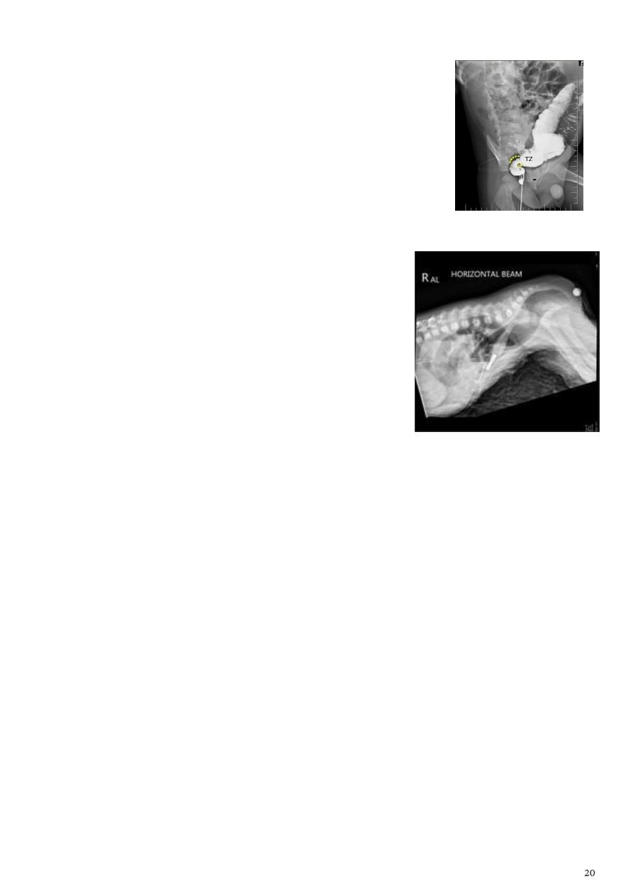
7- Hirschprung disease:
Most common cause of neonatal colonic obstruction.
More in boys.
The affected segment is of small caliber with proximal
dilatation.
Fasculation / saw teeth irregularity of a ganglionic segment.
Most common area is recto-sigmoid.
Transition zone
(could be seen with x-ray).
8- Anal atresia (imperforated anus):
Abdominal radiograph:
o Show multiple dilated bowel loops + absence of
rectal gas.
o Can be variable depending on:
1- Site of atresia (high or low).
2- Level of impaction with meconium.
3- Physiological effects such as straining.
Invertogram
:
o Baby is turned upside down for 3 min.
o Distance of gas bubble in the rectum from the the metal piece less than 3 cm is
low type, more than 3 cm is high type.
For more photos about GIT:
www.muhadharaty.com/lecture/3449
www.muhadharaty.com/lecture/4396

Part2
: Radiology of Renal system
Basic information
Types of study:
Palin kidney, ureter and bladder (KUB).
Intravenous urography (IVU), intravenous pyelography, excretory urography (EU).
Ultrasonography (US).
Magnatic resonance imaging (MRI).
Radionuclide imaging.
More invasive tests.
Benefits:
US - MRI anatomical information.
IVU - CT functional & anatomical information.
Radionuclide scanning functional information.
1- KUB:
Simple procedure.
Requires adequate patient preparation.
Diagnostic vale:
o Radiopaque calculi.
o Calcifications (schistosomiasis of bladder).
o Gas pattern (in intestinal obstruction)
o Organomegaly (liver, spleen).
o Bony abnormality.
2- Intravenous urography (IVU):
Information:
o It means the visualization of kidney parenchyma, calyces and pelvis after
intravenous injection of iodinated contrast medium to the patient, followed by a
series of x-ray films.
o Replaced by US and CT.
o Remains the primary modality for visualization of the pelvicalyceal system and
ureters.
o Providing both functional and anatomical information.

Requirements:
o Fasting 4-6 hours, good hydration is essential.
o Adequate bowel preparation.
o Renal function test beforehand (serum creatinine level below 3).
o Nonionic contrast media are used to guard against contrast nephropathy.
Indications:
o For detailed demonstration of the pelvicalyceal system and ureters.
o Show renal function.
o Suspected acute ureteric colic, hematuria, UTI.
o Renal calculi (stones).
o UT obstruction (hydronephrosis).
o Renal and bladder masses.
o Congenital anomalies.
Contraindication:
o Renal impairment.
o Hypersensitivity to contrast media.
Contrast medium:
o Urographic contrast media are highly concentrated solutions containing iodine,
also known as iodinated.
o Large volume of contrast media (50-100ml) is injected intravenously passes to
glomerular filtrate concentrated in renal tubules passes to pelvicaticeal
systems ureters bladder.
o Contrast media is iodinated solution that make the renal tract white in color so
radiolucent stone appear black (as filling defect).
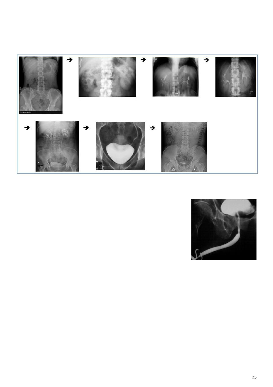
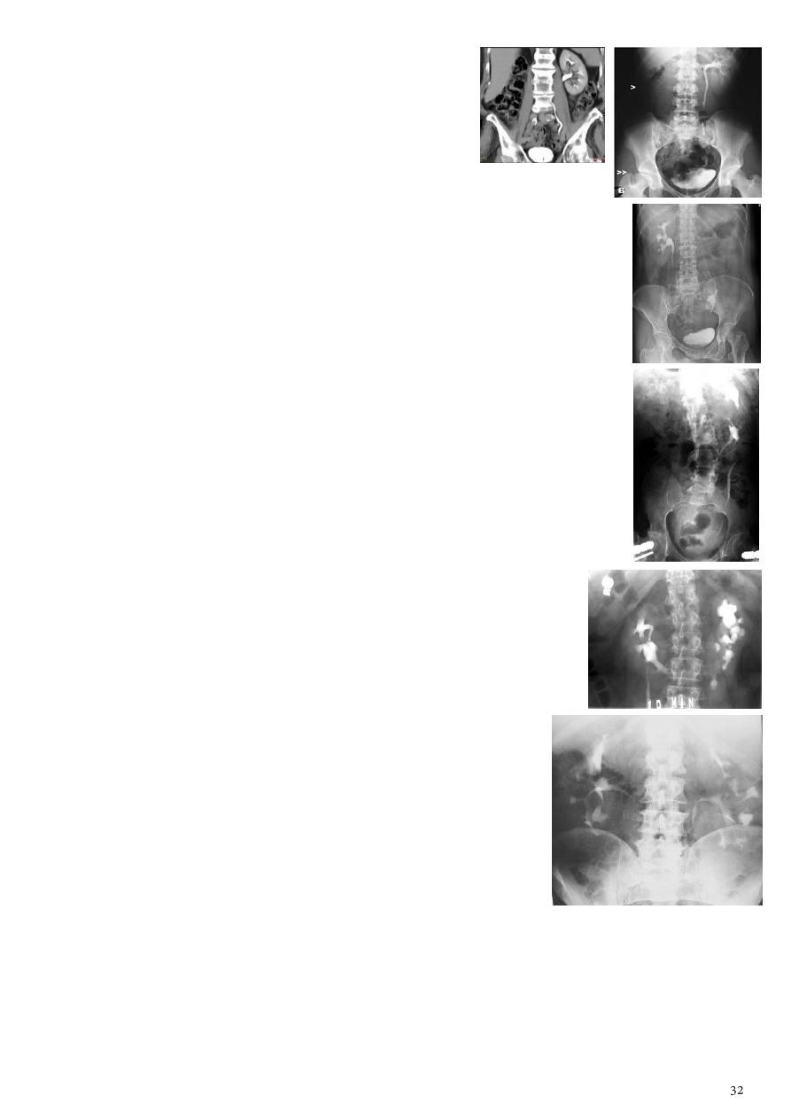
The procedure of performing IVU:
o
KUB
taken before injection of contrast media to see calcification, radiopaque
stones.
o Inject the contrast media.
o
Nephrogram phase
(Immediately after injection of contrast) see parenchyma.
o
Pyelogram phase
(l-5 minutes after injection of contrast) see pelvis.
o Compress on the abdomen by billow.
o After 10 min with compression take the
film
get better distention of pelvis and
calyces and see the upper ureter till the site of compression.
o
Full length film
after the release of compression see the full ureter.
o After 10 min take
full bladder film
the bladder is fully distended with contrast.
o
Post voiding full length film
for reflux, retention, neurogenic bladder.
Interpretation of IVU films:
o Kidney position (left kidney higher), renal parenchymal width (uniform 2-2.5
cm), length of adult kidney (10-16 cm) at IVU but longer in US due to
magnification.
o Calyces symmetrical, cup shape (normal), club shape (dilated).
o Renal pelvis and ureter funnel shaped, ureter not seen by single film due to
peristalsis, filling defect (stone, tumor, blood clot), ectopic ureter.
o Bladder centrally located, smooth outline, smooth indentation, abnormality
(filling defect, wall irregularity, diverticula, trabiculae).
3- Cystourethrography (CUG):
Information:
o Contrast is inserted into the bladder and images are
obtained.
o The patient is then asked to void and images are also
taken.
Benefits:
o Urethral lesions.
o Vesicouretheral reflux.
o Stress incontinence.
4- Ultrasonography (US):
Information about US in general:
o US waves in the 2-10 MHz frequency range.
o The higher the frequency the lees the penetration.
o Air or gas reflect the US waves so put gel.
o US radiation is nonionizing so there is no adverse biological effect.
o US used in obstetrics, gynecology, abdominal US, superficial structure,
pediatrics, pediatric neurology, vascular US (Doppler), cardiology (Echo),
interventional radiology (biopsy, intrathecal injection, aspiration of cyst or
abscess).
Gray scale:
o Echofree or unechoic (black) fluid, edema, cyst, plural effusion, ascities,
bladder, gall bladder, stomach and intestine (if contain fluids).
o Echogenic (white) Bone, calcification, F.B (metallic), stone, gas, pneumothorax.
o Hypoechoic – hyperechoic liver, kidney, pancreas, soft tissue.
Probe (transducer):
o Convex probe (3.5 Hz) for abdomen.
o Linear probe (7.5 Hz) for superficial tissue (thyroid, breast, scrotum, muscle,
soft tissue).
o Endovesiral probe transrectal, transvaginal.
o Biopsy US probe.
o 3D probe used in pregnancy.
Advantages:
o Safe, less expensive, small, available, portable.
o Images in the real time.
o High resolution.
o No adverse effects.
o Noninvasive nature.
o Ultrasound is the tool of choice in obstetrics primarily.
Disadvantages:
o Organs containing gases and bony structures need specialized procedure.
o Only limited window is available.
o It depend on operator skill.
o It is sometimes impossible to obtain good images (in obese patient for example).
o Not detect all diseases.
Renal US:
Requirements:
o Fasting 4-6 hours.
o Not need bowel preparation.
o Not use contrast media.
o No radiation hazards.
Normal kidney:
o Central of the kidney is echogenic (white) because pelvicalyceal system contain
fat and vessels.
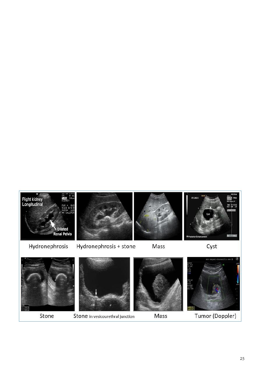
o Peripheral of the kidney is hyperechoic (gray) the parenchyma contain cortex
and medulla.
Abnormalities:
o Echogenic stone, calcifications.
o Echofree cyst, hydronephrosis.
o Hypoechoic tumor, mass.
o Hyperechoic in the parenchyma (RCC), in the pelvicalyceal system (TCC).
Notes:
o Hydronephrosis central (echofree), peripheral (hypoechoic).
o Cyst peripheral (echofree).
o Stone hyperechoic + acoustic shadow.
o Ureter the only abnormality is dilatation.
Renal mass:
o Tumor has blood vessels, fixed, arise from the wall.
o Hematoma not contain blood vessels, mobile.
o Differentiate between them by using Doppler.
Bladder (echofree):
o Stone echogenic + acoustic shadow.
o Mass hypoechoic, use Doppler to see vessels inside the tumor.
o Calcification of wall echogenic plaque.
o Cyst (uretrocele or diverticulum) echofree.
5- Computed tomography (CT):
Information:
o More sensitive.
o Considered with care.
o Plans or views axial, coronal, sagittal.
o Black (hypodense), white (hyperdense).
o Opaque and lucent stones appear hyperdense in CT.
o Types plane CT (without contrast) and contrast CT.
o Oral contrast use gastrographine and the stomach appears white.
o IV contrast the vessels appear white.
o According to the density we can differentiate between fluid, soft tissue, mass.
Requirements:
o Fasting 4-6 hours.
o May need bowel preparation.
o May use contrast media.
o Consider radiation hazards.
Diagnostic value:
o Detection of small radiopaque or radiolucent stone.
o UT obstruction.
o Renal and bladder masses.
o Differentiate cystic form solid masses.
o Congenital anomalies.
o CT angiography.
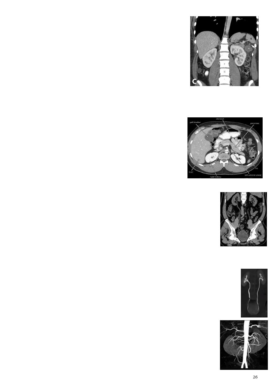
CT urinary tract (CTUT):
o Test for detection of stones.
o No preparations required.
o The patient should stop his breath.
o The patient drink large amount of water to distend the ureter and bladder.
o Take films from the start of the kidney to the end of the bladder every 3 mm.
o Any white dot is stone (sometimes it is phlebolith if this white dot not seen
in the ureter after reconstruction of coronal image of the ureter).
o The two ureters no appear in one reconstructed image (should be 2 images)
CT urography (CTU):
o Show the kidney, ureter, bladder only and remove other structures from
the image.
o There is contrast and multi-detector CT.
o Can give color image (not has any diagnostic value).
CT angiography (CTA):
o Image the renal vessels with the kidney.
o See congenital anomalies, and vascular abnormalities.
6- Magnetic resonance imaging (MRI):
Information:
o Functional imagining.
o Morphological imagining.
o Used when the patient has hypersensitivity to
contrast or has bad renal function.
o T1 weighted image black color of moving fluid
(blood vessels).
o T2 weighted image white color of stable fluid (gall bladder).
Requirements:
o Fasting 4-6 hours.
o No bowel preparation.
o May use contrast media.
o No radiation hazards.
MR urography (MRU):
o No contrast is needed.
o See the flow in the urinary system.
MR angiography (MRA):
o No contrast is needed.
o See the vascularity of urinary system.
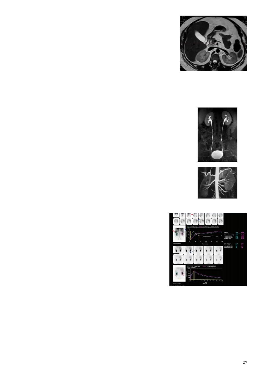
7- Renal scintigraphy (nuclear medicine):
Information:
o Use radiopharmaceuticals (isotopes) IV or orally
o Types of isotopes Tc DMSA or Tc DTPA
o Safe, minimally invasive, expose the patients to
radiation is less.
Uses:
o Evaluating renal function blood flow, GFR,
effective renal plasma flow (ERPF), nephron
uptake and clearance, renovascular hypertension.
o Renal artery stenosis (RAS).
o Acute and chronic renal failure.
o Ischemic nephropathy.
o Pyelonephritis.
o Trauma or surgical complications.
o Renal transplant function, obstruction, and acute or chronic rejection.
o Ureteral obstruction and vesicoureteral reflux.

Risks:
o Diagnostic nuclear medicine procedures result in relatively low radiation
exposure.
o Allergic reactions to radiopharmaceuticals may.
o Injection of the radiotracer may cause slight pain and redness which should
rapidly resolve.
Limitations:
o Cannot reliably differentiate between cysts and tumors.
o Time-consuming.
o Low resolution compared with CT or MRI but more sensitive, and giving functional
information.
Renal pathology
Types renal pathology:
Stone diseases.
UT neoplasms.
UT infection.
UT trauma (hematoma, laceration, avulsion) use CT scan.
Congenital anomalies.
Vesico-ureteric reflux.
Urethral lesions.
Reno-vascular hypertension.
1- Stone diseases:
Causes of urinary tract obstruction:
o Within the lumen Calculi, Blood clot, Sloughed papilla (papillary necrosis).
o Within the wall of the collecting system Tumor (transitional cell carcinoma),
Infective stricture (TB or Schistosomiasis), Intrinsic PUJ obstruction.
o Extrinsic pathology Tumors (CA cervix or recto-sigmoid junction),
Retroperitoneal fibrosis, Aberrant renal artery, retrocaval ureter.
Information:
o Calcium oxalate and phosphate stones (90% of urinary stones) are radiopaque
use KUB, CT.
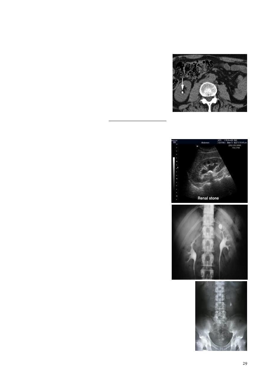
o Pure uric acid and xanthine stones (10% of urinary stones) are radiolucent use
IVU, CT, US.
o Principal feature is dilatation of pelvicalyceal system and ureter, the degree of
dilatation depends on chronicity, and the dilatation is down to the level of
pathology.
o In the renal region
renal stone.
o Outside the renal region
ureteric stone.
o Pelvic rejoin
bladder stone or phlebolith
Methods:
o KUB radiopaque (white).
o IVU Radiolucent (filling defect).
o US hyperechoic (white) + acoustic shadow.
o CT hyperdense (white).
Note: use non-contrast CT.
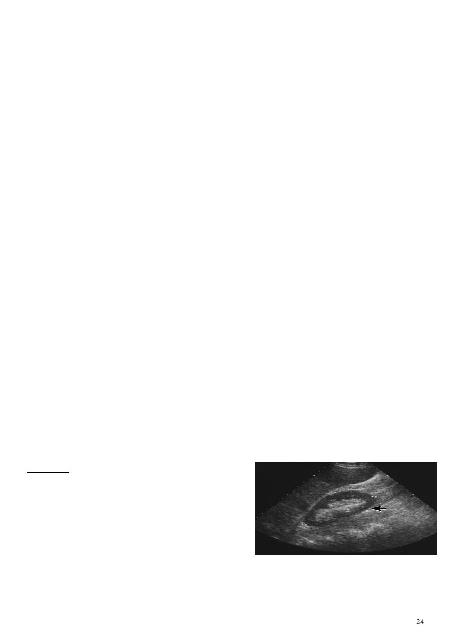
US findings:
o Dilatation of P.C.S.
o Stones larger than 5 mm are easily to seen but
smaller are missed so stones should be large
and no gases.
o
Hyperechoic
+
cast acoustic shadow
.
o Stones may lodged in the VUJ or PUJ are easily to
seen.
o Stones in the middle of ureter are hard to seen.
IVU findings:
o Acutely obstructed kidney show dense
nephrogram.
o
Dilated P.C.S
and
ureter
down to the point of
obstruction (point of hold up).
o Pyeloxinus reflux may result from rapture of fornix
urine and contrast extravasate into the renal
sinus and perirenal space.
o
Filling defect
in IVU could be stone (regular, well
defined) or tumor (irregular, ill defined).
KUB findings:
o Stone oval shape, homogenously radiopaque (white).
o Phlebolith round shape, radiopaque with
radiolucent
center
.
o DDx of stone in KUB
Phlebolith.
Gallstone.
Metallic F.B.
Calcification, calcified LN or cartilage or fibroid.
Stage horn calculus appears radio opaque in KUB.
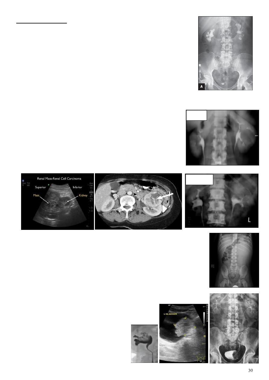
2- Urinary tract neoplasm:
Benign tumors of the kidney:
o IVU:
stretching
and
spraying
of P.C.S.
RCC:
o It account for 85% of malignant tumors of the kidney.
o Arise from the parenchyma, attached to the wall.
o KUB: soft tissue masses.
o IVU: irregular filling defect with
destruction
of calyces.
o Also we can use US (hypoechoic), CT (mass).
Wilm's tumor:
o Most common renal malignancy in children.
o Abdominal x-ray typically reveals a
large soft tissue opacity
displacing bowel shadow.
Urothelial tumors:
o 85-90% of tumors in the collecting systems of kidney are TCC.
o Occur in multiple sites P.C.S, ureter, bladder.
o IVU radiolucent filling defect, projecting into the lumen,
within the collecting system.
o US soft tissue mass lesion irregular in
outline.
o Differentiated from blood clots
and radiolucent stones.
o Use tomography to differentiate
the overlying gas shadow.
Benign
Malignant
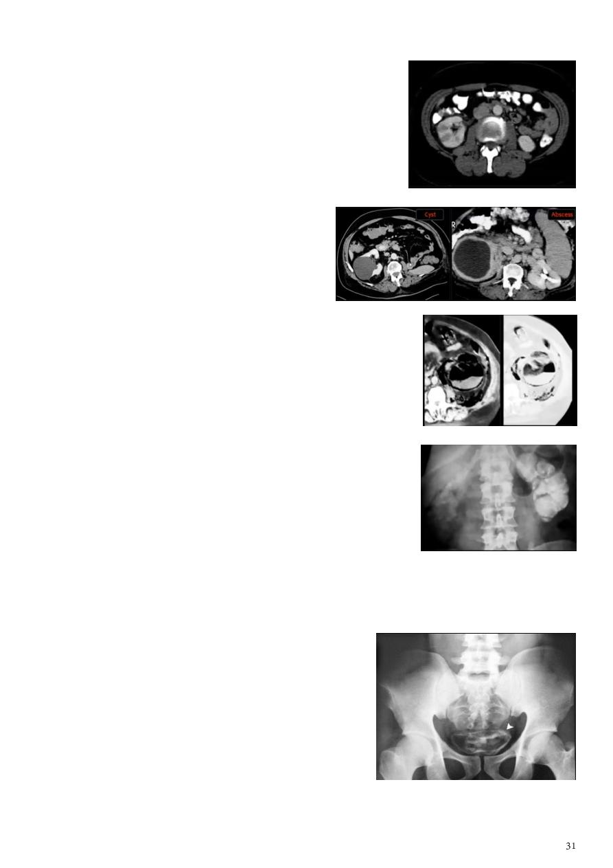
3- Urinary tract infections:
Acute renal conditions:
o Use CT scan.
o Most common infection is acute pyelonephritis
(
striated nephrogram
).
o Findings Swollen kidney, Poor renal function,
Dirty perinephric fat.
Renal abscess:
o Use CT scan.
o Like the renal cyst but it take the
contrast and has
thick wall
.
o Sometimes see air in the abscess.
Emphysematous pyelonephritis:
o
Gas shadow
within and around the kidney.
o Surgical emergency that is lethal if treated medically.
Renal TB:
o KUB large globular amorphous
calcifications
with
destruction of P.C.S.
o IVU cortical scarring, smudged papillae (moth
eaten), infundibular strictures, hydrocalyces,
hydroneprhrosis, autonephrectomy.
o Could be in the kidney and upper or lower third of
ureter and bladder.
o In the late stage it lead to calcification of whole kidney, loss of function.
Schistosomiasis:
o Infestation by S.henatobium.
o
Calcification
(bladder, lower ureter).
o Cobble stone appearance (in early stage).
o Bladder capacity not affected.
4- Congenital anomalies:
Renal agenesis:
o Incidental finding.
o The opposite kidney shows
compensatory
hypertrophy
.
o Can be diagnosed as absent kidney on ultrasound or CT.
o IVU will show a single kidney with active contrast excretion.
Ectopic kidney:
o Incidental findings during routine ultrasound.
o Findings: located in the lower abdomen (
pelvis
), rotated, short
ureter.
o Problems: chronic pyelonephritis, calculi, hydronephrosis.
Crossed ectopia:
o Right kidney in the left side.
o The right ureter is inserted in the right side of bladder.
Horseshoe kidney:
o Incidental finding.
o Caused by fail of separation (usually lower poles fused).
o Use KUB, US, CT, MRI.
o Problems: PUJ obstruction, stone formation.
o IVU shows:
The kidneys at
low position
.
Close to the spine with long axis
parallel
to the spine.
Malrotation
manifested by medially directed calyces.
The renal pelvis and ureters are anterior and lateral in
position.
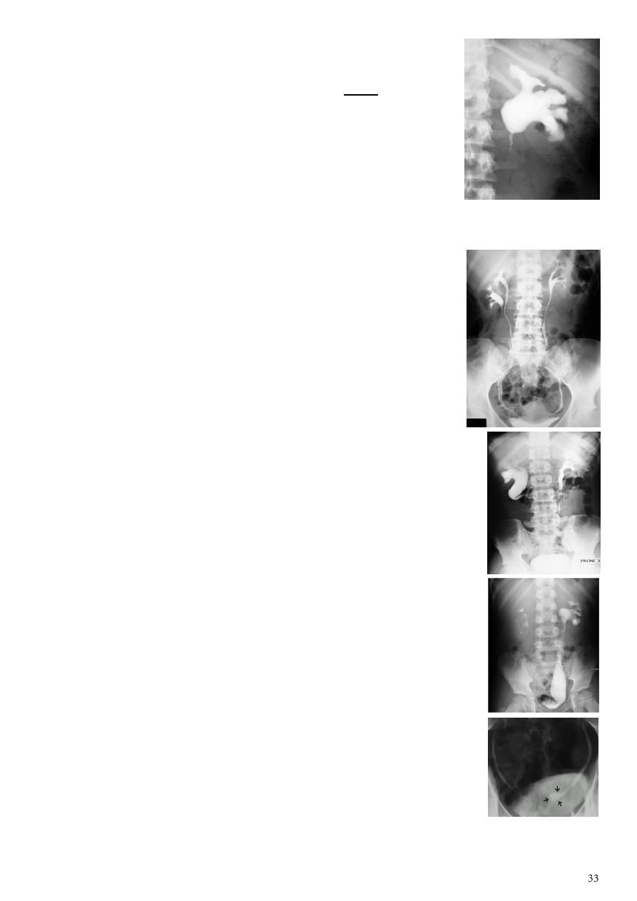
Adult polycystic disease:
o Bilateral numerous cysts of variable size contain fluid.
o Present after the third decade of life, familial.
o Clinically renal colic, loin mass, hematuria,
hypertension, ended with renal failure.
o IVU findings:
Large kidney with
lobulated
outline.
Distortion
of P.C.S.
In advanced cases there is elongation and
stretching of minor and major calyces (
spider leg
) + non-functioning kidney.
Infantile polycystic disease:
o Usually affect liver, spleen, pancreas Incompatible with life.
o Bilateral large kidney due to
numerous small cysts
(1-2 mm size).
o The outline is not lobulated as in adult.
o I.V.U, may be normal.
o Nephrogram shows minute filling defects.
PUJ obstruction (infantile hydronephrosis):
by IVU
o Marked dilatation of pelvis and may be extra-renal.
o Calyceal dilatation is late and in advanced cases form
foot
shape P.C.S.
o The ureter is normal or not seen.
o Delayed film with
I.V. diuretic
produce gross dilatation.
Q: why not hydronephrosis? Infant patient, no cause of hydronephrosis like mass or stone,
there is PUJ obstruction.
Bifid collecting system and ureter:
o Unilateral or bilateral.
o Partial only P.C.S is bifid, 10 %of population.
o Complete two ureters may be separate down to their
insertion into the bladder, 1-2 % of population.
o Upper moiety ureter inserts inferior and medial to its
normal site, or ectopically to vagina or urethra leading to
urine incontinence, if beyond urethral sphincter, may
associate with obstruction or uretrocele.
o Lower moiety ureter inserts into normal anatomical
position, usually associated with reflux.
Retrocaval ureter :
o In the right ureter only.
o Behind inferior vena cava.
o Lead to obstruction of upper third of ureter and
hydronephrosis.
Mega ureter:
o Cause is unknown.
o Unilateral or bilateral dilatation of the ureter.
o No evidence of organic obstruction (stone, mass).
o Should exclude stone, then say mega ureter.
Ureterocele:
o Congenital cystic dilatation of lower end of ureter (intra-mural
part) due to pin-hole meatus.
o Simple the orifice is in proper position of bladder.
o Ectopic in bladder neck, urethra, uterus, vagina.
o IVU findings :
Cobra head appearance
rounded or elliptical dilatation
of lower end of ureter with thin lineal filling defect around it.
Proximal dilatation of rest of ureter.
In advanced cases hydronephrosis.
In obstructed ureterocele filling defect in the bladder.
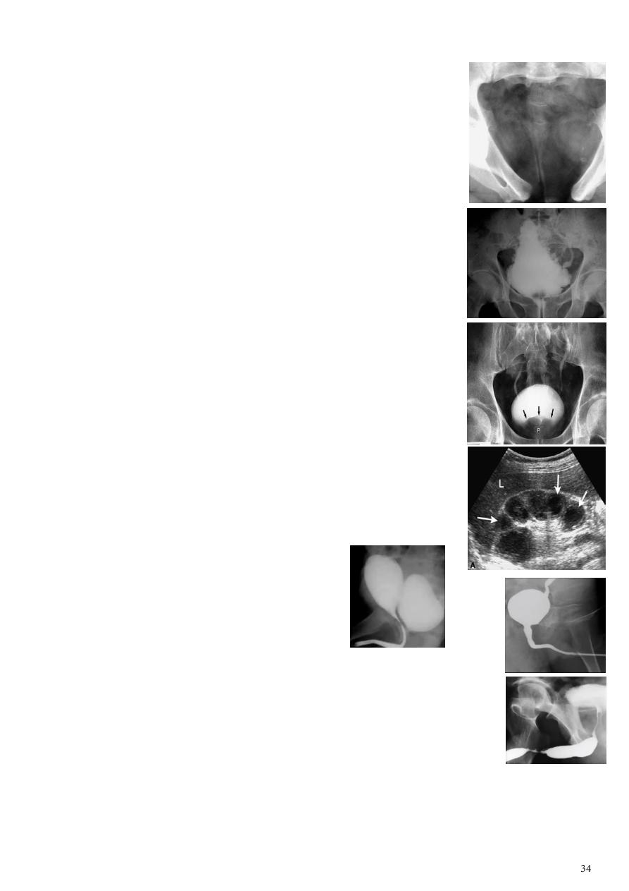
Ectopia vesicae:
o Bladder located at low position.
o Plain x-ray shows
separation
of symphysis pubis.
o Bladder exstrophy (also known as ectopia vesicae) refers to
a herniation of the urinary bladder through an anterior
abdominal wall defect.
5- Other conditions:
Neurogenic bladder:
o Irregular shape of the bladder called
christmas tree sign
.
Benign prostatic hyperplasia:
o IVU show filling defect in the bladder which could be stone,
tumor or BPH and we can differentiate between them by
clinical picture.
o In male BPH lead to
filling defect in the base
on bladder.
o In female uterus compress the bladder from above.
Multicystic dysplastic kidney:
o Hypoechoic cysts of variable sizes and shapes.
o Interfaces between cysts.
o Absence of an identifiable renal sinus.
o Lack of communication between cysts on sonograms.
o Minimal surrounding parenchyma.
Vesical diverticulum:
o Filled with contrast.
o Connected to the bladder posteriorly.
Vesicoureteric reflux:
o Detected by voiding cystourethrogram.
o The contrast ascending to the lower ureter (unilateral or bilateral).
Urethral stricture:
o Use ascending cystourethrogram.
o Area of narrowing in the urethra.
For more photos about renal system:
www.muhadharaty.com/lecture/4213
www.muhadharaty.com/lecture/3340

Part3
: Radiology of musculoskeletal system
Basic information
Types of study:
Plain bone radiographs.
Radionuclide bone scan.
CT scan.
MRI.
1- Plain bone radiographs:
Radiological signs of bone diseases need longer time to develop in adults than in
children.
Normal X-ray of the bone does not exclude the presence of pathology for example
osteomyelitis in children and scaphoid fracture in the first week.
Conventional radiological signs of bone diseases:
Decreased bone density:
o Focal (lytic area or area of bone destruction).
o Generalized (osteopenia either osteoporosis or
osteomalacia).
Increase bone density: sclerosis (focal or generalized).
Periosteal reaction:
o Definition: new bone formation by the periosteum.
o Normal periosteum is not visible.
o Types:
Solid periosteal reaction e.g. osteoid osteoma
(A).
Single laminated periosteal reaction e.g. Brodie's abscess
(B).
Multi-laminated periosteal reaction e.g. Ewing's sarcoma
(C).
Hair-on-end periosteal reaction e.g. Ewing's sarcoma
(D).
o Causes:
Trauma, Tumors, Infection, Inflammation.
Metabolic: thyroid achropachy, hypertrophic osteoarthropathy,
hypervitaminosis A.
Vascular: venous stasis.
Physiological: in 35% of infants between 1-6 months.
Congenital: syphilis, osteogenesis imperfecta.
Cortical thickening:
o Caused by lying down of new bone by the periosteum for long duration of time.
o It indicates slow process.
o The thickened cortex appears irregular and dense.
o Causes: chronic osteomyelitis, stress fracture, healed trauma, bone tumors as
osteoid osteoma.
Alteration of trabecular pattern:
o Definition: reduction in the number of trabeculae with alteration in the remaining
trabeculae.
o In osteoporosis there is cortical thinning and trabeculae that remain become
more prominent than usual.
o In Paget's disease the trabeculae are thickened and extend into the compact
cortex that normally devoid of trabeculae.
Alteration in the shape of the bone:
o As in osteogenesis imperfecta, acromegaly & expanding bone tumors.
Alteration of bone age:
o The best site of assessment of bone age is at the wrist, hands, and in newborn the
knee joint.
o In cretinism there is delayed appearance of the epiphyseal bone centers.
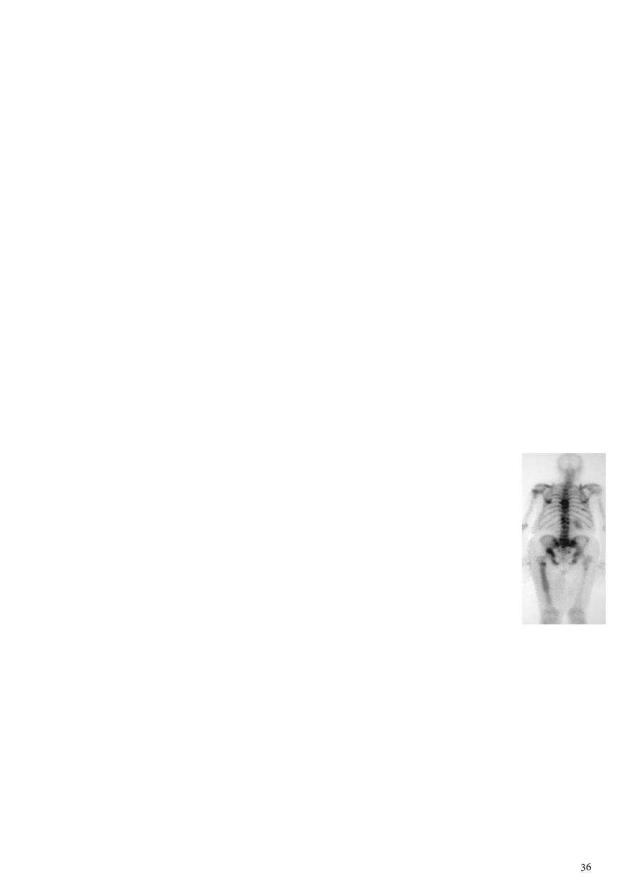
2- Radionuclide bone scan:
Tc99m- labelled with phosphate complex is a bone seeking agent.
Given IV and excreted in urine.
Advantages any lesion in the bone will take it and appear as hotspot
area in the bone.
Disadvantages it is not specific and cannot differentiate between
different bone diseases because it is taken up by soft tissue
calcifications, areas of tissue damage, soft tissue tumors.
Positive scan shown as increased uptake (hot areas) seen in trauma,
tumor, infection, infarction, Paget's disease.
Indications:
o Detection of metastases.
o Detection of osteomyelitis.
o Determination if the lesion is solitary or multiple.
o Determination (in equivocal cases) of whether an abnormality seen on radiograph
is significant or not.
o Investigation of clinically suspected bone lesion despite normal radiographs.
o Investigation of painful hip prosthesis.
3- CT scan:
CT scan in bone imaging is similar to x-ray but with high level of
radiation emitted.
Bone window setting is required (Window width \window
level 3000\450 ) for optimum results.
Indications:
o Abnormality in complex bones (spine, pelvis, face, skull).
o In orthopedic (compound fractures, gunshot injury, knee joint injury, shoulder
dislocation).
o Determination of the local extent of bone tumor (within, outside the bone, soft
tissue invasion).
o Planning for surgery (3D CT in planning corrective surgery for fracture & bone
deformity).
o Guide for bone biopsy.
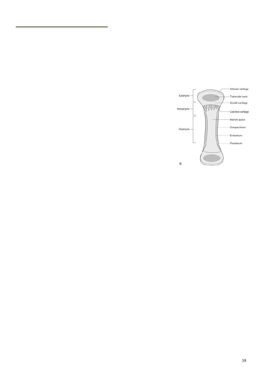
Bone:
o Cortex need time for H+ molecule to enter and give signal.
o Medulla bone marrow / fat (not need time to give signal because H+ molecule
enter through it easily).
Use US for:
o Abscess.
o Cellulitis.
o Sub-periosteal reaction.
4- Magnetic resonance imaging (MRI):
Calcified structures produce signal void areas on MRI.
Indications:
o Showing the intra and extra-osseous extent of bone pathology.
o Investigation of disc (bulging, herniation, prolapse) and spinal stenosis.
o Grading of disc lesions.
o Assessment of soft tissue masses, cartilage, ligaments, meniscal injury, avascular
necrosis.
o Used when nerve being compressed.
Solitary Bone Lesion:
Causes:
Bone tumors (benign, malignant).
Tumor like conditions (fibrous cortical defect, fibrous dysplasia, bone cyst).
Osteomyelitis.
Conditions of uncertain origin (Langerhans histiocytosis, osteoid osteoma).
Assessment of radiological findings in bone lesion:
Check the age and sex of the patient:
o Osteosarcoma (10-35 years).
o Ewing's sarcoma (3-5 years).
Site:
o Metaphyseal lesion (osteomyelitis).
o Sub articular (giant cell tumor).
o Appendicular skeleton (primary bone tumor).
o Axial skeleton (multiple myeloma, metastases).
Edge (zone of transition):
o Well defined clear cut with narrow zone of transition benign or slowly growing
lesion.
o Ill-defined wide zone of transition aggressive rapidly growing lesion
(osteomyelitis, malignant tumors).
o Well defined lytic lesion with no sclerotic margin metastases, myeloma.
Adjacent cortex:
o Cortical destruction (osteomyelitis, malignant tumors).
o Cortical expansion with no destruction (fibrous dysplasia, enchondroma).
Periosteal reaction:
o Osteomyelitis.
o Malignant tumors (osteosarcoma, Ewing's sarcoma).
o Metastasis (Neuroblastoma).
Calcification:
o Well defined, patchy popcorn (cartilaginous origin).
o Ill-defined speckles (osteoid forming tumors as in osteosarcoma).
Soft tissue swelling:
o Ill-defined swelling with blurring of the tissue fat planes due to edema
inflammation as osteomyelitis.
o Well defined swelling with displacement of clear cut fat planes tumors.
Malignant bone tumors:
Information:
Secondary bone tumors (metastases) are the commonest malignant tumors affecting
the bone.
Osteosarcoma is the commonest primary malignant bone tumor in young adults.
Use conventional X-ray, CT, MRI (show the extent within the bone marrow, soft
tissue involvement).
General features on X-ray are:
Area of bone destruction or sclerosis
With ill-defined margins.
Wide zone of transition.
Periosteal reaction.
With or without cortical destruction.
Soft tissue swelling.
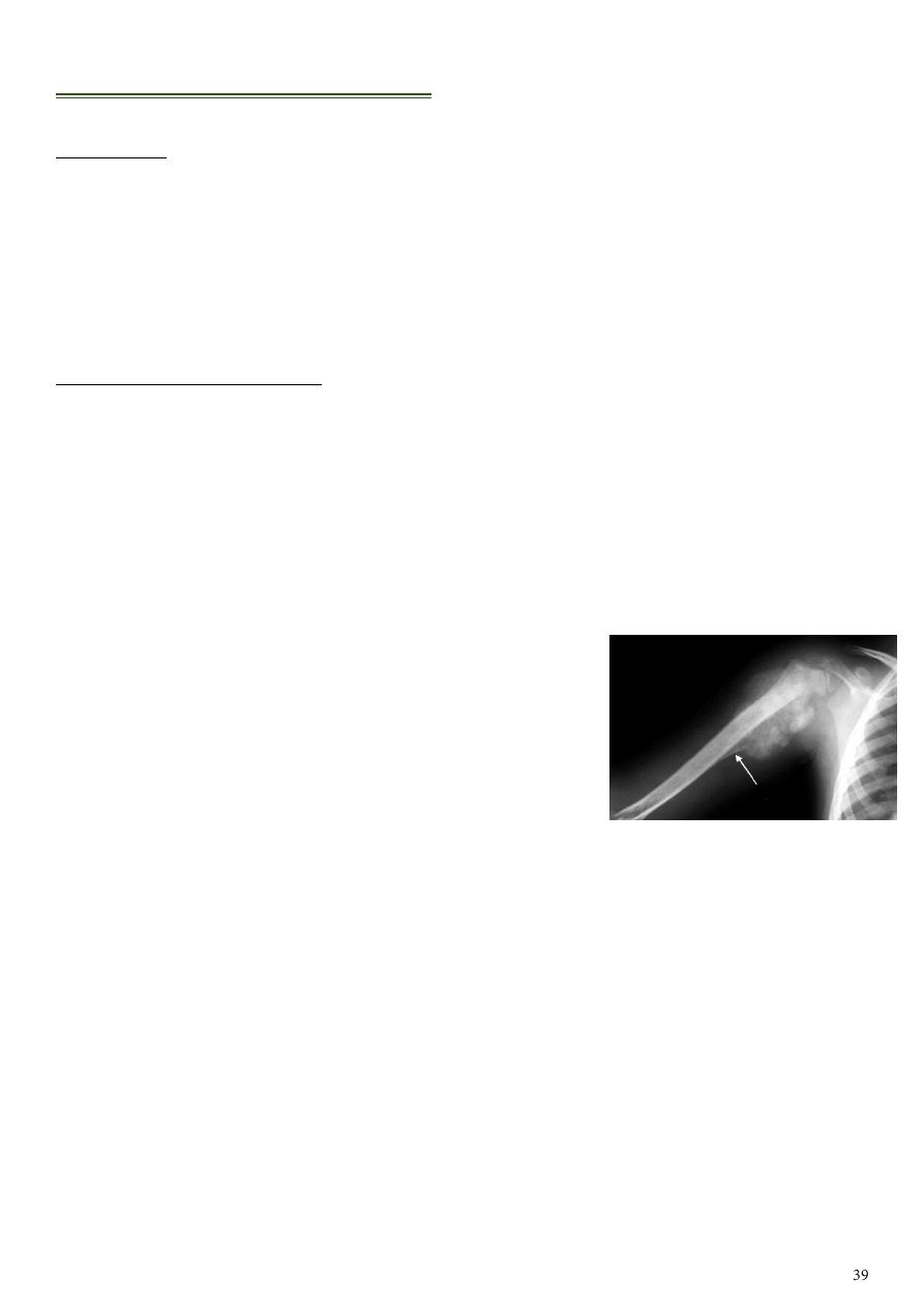
1- Osteosarcoma:
Age: 5-20-years or in elderly with Paget's disease.
Site: metaphyseal around the knee joint.
Presentation: history of trauma, pallor, fever, anorexia.
Types:
o Lytic destruction of cortex, multiple lesions.
o Blastic new bone formation in dense form along with excessive area of
sclerosis.
o Mixed there is destructive lesion in the center surrounded by area of sclerosis.
Plain x-ray findings:
o Poorly defined bony destruction.
o
Sun ray speculation
periosteal reaction.
o
Codman's triangle
elevation of the periosteum at the margin.
o Cortical destruction.
o Soft tissue swelling.
2- Chondrosarcoma:
Age: 30-50 years.
Site: pelvic bones, scapula, humerus, femur.
Findings:
o Ill-defined expanding lytic lesion.
o Flecks of calcification.
o Periosteal reaction.
o
Large extra osseous
component.
3- Ewing's sarcoma:
Highly malignant with tendency to metastasize.
Age: children.
Site: shaft of long bone.
Findings: ill-defined destruction with
onion peal
periosteal reaction
.
Note: sometimes we find
sunray speculation
in
Ewing's sarcoma.
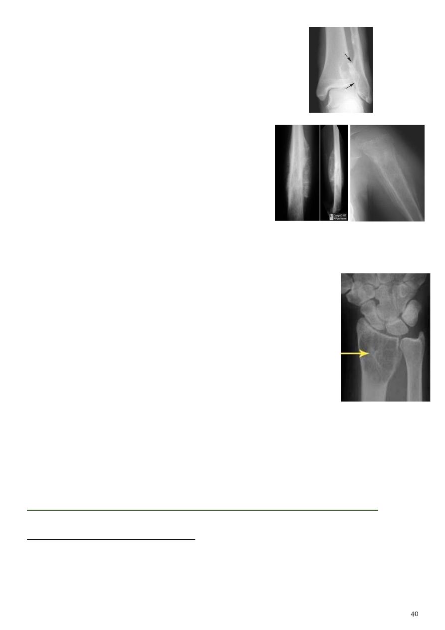
4- Giant cell tumor:
Slowly growing, locally invasive, rarely metastasize.
Age: after closure of epiphysis (20-40 years).
Site: around knee & wrist joints.
Findings:
o Lytic expansile lesion (
soap bubble appearance
).
o
Sub articular in location
.
o Not clearly defined margin.
o Thinning of the cortex (sometimes with destruction of cortex).
o No soft tissue affection.
o It is centric or peri-centric.
o Not pass through the diaphyseal plate.
Benign bone tumors and tumor like conditions:
Features of benign tumors in X-ray film:
Well demarcated.
Cortical expansion but no destruction (unless pathological fracture occurred).
No periosteal reaction (unless pathological fracture developed).
Sunray speculation onion peal
No soft tissue mass.
No or little increase in uptake on bone scan (unless pathological fracture developed).
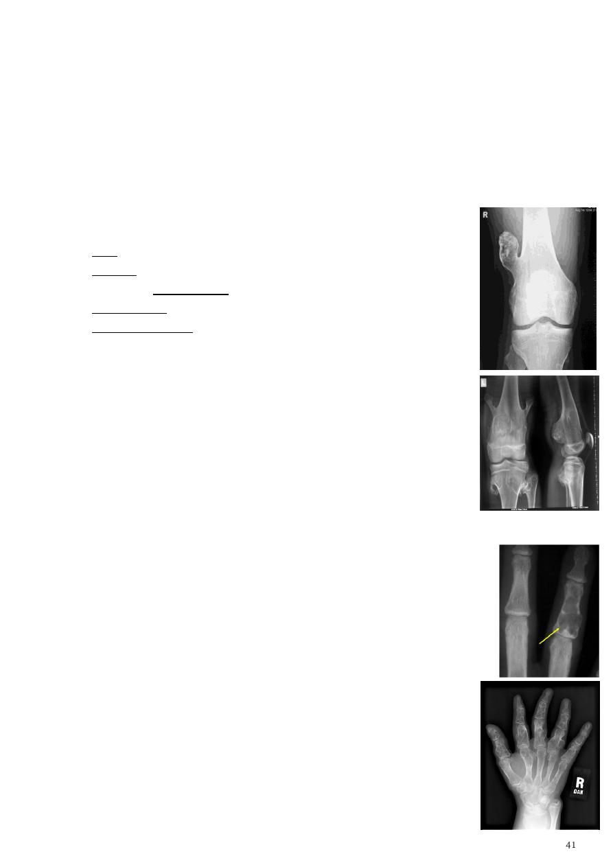
1- Osteochondroma (exostosis):
Exostosis
is cartilage-covered bony projection on the external surface of a bone.
Most common benign bone lesion.
Age: < 20 years.
Location: tibia, femur, humerus.
Malignant transformation (in < 1%) if:
o Pain in the absence of fracture, bursitis, nerve compression.
o Growth of lesion after skeletal maturation
o Dispersed calcifications in the cap.
o Enlargement of lesion.
o Increased uptake on bone scan.
Types:
o Pedunculated: slender pedicle directed away from growth plate.
o Sessile: broad base.
Characteristic findings:
o Metaphyseal location (cartilaginous origin).
o Continuous with parent bone.
o
Cauliflower-like calcification
in the chondrous portion of cap.
o Lesion
grows away
from joint.
Multiple osteochondromas called
Diaphyseal aclasia
.
If the thickness > 1 cm of cartilaginous cap by CT, > 2 cm by MRI give
high possibility of malignant transformation.
2- Enchondroma:
Site: small bones of the hands & feet.
Clinical features: painless asymptomatic swelling.
Findings:
o Lytic lesion.
o Expansion &thinning of the cortex.
o No periosteal reaction (unless pathological fracture develops).
1 % risk of malignant transformation in solitary type
Multiple enchondromatosis (
Ollier's disease
) affect long bones &
carry 10% risk of malignant transformation.
3- Osteoma:
It is excess lying down of normal bone (not tumor).
Localized masses of mature bone on the endosteal or
periosteal surface of cortex.
Commonly in the skull or paranasal sinuses.
Could be single or multiple.
Multiple osteoma associated with
Gardner's syndrome
(familial colorectal polyposis).
4- Fibrous cortical Defect & Non ossifing Fibroma (NOF):
Common incidental findings in children.
Site: affect diaphysis of long bone.
Findings: well defined lucent areas in cortex with sclerosed margin.
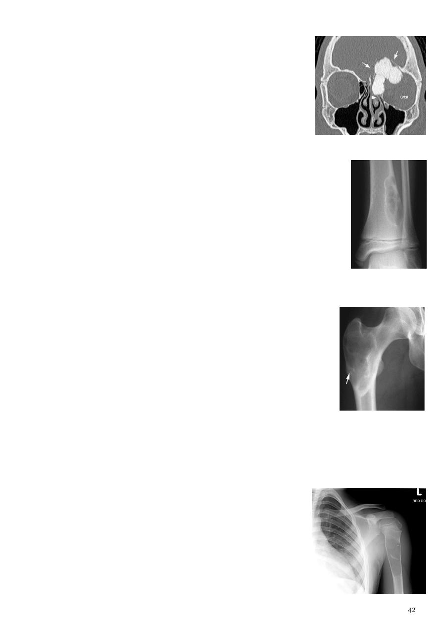
5- Fibrous Dysplasia:
Defect in the osteoblastic development and maturation as a result of mutation.
Monostotic:
o Age: 10~30yrs.
o Site: ribs, proximal femur, craniofacial bones.
o Usually asymptomatic.
Polystotic:
o Age: at first decade.
o Site: femur, tibia, pelvis, cranial bones, spine, feet.
o Usually unilateral, asymptomatic.
It causes leg length discrepancy,
Findings:
o
Shepherd crook deformity
(coxa varus angulation of the proximal femur).
o Facial asymmetry, rib deformity, tibial bowing associated with
hyperparathyroidism, acromegaly, DM.
o Lytic expansile lesion.
o
Ground-glass matrix mineralization
.
o Sclerosed margin.
6- Solitary bone cyst:
Age: young adults & children.
Site: long bones.
Findings:
o Well-defined expanding lytic lesion.
o
Fallen fragment sign
a piece of cortical bone has broken off and descended
through the serous fluid contained within the lesion and can be seen in the
dependent portion of the lesion.
o A fallen fragment sign is said to be pathognomonic for a unicameral bone cyst.
7- Aneurysmal bone cyst:
Benign but may be aggressive in appearance.
Age: children, young adults.
Site: spine, long bones, pelvis.
Occur in the metaphyseal area.
DDx: giant cell tumor.
Findings:
o Purely lytic lesion (
soap bubble appearance
).
o Massive cortical expansion (can expand upward with expansion).
o CT and MRI show blood pools (
fluid -fluid levels
within the cyst).
8- Osteoid osteoma:
Age: young adults.
Site: tibia, femur.
Presentation: benign, fever, sweating, painful escpically at night.
It is excessive lay down of bone inside the cortex + sclerosis.
Managed by aspiration to relieve the pain.
Findings:
o Lucent area surrounded by calcification.
o
Nidus
surrounded by
sclerotic rim
with or without periosteal reaction.
o Radionuclide bone scan: area of increased uptake.
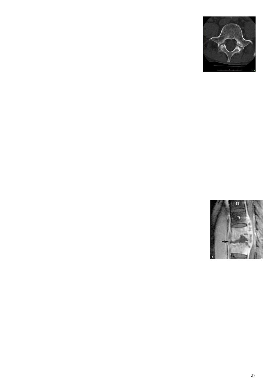
9- Hemangioma:
Occur in the vertebra.
It is asymptomatic condition.
No change in the size of vertebral disc.
Fat tissue in the vertebra.
Incidental finding.
MRI show
vertebral bodies high lightened
.
Multiple focal bone lesions:
1- Metastases:
Seen in bone with active haemopoiesis (spine, skull, ribs, pelvis, humeri, femora).
Metastases site of predilections:
o Vertebral metastases (94%) causing primarily affection of the pedicle.
o Intradural extramedullary metastases (5%)
o Intramedually metastases (1%)
Types of metastasis:
o Osteolytic (most common causes) neuroblastoma (in children), breast (adult
female), bronchus (adult male), thyroid, kidney, colon, vertebral pedicles.
o Osteoblastic prostate, breast, carcinoid, TCC of bladder, neuroblastoma.
o Mixed breast, prostate, lymphoma.
o Solitary expansile bubbly metastases with soft tissue involvement thyroid,
kidney.
o Bone metastases with sun burst periosteal reactions prostate, retinoblastoma,
neuroblastoma.
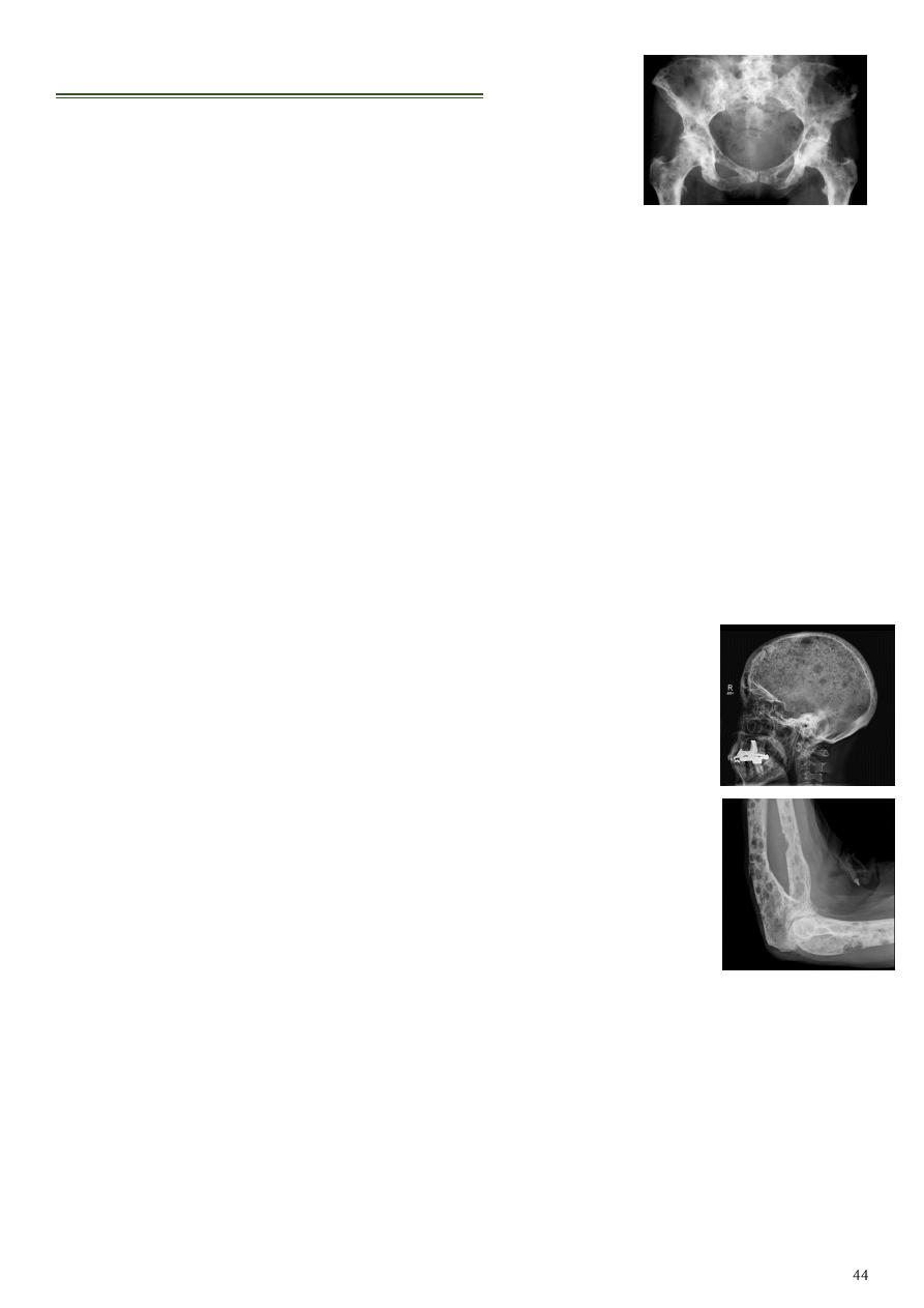
2- Multiple Myeloma:
Site: axial skeleton.
Findings:
o Well demarcated lytic lesion occasionally with expansion of the
bone.
o
Rain drop appearance
in the skull.
o In the spine Generalized form can resemble osteoporosis.
Solitary type (
plasmacytoma
):
o Represent early stage of MM; precede it by 1-20 years.
o Negative IgG spike in the serum.
o It affects the thoracic, lumbar spine, pelvis, ribs, femora.
o It is seen as expansile lytic ill-defined lesion with soft tissue mass.
Differences:
o Secondary metastasis multifocal lesion, expansile or not, bence jones protein
not present.
o Multiple myeloma multiple, expansile, lytic lesion, bence jones protein is
present.
o Myeloma resembles metastases in everything except it's more well defined,
cause bone expansion and spares vertebral pedicle.
Bone infections:
1- Osteomyelitis:
Information:
o Osteomyelitis refers to inflammation of bone that is almost always due to
infection (staphylococcus aureus 80-90%).
o Osteomyelitis can occur at any age (commonly 2-12 years of age).
o More common in males (M:F of 3:1).
o Osteomyelitis results from hematogenous spread, direct extension from trauma
or ulcers.
o Metaphysis is the most common site of bone infection because this area is highly
vascularized so there is hematogenous spread of the infection.
Location:
o Neonates metaphysis, epiphysis.
o Children metaphysis.
o Adults epiphyses, subchondral regions.
The earliest changes:
o Are seen in adjacent soft tissues +/- muscle outlines.
o Swelling and loss or blurring of normal fat planes.
o Changes may not be obvious until 5 to 7 days in children and 10 to 14 days in
adults.
Acute osteomyelitis:
o
o Periosteal irritation and reaction
Moth eaten appearance
.
o Focal bony lysis or cortical loss.
o Endosteal scalloping.
o Loss of bony trabecular architecture.
o New bone apposition.
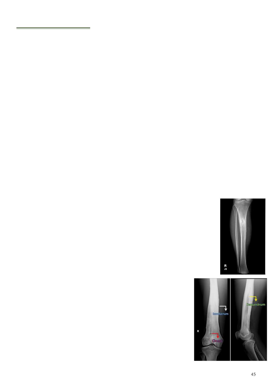
Chronic or untreated osteomyelitis:
o
dead bone represents devascularization
of a portion of bone.
o
new bone formation represents a thick
sheath of periosteal new bone surrounding sequestrum.
o
an opening in a involucrum which allows
drainage of purulent and necrotic material out of the
dead bone.
o
it is portion of tract extending beyond the
involucrum to the skin surface.
Special types of chronic osteomyelitis
(depend on type of organism and immunity)
:
o
Brodie’s abscess
a localized osteolytic lesion at the end of the long
bone surrounded by sclerosis.
o Gary’s osteomyelitis (
Sclerosing osteomyelitis
) Characterized by
localized sclerosis in the shaft of long bone, and it typically affect the
mandible. DDx ostiod osteoma.
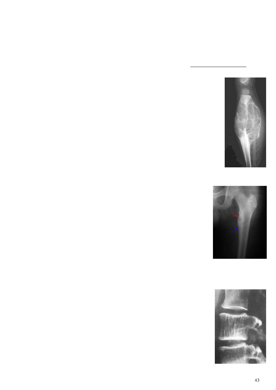
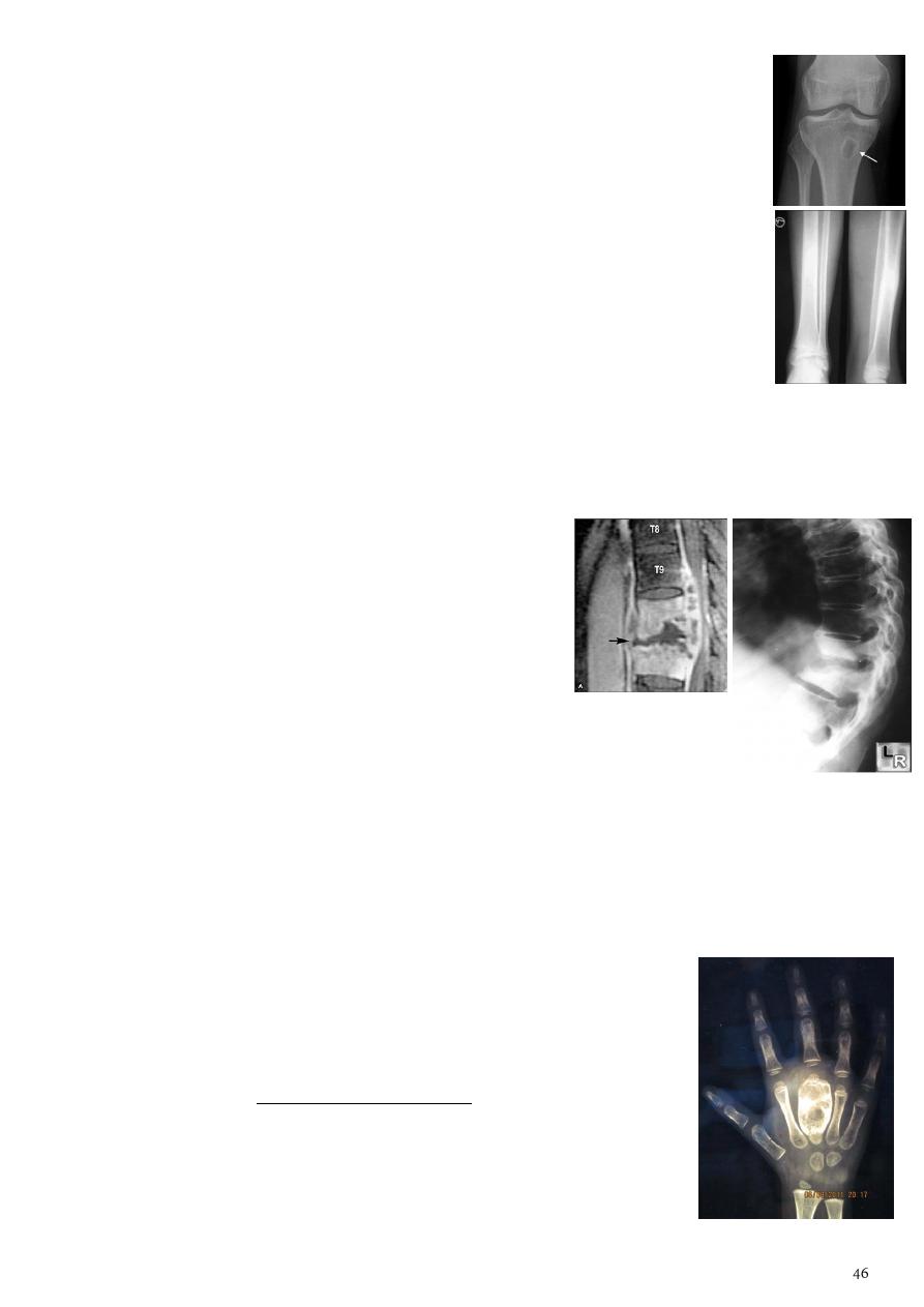
2- Tuberculosis of the bone:
Spread from infected joint.
Most common site dorsolumber part of spine.
More than 90% of adult (40 years) T.B infection located in vertebra.
Osteomyelitis in adult is less than in children because in adult there is complete
sclerosis but in children this area still vascularized / but TB in adult is more common
and can still in area of low vascularity (low O2) so T.B in adult is subarticular or
subchondral in long bones.
TB of long bones (early findings):
o Osteopenia in proximal area.
o Soft tissue swelling.
o Destruction of articular surface.
o Moth eaten not occur / sclerosis only slightly.
Pott’s Disease
(findings in the spine):
o Affect two adjacent vertebrae.
o Erosion of the superior and inferior end plates.
o Irregular narrowing of the joint space.
o Para-spinal cold abscess (seen as a fusiform para spinal soft tissue on AP or PA
view of the chest).
o End with wedging of the vertebra (anterior loss of vertebral height) lead to
angular kyphosis.
o
Gebus deformity
(wedging of the vertebra, sign of old T.B, occur after treatment
and healing).
T.B Dactylitis or spina ventosa
:
o T.B of peripheral bone is not common and usually affect the
small bones (phalanx)
o There will be destruction of the phalanx.
o Expansion of middle metacarpal bone + dense sclerosis.
o Expansion with soft tissue shadow around it.
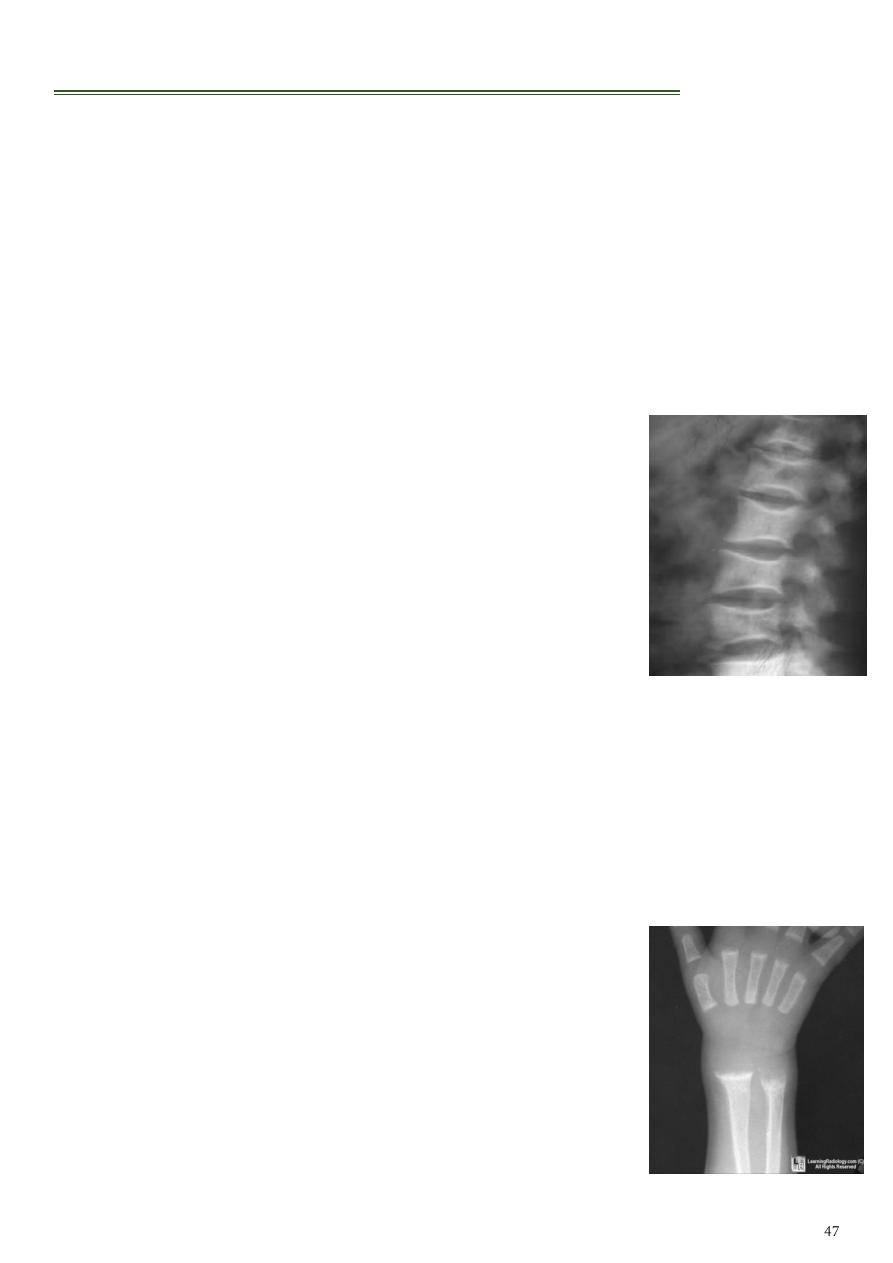
Generalized decrease in the bone density:
1- Osteoporosis:
Reduction in the bone matrix that subsequently results in reduced calcium contents.
Causes:
o Idiopathic: juvenile, senile and postmenopausal.
o Cushing syndrome & steroid therapy.
o Disuse (immobilization for fracture treatment or local pain).
o Sudeck's atrophy (disorder of the sympathetic nervous system where sever
osteoporosis and soft tissue edema occur disproportionate to the trauma or the
degree of disuse).
Findings:
o The changes are best seen in the spine.
o
Wedged
vertebra with
widening
of the disc space.
o Overall reduction in bone density.
o
Clear penciled
in cortex.
o Long bones (thin cortex, resorption of many trabeculae
but those that remain stands out clearly).
Other causes of reduced bone density:
o Metastatic carcinoma.
o Multiple myeloma.
o Hyperparathyroidism.
o Osteomalacia.
Bone mass assessed by quantitative CT or by dual energy X-ray (DEXA scan).
2- Rickets and Osteomalacia:
Poor mineralization of osteoid (decrease in number of osteoid tissue in the matrix).
If occur before epiphyseal closure then it is rickets, after that called osteomalacia.
Causes:
o Dietary deficiency of Vit.D.
o Lack of exposure to sun light.
o Malabsorption.
o Renal disorders.
Findings in rickets:
o The findings are best seen at the knee, wrist, ankles.
o
Frayed
,
cupped
,
splaying
of the metaphysis.
o Increased distance between the growing epiphysis &
metaphysis.
o Generalized decrease in bone density.
o
Bossing
and
bowing
deformity (due to softening).
o
Greenstick fractures
.
o
Rackety rosary
of the ribs.
Findings in osteomalacia:
o Decrease bone density (osteopenia).
o Thin cortex and trabeculae.
o
Looser's zones
:
These are thin short lucent lines with sclerotic margins running
across the cortex at right angle.
Best seen in the scapula, medial aspect of the femoral neck,
pubic rami.
o
Codfish appearance
vertebral collapse resulting in biconcave
vertebra with widened disc.
o
Bowing
of the femur.
o
Triradiate pelvis
in severe cases the pelvic side walls bend
inwards, lead to waddling gait.
After treatment with Vit.D = appearance of dense line of calcification
(dense sclerosis).
3- Hyperparathyroidism:
Primary hyperparathyroidism (tumors of the parathyroid glands).
Secondary hyperparathyroidism (with chronic renal failure).
Findings in hand:
o
Subperiosteal bone resorption
(in the radial aspect
of the middle phalanges).
o Resorption of the terminal tuft of terminal phalanges
and outer end of the clavicle.
o Decrease bone density with loss of corticomedullary
differentiation.
o Vascular calcification, soft tissue calcification, multiple callus
formation (
chondrocalcinosis
).
o Brown tumor (lytic expansile lesion particularly in mandible &
pelvis).
Findings in skull:
o
Salt and pepper sign
(multiple tiny hyperlucent areas in the
skull vault caused by resorption of trabecular bone).
o Loss of definition between the inner and outer tables of the skull.
o Ground-glass appearance.
o In multiple myeloma (dots = 3-4 mm), in hyperparathyroidism (dots = 1-2 mm).
4- Renal Osteodystrophy:
Occurs in patients with chronic renal failure.
Findings:
o Features of osteomalacia in adults & rickets in children.
o Features of hyperparathyroidism.
o Sclerosis: bands of increased density in the spine named as
rugger jersey spine
& across the metaphysis of long bones.
5- Osteogenisis imperficta:
Imperfect bone formation with multiple callus formation and
multiple bone fractures.
Die within 2-3 years or still birth due to associated CHD.
Findings:
o Generalized decrease in bone density
o The medulla is very thin.
o Multiple bone deformity (large, small, flat bones) due to multiple
fractures and bizarre healing.
o Widening of the sutures and
wormmian bone
formation.
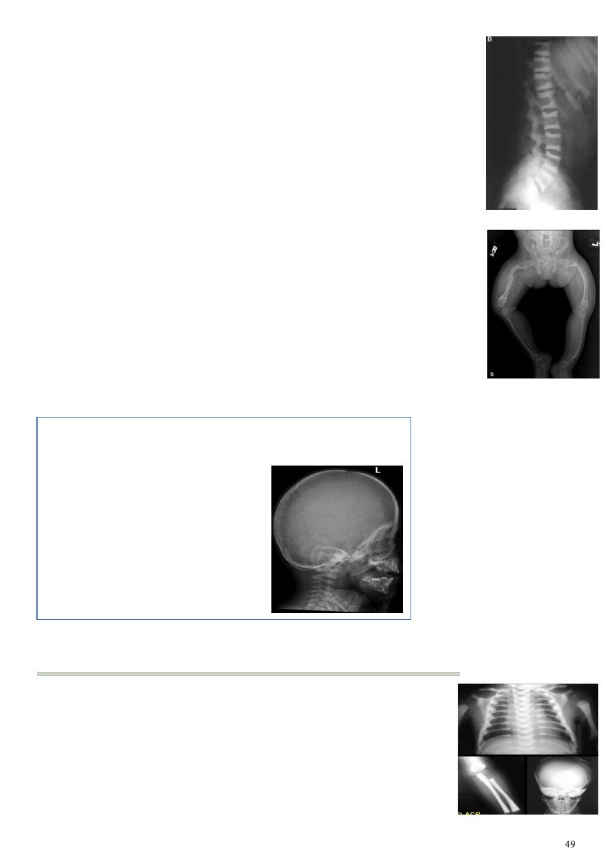
Generalized increase in the bone density:
1- Sclerotic metastases.
2- Osteopetrosis (Marble bone disease): Congenital condition, the
bone is brittle & easily fracture but heals normally.
3- Myelosclerosis: Replacement of the bone marrow by fibrous tissue
& progress to lay down new bone, splenomegaly is invariably present.
4- Fluorosis.
Wormmian bone Are a subset of the small intra sutural
bones that lie between the cranial sutures mainly seen
around the lambdiod sutures.
Causes:
Osteogenesis imperfecta.
Rickets.
Cleidocranial dysostosis.
Hypothyroidism.
Down syndrume.
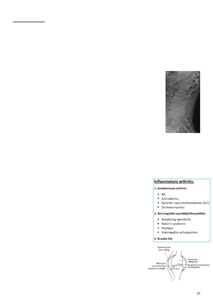
Arthritis:
1- Osteoarthritis (OA):
It is type of degenerative joint disease (DJD).
80% of population > 50 years have radiological evidence of OA.
Types:
o Primary OA no underlying local etiological factors, abnormally high mechanical
forces on normal joint, age related.
o Secondary OA trauma, inflammatory arthritis,
hemochromatosis, acromegaly, congenital hipdysplasia,
osteonecrosis, loose bodies, normal forces on abnormal joint.
Radiographic features (OA changes):
o Narrowing of joint space (usually asymmetrical).
o Subchondral sclerosis.
o Subchondral cysts (true cysts or pseudocysts).
o Osteophytes.
o Lack of osteoporosis.
In the Spine:
o Lower cervical and lower lumbar spine are most commonly affected.
o Osteophytes may encroach on neural foramina (best seen on oblique views).
o Vacuum phenomenon: gas (N2) is pathognomonic of the degenerative process.
o OA of the spine occurs in the apophyseal joints.
o
Degenerative spondylolisthesis
(pseudospondylolithesis).
o No wedging in vertebra like that occur in T.B.
In the knee OA changes + elongation and spike formation
of tibial space (tibial sper).
2- Rheumatoid arthritis (RA):
It is type of inflammatory arthritis.
F:M = 3:1.
Start at peri-articular area at site of attachment of ligaments.
Early changes:
o Peri articular soft tissue swelling (edema, synovial
congestion)
o Peri articular osteoporosis in symmetrical distribution
(hallmark)
o Preferred sites of early involvement:
Hands:
2
nd
and
3
rd
MCP joint.
Feet:
4
th
and
5
th
MTP joint.
Late changes
o Erosions (
pannus formation
, granulation tissue).
o Erosions of the ulnar styloid and triquetrum are characteristic.
o Subchondral cysts formation (results from synovial fluid).
o Subluxations.
o Carpal instability.
o Ulnar deviation.
o Fibrous ankylosis (late finding).
o Narrowing or loss of joint space between metacarpal bones.
o Boutonniere deformities.
o Swan-neck deformity.
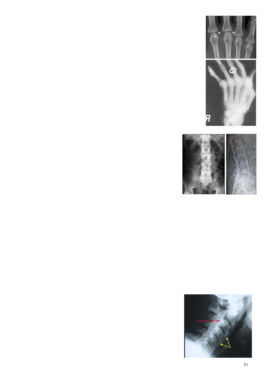
3- Ankylosing spondylitis (AS):
Information:
o Seronegative spondyloarthropathy of the axial
skeleton and proximal large joints.
o Clinical: males >> females.
o HLA-B27 in 95%.
o Insidious onset of back pain and stiffness.
o Onset: 20 years.
Radiographic features:
o
Sacro-iliac joint
start as inflammation then destruction then sclerosis then
ankylosing.
o Thoracolumbar spine bridging osteophyte or called syndesmophytes (lateral
view:
squaring
) (PA view:
Bamboo spine
).
o Vertebra filled with osteoid tissue lead to sclerosis.
o Ligamentous ossification calcification of anterior spinal ligament.
o Ankylosed spine (fracture).
o Enthesopathy (whiskering of tuberosities).
o Arthritis of proximal joints (hip > shoulder).
o Widening of joint space, erosions, osteophytes.
4- Diffuse idiopathic skeletal hyperostosis (DISH):
Radiographic features:
o Flowing osteophytes (at least
four contiguous
vertebral
bodies).
o Preserved disk height.
o No sacroiliitis or facet ankylosis.
o Calcification of ligaments and tendons.
o Associated with hypertrophic DJD.
It is one of the DDx of ankylosing spondylitis:
o Differentiate between them by the changes in sacro-iliac joint.
o Spinal changes are similar in both.
5- Erosive osteoarthritis:
Characteristically affects middle-aged women.
Radiographic features:
o Erosive and productive changes of DIP and PIP.
o
Gull-wing pattern
: secondary to central erosions,
occur in distal and proximal phalanges.
o Marginal proliferation osteophytes.
o Interphalangeal fusion may occur.
Typical involvement of
first CMC
may help distinguish
erosive OA from RA, psoriatic arthritis, and adult Still's disease.
No affection to metacarpal joints (RA affect them).
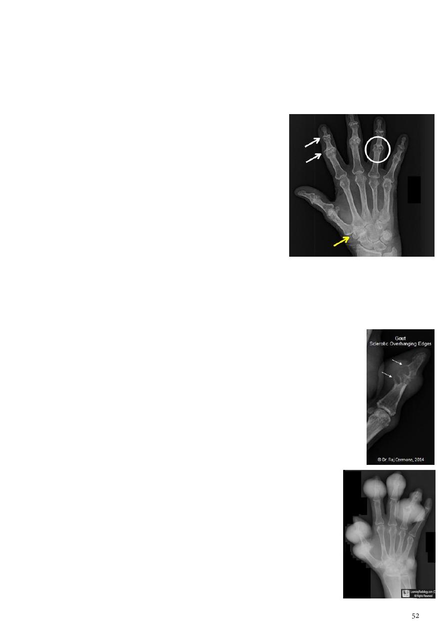
6- Gout:
Information:
o Site of predilection is big toe (First MTP).
o Lower extremity > upper extremity.
o Small joints > large joints.
o Presence of tophi (excessive amount of uric acid).
o Erosions and tophi only seen in longstanding disease.
Radiographic features:
o Soft tissue and bursa deposition:
Tophi: juxtaarticular, helix of ear.
Bursitis: olecranon, prepatellar.
o Tophi lead to
punched-out lytic lesion
+
overhanging edge
(seen in lateral view as sclerosis at the base of punched out
lesion).
o Tophi calcification
o Marginal, peri articular erosions: overhanging edge
o Erosions may have sclerotic borders.
o Joint space is preserved.
o Chondrocalcinosis.
o
Snow-night appearance
.
7- Septic arthritis:
Cause:
o Organism Staphylococcus aureus (most common), B-Streptococcus in infants,
Salmonella in sickle cell patients.
o Hematogenous spread to synovium and subsequent
spread into the joint.
o The diagnosis is made by joint aspiration.
Radiographic features:
o Joint effusion.
o Juxtaarticular osteoporosis.
o Destruction of subchondral bone on both sides of the joint.
Same as TB arthritis but joint space is preserved and it healed by sclerosis.
8- Neuropathic arthritis (Charcot's joint):
Causes:
o Diabetes neuropathy: usually foot.
o Tertiary syphilis: usually knee.
o Syringomyelia: usually shoulder.
o TB, immobility, long standing polio.
Radiographic features:
o Joint instability: subluxation or dislocation.
o Prominent joint effusion.
o Hypertrophic type (20%) marked fragmentation of articular bone, much
reactive bone.
o Atrophic type (40%) bone resorption of articular portion.
o Combined type (40%).
Other changes:
o Reduction of joint space.
o Osteophytes.
o Osteopenia.
o OA changes.
o Joint deformity.
Other conditions:
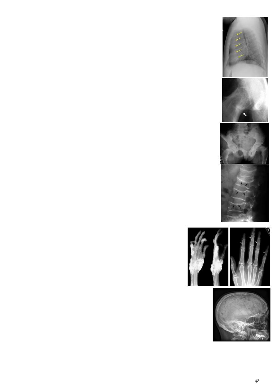
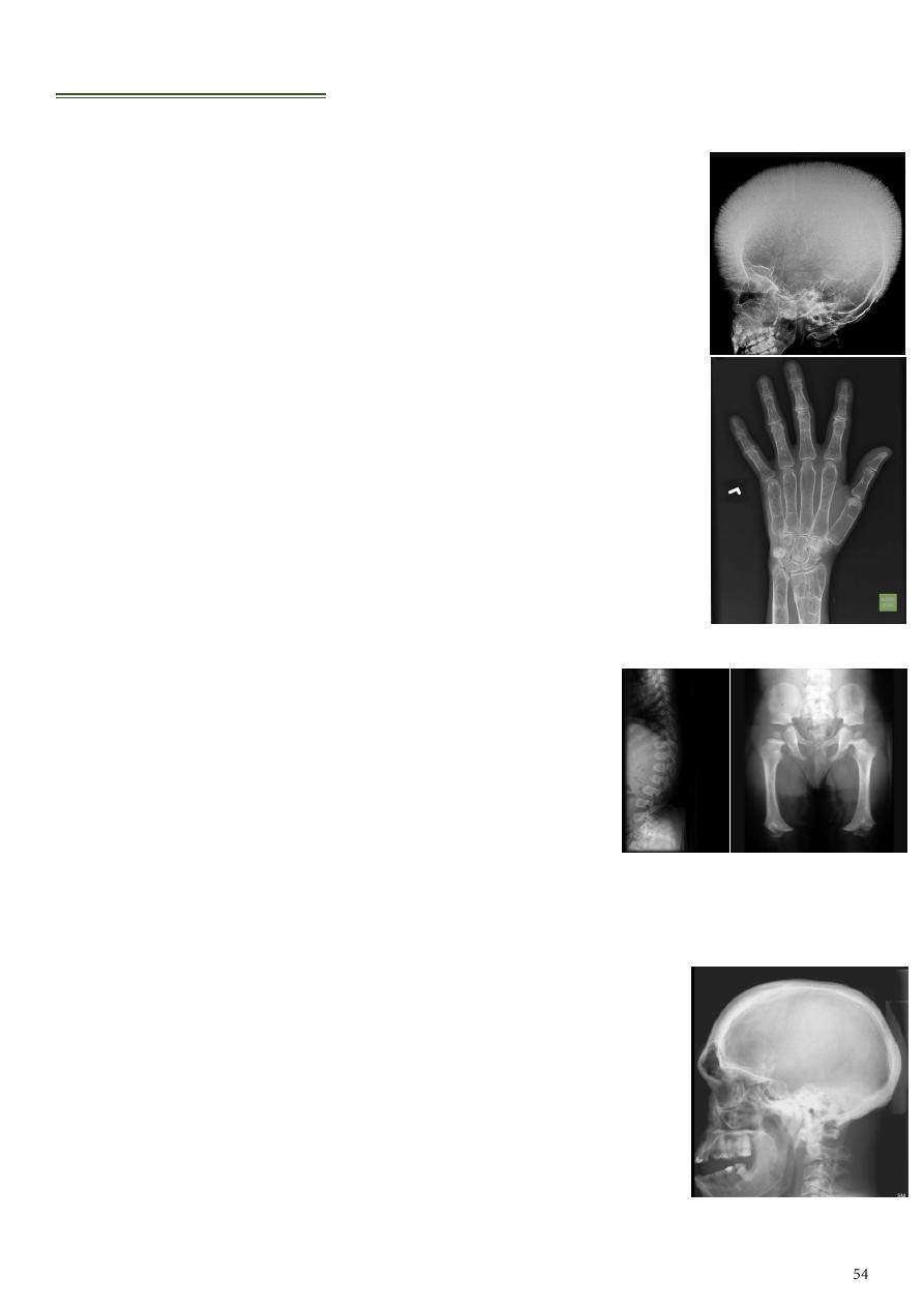
1- Alteration in the Trabecular Pattern in Hemolytic anemia:
Thalassemia and sickle cell anemia result in bone hyperplasia.
Sickle cell anemia in addition causes infection & infarction.
Bone marrow hyperplasia:
o Thinning of cortex.
o Increase thickness of bone with resorption of some trabeculae
and increase thickness of the remaining.
o Increase diploic space thickness with vertical striation resulting
in
hair on end appearance
.
o Enlargement of ribs.
o Widening of the phalanges (
cylindrical shape appearance
of
the hand).
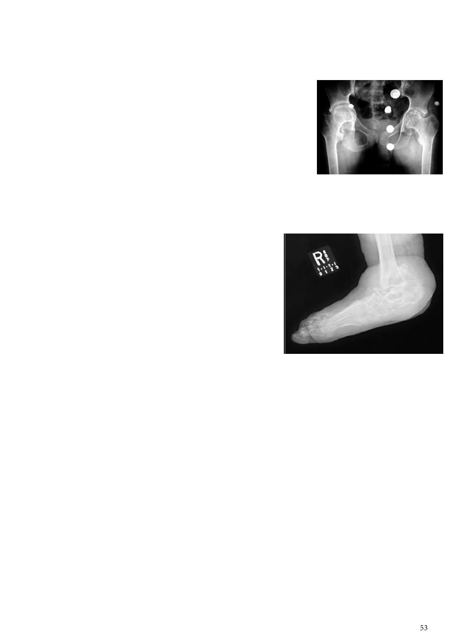
Infarction:
o Infarction of the bone ends results in sclerosis and flattening of
the femoral & humeral heads
o Medullary infarction appears as lytic areas with or without
periosteal reaction.
o Healing appears later as areas of irregular medullary calcification.
2- Changes in Bone Shape:
Diaphyseal achalasia
(in page 39)
.
Achondroplasia:
Defective ossifications of bones formed in cartilages.
Shortening of the shaft of the long bones with
distal metaphyseal flaring
.
Deformity of the pelvis (
contracted pelvis
).
Bullets like vertebrae
due to anterior beaking.
Acromegaly:
The bone changes are maximum at the hands, feet & face.
Increase joint space due to overgrowth of cartilage.
Enlargement of the tufts of the terminal phalanges.
Enlargement of the pituitary fossa
double floor sign
.
Thickening of occipital tubercle
.
Widening of the skull vault and
frontal bone posing
.
Enlargement of the sinuses & mastoid air cells.
Prognathisim
: widening of angle between the body and ramus of the mandible.
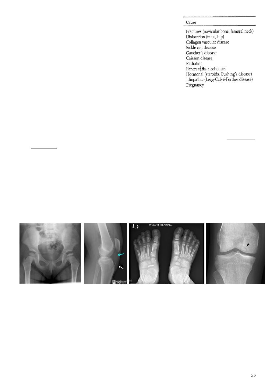
3- Osteonecrosis:
Other names: avascular necrosis, ischemic necrosis,
aseptic necrosis.
Causes: Interruption of arterial supply,
Intra/extraosseous venous insufficiency.
MRI Most sensitive imaging modality: 95%-100%
sensitivity.
Plain film findings:
o Areas of radiolucency, fissuring, fragmentation, bone collapse and condensation.
o End with dense and flat bone with loss of bone contour and secondary
osteoarthritis.
4- Osteochondrosis:
Legg-Calve-Perthes disease
osteochondrosis of the femur head, lead to mushroom
deformity, for early OA changes.
Scheuermann's disease (adolescent Kyphosis) osteochondrosis of the vertebral end
plates, lead to vertebral wedging ending with kyphosis.
Osgood-Schlatter
osteochondrosis of the tibial tubercle.
Blount's disease tibial epiphysis.
Kohler's
osteochondrosis of the Navicular bone (flat disk like shape).
Kienbock's osteochondrosis of the lunate bone.
Osteochondritis dissecans medial femoral condyle, talus, trochlea, resulting in
intra-articular loose body.
5- Developmental dysplasia of Hip (congenital dislocation of the hip):
Information:
o An abnormally lax joint capsule allows the femoral head to fall out of the
acetabulum, leading to deformation.
o Predisposing factors for the development of CDH Abnormal ligamentous laxity,
Acetabular dysplasia.
o CDH occurs most commonly (70%) in the left hip, bilateral involvement is seen in
5%.
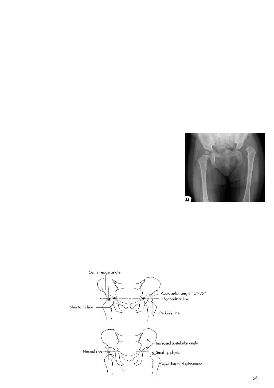
Radiographic features:
o 1-3 months:
Best diagnosed by US (safe).
In CDH < 50% of femoral head is covered by acetabulum (normally = 50%).
o At 3-6 months :
Plain x-ray with Von Rosen view (abduction of the thigh 45 degree and internal
rotation).
In DDH the lines that drown through the femura will meet in higher level than
the normally should at lumbosacral joint.
Never crossed at lumbo-sacral joint.
o 6 months and later:
Use AP view to visualize femoral epiphysis.
Superolateral displacement of proximal femur (disturbed shenton’s line)
Femoral head is located lateral to Perkin's line.
Increase in acetabular angle.
Small capital femoral epiphysis.
Retardation of epiphyseal plate (tubercle).
o Other features that are sometimes present:
Abnormal sclerosis of the acetabulum.
Shallow acetabulum.
Formation of a false acetabulum.
Delayed ossification of femoral head.
Old neglected CDH pseudoathrosis of femoral head with iliac bone.
Shenton line is drawn along the inferior border of the superior pubic ramus and
should continue laterally along the infero medial aspect of the proximal femur as a
smooth line. If there is supero lateral migration of the proximal femur due to DDH
then this line will be discontinuous.
Perkin line is drawn intersecting the lateral most aspect of the acetabuler roof &
iliac crest.
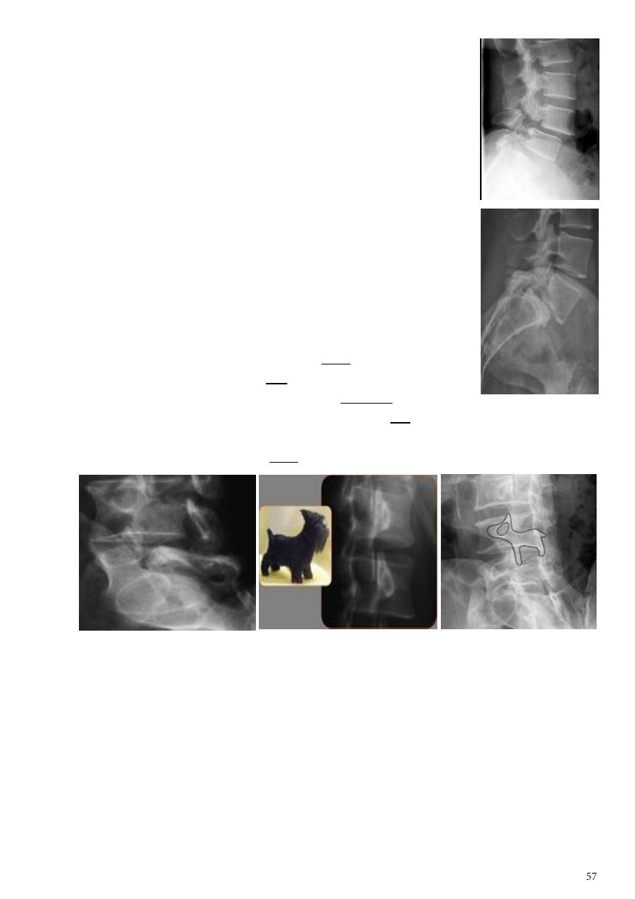
6- Spondylolysis and spondylolisthesis:
Spondylolisthesis:
o Is a term denoting forward or backward movement of a
vertebra relative to the vertebral segment below.
o Typically due to spondylolysis (pars interarticularis defects).
Spondylolysis:
o Is a defect in the pars interarticularis of the neural arch.
o The portion of the neural arch that connects the superior and
inferior articular facet in which cause defect in the neck of
scotty dog.
Scotty dog sign:
o Refers to the normal appearance of the lumbar spine when
seen on oblique radiographic projection.
o On oblique views, the posterior elements of vertebra form
the figure of a Scotty dog with:
The transverse process being the nose.
The pedicle forming the eye.
The inferior articular facet being the front leg.
The superior articular facet representing the ear.
The pars interarticularis (the portion of the lamina that lies between the
facets) equivalent to the neck of the dog.
For more photos about musculoskeletal system:
www.muhadharaty.com/lecture/4214
www.muhadharaty.com/lecture/4400
www.muhadharaty.com/lecture/4215
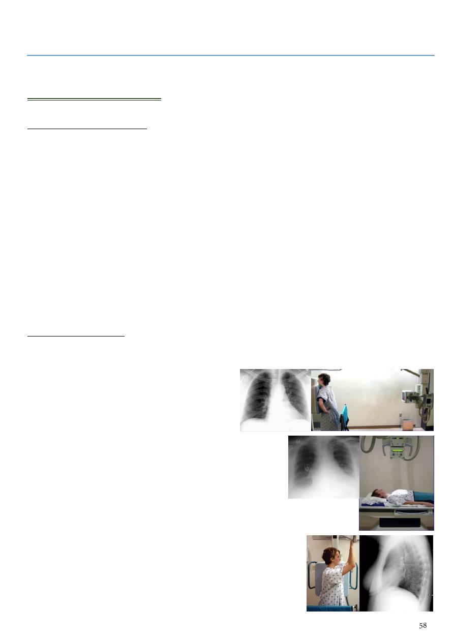
Part4
: Radiology of the chest
Basic information
Type of imaging modalities:
Plain chest x-ray.
Fluoroscopy.
Computerized tomography CT.
MRI.
Radionuclide lung scan.
Ultrasound.
Pulmonary angiography.
Positron emission tomography (PET).
ECHO.
Doppler.
Magnetic resonance angiography (MRA).
Plan chest radiograph:
1- Standard views:
Postero-anterior (P/A) view:
o The x-ray tube is 72 inches away from
the patient.
o The patient is in the standing
position.
o Most common used view.
Anterio-posterior (A/P) view:
o The x-ray tube is 40 inches away from the
patient.
o The patient is in the supine position.
o It shows magnification of the heart and widening of the
mediastinum.
o AP view is less useful so used in very ill patient who
cannot stand erect.
Lateral (Right or left) view:
o Used to see the site of the mass.
o Sometimes used in pleural effusion.
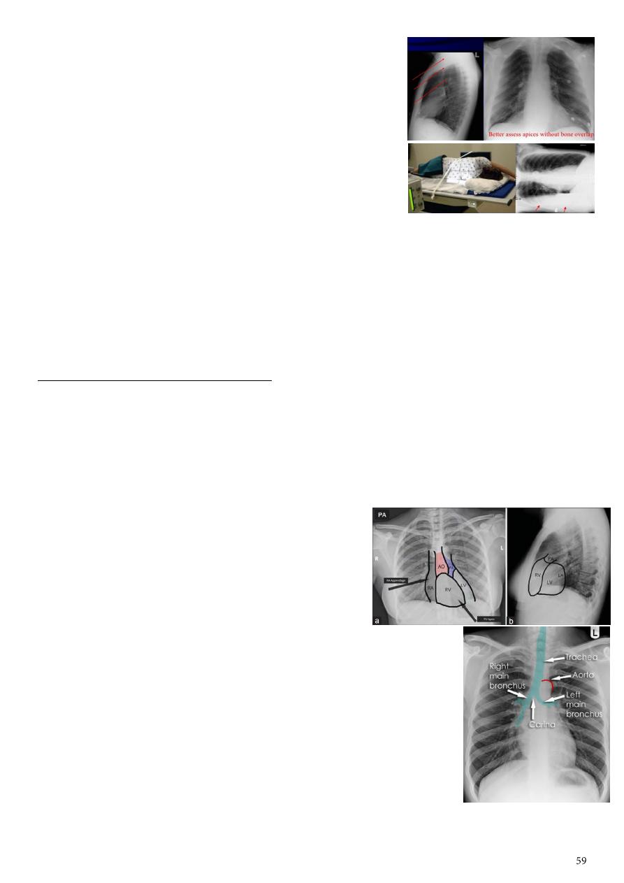
2- Additional views:
Oblique view (ribs).
Apical lordotic view:
o Used to get clear view due to presence of clavicle.
Expiration view.
Decubitus view:
o Used to detect small amount of pleural effusion.
Notes:
Correct film: distance from vertebra to clavicle is same at both sides, if not called rotated film so the
patient should repeat the film.
Posterior ribs appear horizontal, and anterior ribs appear oblique.
The film should be taken in inspiratory phase, If the image is acquired in the expiratory phase, the
lungs are relatively airless and their density is increased. Also, the raised position of the diaphragm
leads to exaggeration of heart size, and obscuration of the lung bases.
Diaphragm level anteriorly (till 6
th
rib), posteriorly (till 10
th
rib).
Systemic approach in chest radiograph:
Bone and soft tissue:
o Look at the soft tissues, especially around the neck, the thoracic wall, and the
breasts.
o Fat is less dense than muscle and so appears blacker.
o Note that the edge of fat is smooth.
o Irregular areas of black within the soft tissues may represent air tracking in the
subcutaneous layer "surgical emphysema".
Heart:
o The heart has a conical form and is enclosed
by pericardium.
o It is positioned posteriorly to the body of the
sternum.
o With one-third of it is situated on the right and two-thirds
on the left of the midline.
o Right border: IVC, right atrium, SVC.
o Left border: left ventricle, left atrium, pulmonary trunk and
arch of aorta.
o Inferior border: right ventricle.
o Superior border: right and left atria, SVC, ascending aorta
and pulmonary trunk.
Mediastinum:
o It lies in the midline of the chest between the pleural
surfaces of each lung and extends from the sternum to the vertebral column.
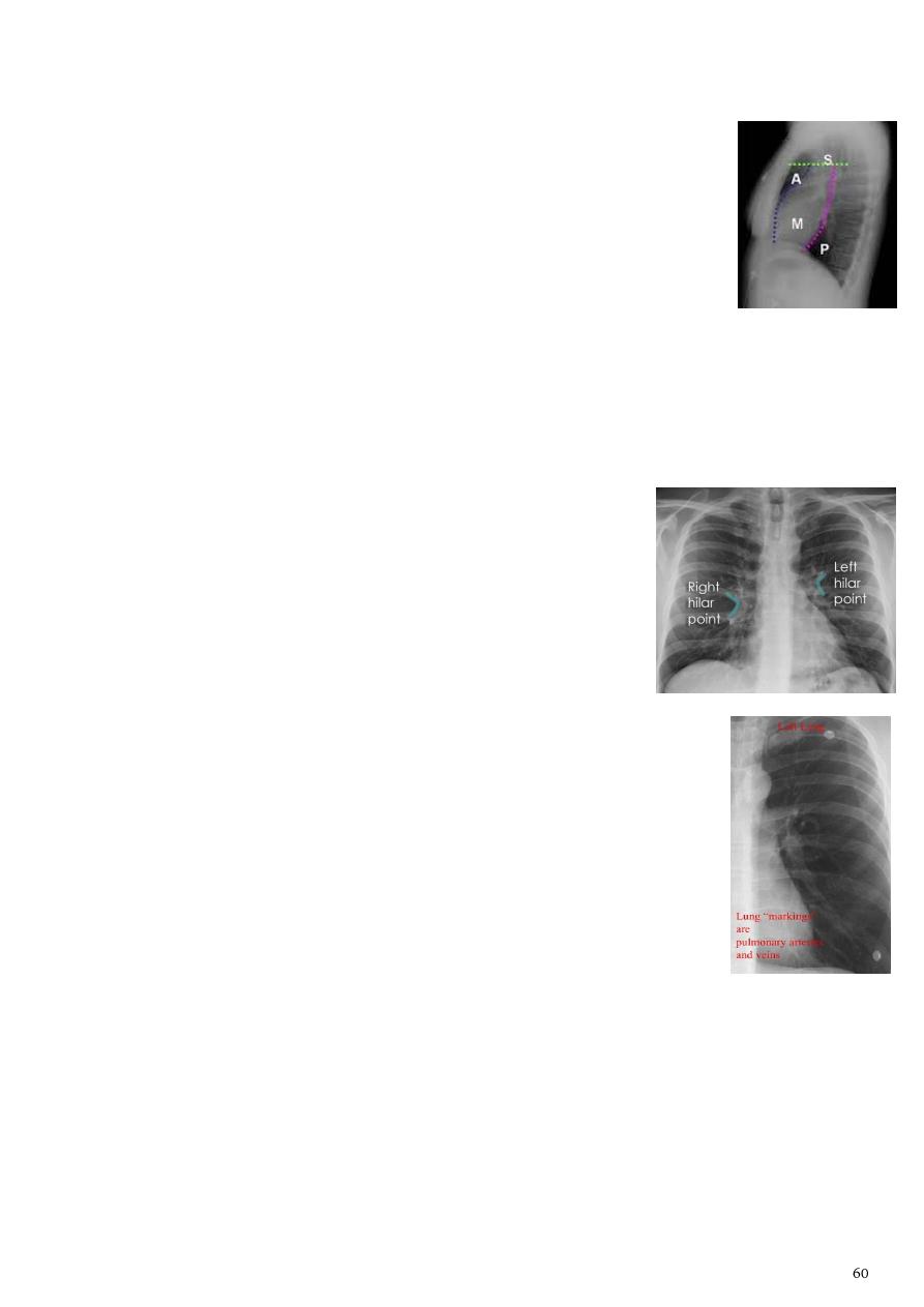
o The mediastinum contains heart, great vessels, esophagus, trachea, phrenic
nerve, cardiac nerve, thoracic duct, thymus, mediastinal lymph nodes.
o Anatomical division:
– Superior mediastinum: located between the thoracic
plane inferiorly and the thoracic inlet superiorly.
– Anterior mediastinum: anterior to the pericardium.
– Middle mediastinum: within the pericardium.
– Posterior mediastinum: posterior to the pericardium.
o Relations:
– Superiorly: continuous with the loose connective tissue of the neck.
– Anteriorly: chest wall.
– Laterally: lungs and pleura.
– Posteriorly: thoracic spine.
– Inferiorly: diaphragm.
Hila:
o The hila (lung roots) consisting of the major bronchi and
the pulmonary veins and arteries.
o There are also lymph nodes on each side (not visible
unless abnormal).
o The left hilum is often higher than the right.
o The hila are not symmetrical but contain the same basic
structures on each side.
o Both hila should be of similar size and density.
o If either hilum is bigger and denser, this is a good indication that
there is an abnormality.
o Lateral surface of the hilum is concave "if convex it means early
problem".
Pulmonary vasculature:
o Lung markings reflects pulmonary vasculature.
o When this normal markings disappear it means pneumothorax.
Pleura.
Lungs:
o The left lung upper lobe, oblique fissure, lower lobe.
o The right lung upper lobe, horizontal fissure, middle lobe, oblique fissure,
lower lobe.
o Upper zone in the right lung = upper and middle lobe / in the left lung = upper
lobe.
o Lower zone in the right lung = lower lobe / in the left lung = lower lobe.
o Each lobe has its own pleural covering
o The horizontal fissure (right) is often seen on a normal frontal view.
o The oblique fissures are often seen on a normal lateral view.
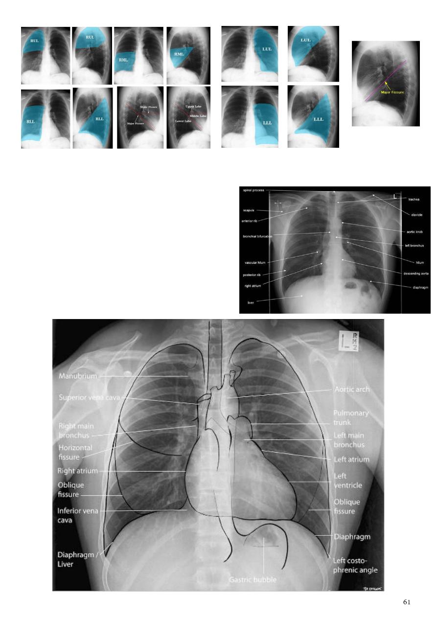
In summary - look to the structures in the CXR by the following order:
1- Trachea (position).
2- Heart (position, size, shape, contour).
3- Diaphragm (normal, elevated, gas under it).
4- Costo-phrenic angle (acute, meniscus sign).
5- Lung field (pulmonary markings, patch,
nodule, mass, cavity).
6- Ribs and bones.
7- Soft tissues (surgical emphysema).

The heart
1- Cardiothoracic ratio (CTR) = Cardiac Width = Thoracic Width:
For correct measuring of CTR the film should be PA view, no
rotation of the patient, should be complete inspiration.
CTR greater than 1:2 (50%) considered abnormal.
Enlargement of the cardiac silhouette on chest x-ray can be due to:
o Cardiomegaly (most common):
Mitral valve disease.
Congestive heart failure.
Congenital heart disease:
– Tetralogy of Fallot (boot shape).
– Ebstein anomaly (box shape).
o Pericardial effusion.
o Anterior mediastinal mass.
o Prominent epicardial fat pad.
2- Mitral valve disease:
Typical radiographic features of mitral regurgitation include:
Left atrial enlargement: convexity or straightening of the left atrial appendage just
below the main pulmonary artery (along left heart border).
Straight left cardiac border (
mitralization
).
Left ventricular enlargement (volume overload).
: the right side of the enlarged left atrium pushes into the
adjacent lung and creates an addition contour superimposed over the right heart.
Elevation of the left main bronchus.
Splaying and widening of the carina (above 90 degree).
Upper zone venous enlargement (pulmonary venous hypertension).
Features of pulmonary edema.
Cardiomegaly.
Cardiomegaly Normal CTR
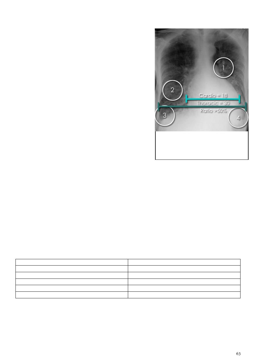
3- Congestive cardiac failure (CCF):
It can affect the left (common) or right cardiac
chambers or both.
Left sided congestive cardiac failure = give the
features of pulmonary edema which includes:
o Prominent hilum due to central
o
Cephalization of pulmonary veins
lobe pulmonary venous diversion.
o Pulmonary interstitial edema.
o Pulmonary alveolar edema.
o Cardiomegaly (CTR = 18/30).
o Pleural effusion.
Clinical features:
o Worsening exercise tolerance.
o Chronic uncontrolled hypertension.
o Rapid onset of dyspnea.
o Atrial fibrillation.
4- Pulmonary edema:
Defined as an abnormal accumulation of fluid in the extra-vascular compartments of
the lung.
Radiographic features when pulmonary capillary wedge pressures = 20-25 mmHg:
Septal lines (
Kerley lines
):
o Are seen when the interlobular septa in the pulmonary interstitium become
prominent.
o This may be because of lymphatic engorgement or edema of the connective
tissues of the interlobular septa.
Kerley A lines
Kerley B lines
2-6 cm long
1-2 cm long
<1 mm thick
Thin
Oblique
Perpendicular
Course towards the hila
In the peripheries of the lung (seen at lung bases)
Represent thickening of the interlobular septa
Represent thickened sub pleural interlobular septa
Radiographic features when pulmonary capillary wedge pressures > 25 mmHg:
o Cardio-thoracic ratio (If increased it indicate cardiogenic cause).
o
o Peri-bronchial cuffing.
o Kerley lines become more prominent.
1- Pulmonary venous hypertension.
2- Pulmonary edema.
3- Kerley B lines.
4- Pleural effusion.
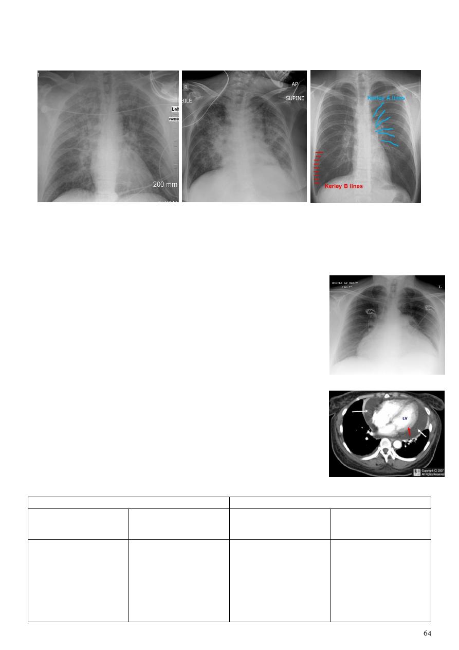
o Pleural effusions.
o Upper lobe pulmonary venous diversion.
Note:
Interstitial pulmonary edema (Kerley line).
Alveolar pulmonary edema (Bat wing sign).
5- Pericardial effusion:
Normal pericardial sac contains approximately 30-50 mL of
fluid.
Pericardial effusion occur when fluid collects in
Plain radiograph:
o Very small pericardial effusion can be occult on plain film.
o
Globe shape heart
or
pumpkin shape heart
it is
globular enlargement of the cardiac shadow giving
a water bottle configuration.
Causes of congenital heart disease:
Cyanotic CHD
Acyanotic CHD
increased pulmonary
vascularity
decreased pulmonary
vascularity
increased pulmonary
vascularity
normal pulmonary
vascularity
TAPVR
TGA
truncus arteriosus
large AVSD
single
entricle without
pulmonary stenosis
TOF
pentalogy of
Ebstein
anomaly with atrial
septal defect
VSD
ASD
AVSD
PDA
small shunts
aortic valve stenosis
aortic coarctation
pulmonary stenosis
Interstitial pulmonary edema
Alveolar pulmonary edema
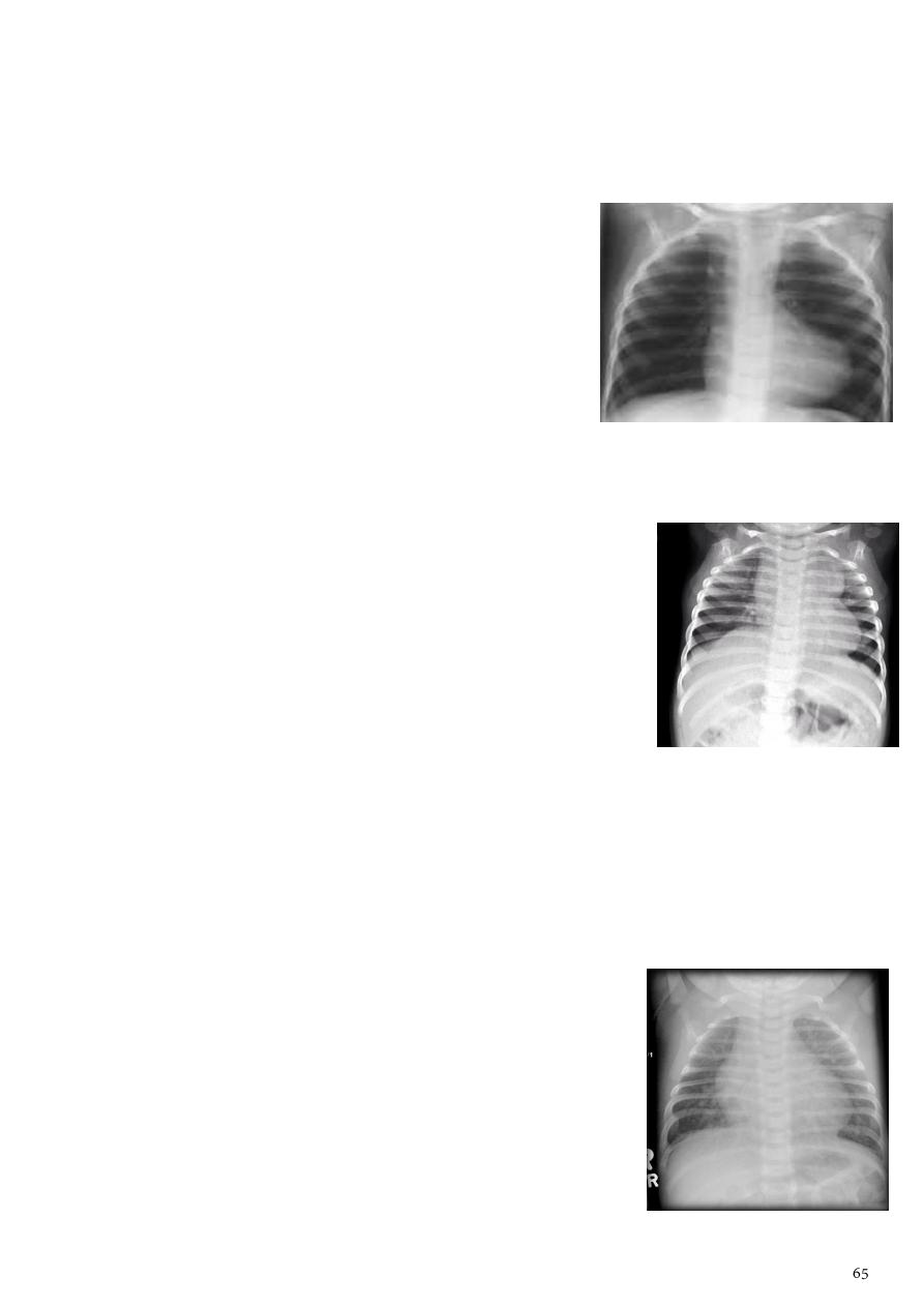
6- Tetralogy of Fallot (TOF):
It is one of the most common cyanotic CHD.
Has four features VSD, overriding aorta, right ventricular hypertrophy, pulmonary
artery stenosis
Plain films show:
o
Boot shaped heart
(apex is rounded-tenting from
the head).
o Upturned cardiac apex (due to right ventricular
hypertrophy).
o Concave pulmonary arterial segment.
o Pulmonary oligaemia (due to decreased
pulmonary arterial flow).
o Right sided aortic arch (seen in 25%).
7- Transposition of the great arteries (TGA):
It is the most common cyanotic CHD (7% of all CHD).
Cyanosis in first 24 hours of life.
Frontal chest radiograph classically shows:
o Cardiomegaly.
o Cardiac contours appearing like an
Egg on a string sign
.
o Apparent narrowing of the superior mediastinum (result
of the aortic and pulmonary arterial configuration).
8- Ventricular septal defects (VSD):
It is the most common CHD in children.
It is the second most common CHD in adults
It typically results in a left to right shunt.
The chest radiograph can be normal with a small VSD.
Larger VSDs may show:
o Cardiomegaly (particularly left atrial
enlargement although the right and left ventricle can
also be enlarged).
o Features of pulmonary arterial hypertension.
o Pulmonary edema.
o Pleural effusion.
o And/or increased pulmonary vascular markings.
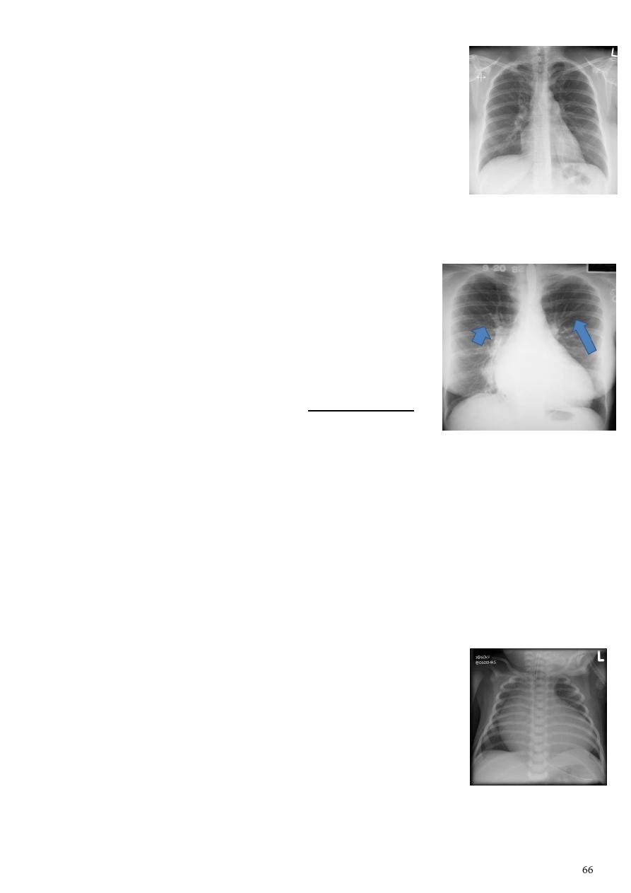
9- Pulmonary arterial hypertension:
Results from elevation of the resistance in the pulmonary
arterial bed, usually at the arteriolar level.
Plain radiograph:
o Elevated cardiac apex (due to right ventricular hypertrophy).
o Enlarged right atrium.
o Enlarged right and left main pulmonary arteries.
o Prominent pulmonary outflow tract.
o
Pruning
of peripheral pulmonary vessels.
10- Pulmonary venous hypertension (PVH):
Results from an increase in pressure in the pulmonary
veins, usually as a result of left atrial hypertension.
This is measured clinically as an increase in the pulmonary
capillary wedge pressure (PCWP) over the normal 12 to 14
mmHg.
As the pressure approaches 20 mmHg, interstitial edema
develops.
Common causes of PVH obstruction to LV inflow, LV systolic dysfunction, severe
mitral regurgitation, acute pulmonary and systemic volume overload.
Eisenmenger syndrome:
It is a complication of an uncorrected high-flow, high-pressure CHD like VSD.
Leading chronic pulmonary arterial hypertension and shunt reversal (L to R becomes R
to L).
Important signs:
Figure of 3 sign contour abnormality of the aorta with
inferior rib notching.
Roesler sign coarctation of the aorta.
Box shape heart Ebstein anomaly.
Boot shaped heart TOF.
Egg-on-a-string sign TGA.
Figure of 8 heart or cottage loaf heart TAPVR.
Box shape heart
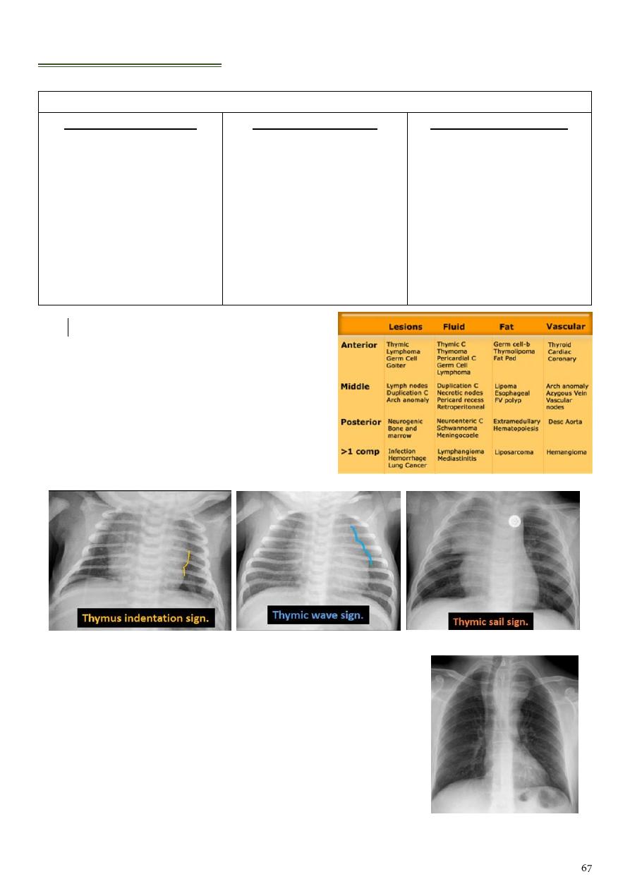
The mediastinum
Differential diagnosis of mass in the mediastinum
Anterior mediastinum
Thymoma.
Thymic cyst.
Thyroid neoplasms.
Thyroid goitre.
Parathyroid neoplasms.
Lymphoma (HL, NHL).
Germ cell tumors.
Mediastinal teratoma.
Teratocarcinoma.
Middle mediastinum
Malignancy.
Lymphadenopathy.
Hiatus hernia.
Thoracic aortic
Thyroid mass.
Bronchogenic cysts.
Posterior mediastinum
Nerve sheath tumors.
Neurofibroma.
Schwannoma.
Esophageal neoplasm.
Lymphoma.
Metastasis.
Paraspinal abscess.
Mediastinitis.
Sarcoidosis.
Note
:
Thymoma is most common primary
neoplasm of the antero-superior
mediastinum.
Sarcoidosis cause bilateral hilar
lymphadenopathy.
1- Normal thymus gland:
2- Retro sternal goiter:
Chest radiograph:
Inverted cone shape of homogenous opacity in the
superior mediastinum.
Narrowing and deviation of trachea to opposite site.
: opacity present in upper chest and
lower neck.
The superior margin of the radio-opacity/mass is
untraceable.
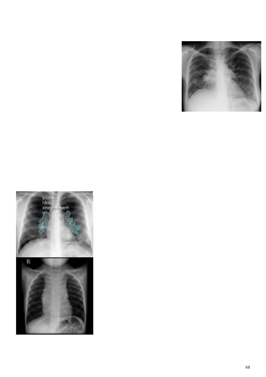
3- Lymphadenopathy:
Conditions than can result in mediastinal lymph adenopathy:
o Primary lung lung cancer.
o Metastatic malignancies (esophageal, breast, thyroid
o Mediastinal lymphoma (large B-cell lymphoma).
o Sarcoidosis.
o Infective (acute suppurative).
o Reactive (follicular hyperplasia, granulomatous TB,
fungal infection, neoplastic, drugs like cyclosporine-
phenytoin-methotrexate).
Lymphoma is a malignancy arising from lymphocytes or lymphoblasts.
Lymphoma can be restricted to the lymphatic system or can arise as extra nodal
disease.
Nodal disease Hodgkin's disease is almost entirely confined to the lymph nodes.
Extra nodal disease Extra nodal HD although uncommon may be found in any
organ system, either as a primary manifestation or as dissemination of systemic
disease.
Non-Hodgkin lymphoma located inside L.N and extra-nodal.
Bilateral hailer lymphadenopathy:
Well defined mass, lobulated, opaque.
Causes: sarcoidosis, TB, lymphoma, metastasis.
Hodgkin lymphoma:
Widening of mediastinum.
Soft tissue opacity.

The lung
Types of pathology that can be detected by CXR:
Patch or opacity:
o Consolidation (pneumonia/TB) contain air bronchogram.
o Collapse loss of volume, deviation of the trachea and mediastinum.
Mass:
o Single cancer, hydatid cyst.
o Multiple metastasis (solid content by CT), multiple hydatid cyst (fluid content
by CT).
o Note
Less than 3 cm = nodule / more than 3 cm = mass.
Cavity:
o Abscess A/F level.
o Raptured hydatid cyst water lilly sign.
o TB cavity in upper lobe, no A/F level, clinical picture of TB.
Lesions in the pleura:
o Pneumothorax.
o Pleural effusion.
o Hydropneumothorax.
o Empyema.
1- Consolidation:
It is a radiological sign that refers to non-specific air-space opacification on a chest
radiograph or chest CT appear radio-opaque on chest radiograph and chest CT.
Many things can fill the alveolar spaces:
o Fluid (heart failure).
o Pus (pneumonia).
o Blood (pulmonary hemorrhage).
o Cells (lung cancer).
Loss of volume:
o Pull the fissure.
o Pull the bronchus.
o Pull the mediastinum.
Lobar consolidation:
o Increased density/opacity is seen in individual lung lobes.
o Not cross the fissure.
o Air bronchograms can be seen.
o Volume loss not seen.
1- Cavity = wall + air.
2- Without wall = emphysematous ballue.
3- Cavity only air = TB.
4- Cavity + horizontal A/F level = abscess.
5- Cavity + water lilly sign = hydatid cyst.
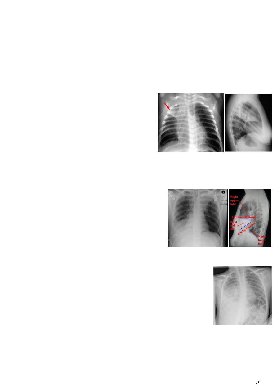
Multi-focal consolidation:
o Multiple areas of opacity seen throughout the lung.
o Usually ill-defined with peripheral distribution (Starting from bronchi and
spreading outwards).
o Causes bronchopneumonia, primary malignancy, metastasis.
- Right upper lobe consolidation:
Increased opacity within the right upper lobe.
Some loss of outline of the upper right heart
border may be apparent.
Dense opacity seen above the horizontal
fissure.
Air-bronchogram line.
The lower border of the consolidation is
sharply delinated by the horizontal fissure
suggesting it lies in the anterior segment of the RUL.
- Right middle lobe consolidation:
Opacification of the RML bordered superiorly by
the horizontal fissure, and medially by the right
heart border.
Indistinct right heart border.
Loss of the medial aspect of the right
hemidiaphragm.
- Right lower lobe consolidation:
Airspace shadowing that abuts the right hemidiaphragm.
Obliterating the crisp margin of the hemidiaphragm and normal
aerated lung.
- Silhouette sign:
In PA view of right lung:
Middle lobe pneumonia heart border not appear but diaphragm border appear.
Lower lobe pneumonia heart border appear but diaphragm not appear.
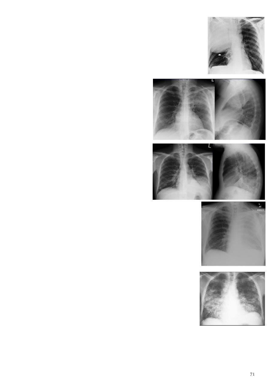
- Bulging fissure sign:
Refers to lobar consolidation where the affected portion of the
lung is expanded.
It is now rarely seen due to the widespread use of antibiotics.
Most commonly seen in Klebsiella (Friedlander's) pneumonia.
- Left upper lobe consolidation:
- Left lower lobe consolidation:
- Left total lung consolidation:
- Bronchopneumonia (lobular pneumonia):
Associated with suppurative peribronchiolar inflammation.
Subsequent patchy consolidation of one or more secondary
In response to a bacterial pneumonia (child, fever, cough).
CXR description:
o Multiple ill-defined non homogenous patchy opacification.
o In upper zone only TB bronchopneumonia.
o In middle or lower zone Bronchophrenic shadow.
o In middle or lower zone + cardiomegaly pulmonary edema.
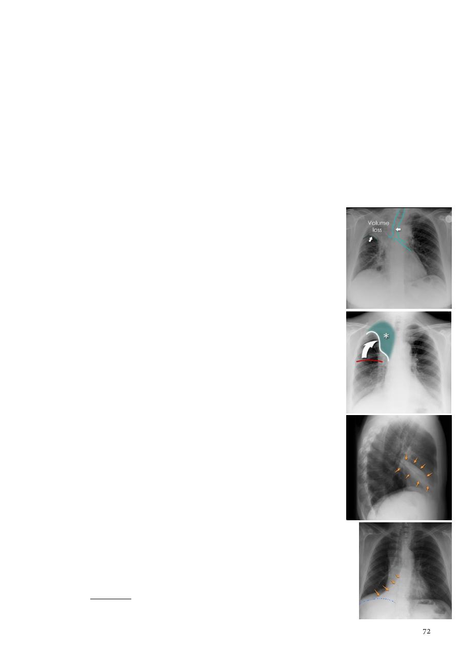
2- Collapse:
Lobar collapse refers to the collapse of an entire lobe of the lung.
Atelectasis more generic term for 'incomplete expansion'.
Individual lobes collapse due to obstruction of the supplying bronchus that caused by:
o Luminal aspirated foreign material, mucous plugging.
o Mural bronchogenic carcinoma.
o Extrinsic compression by adjacent mass.
Radiological features for all types of collapse = generic markers of volume loss:
o Elevation of the ipsilateral hemidiaphragm.
o Shift of the mediastinum towards the side of atelectasis.
o Crowding of the ipsilateral ribs.
o Crowding of pulmonary vessels or air bronchograms.
- Right upper lobe collapse:
Use frontal radiographs.
Increased density in the lobe.
Elevation of the horizontal fissure.
Elevation of the right hilum.
Loss of the normal right medial cardiomediastinal contour.
Hyperinflation of the right middle & lower lobe (Lead to
increased translucency).
Right juxtaphrenic peak.
Generic markers of volume loss.
common cause of lobar collapse is a hilar mass.
When a right hilar mass is combined with collapse of the right
upper lobe, the result is an S shape to elevated horizontal fissure.
- Right middle lobe collapse:
Frontal CXR opacity cause obscuration of the right cardiac
border.
Lateral CXR opacity is
tongue like shape
and diaphragm is
elevated (
triangular in shape
in right middle lobe consolidation
and diaphragm not elevated).
- RT lower lobe collapse:
The medial aspect of the dome of right hemidiaphragm is lost.
The right hilum is depressed.
The right heart border which is contacted by the right middle lobe
remains well seen.
Generic markers of volume loss.
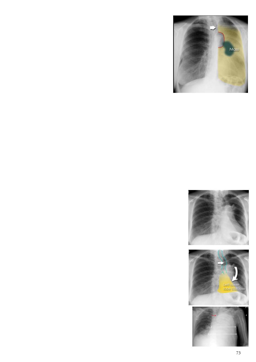
- Left upper lobe collapse:
Note: The left upper lobe collapses anteriorly becoming
a thin sheet of tissue apposed to the anterior chest wall,
and appears as a hazy or veiling opacity extending out
from the hilum and fading out inferiorly. It thus reverses
the normal slight increase in radiographic density seen as
you move down the lung (due to increased thickness of
the chest soft tissues).
Parts of left cardiomediastinal contour may be
obliterated by the the lingual.
The anterior parts of the aortic arch are also often obliterated from view.
the hyper-expanded superior segment of the left lower lobe
insinuates itself between the left upper lobe and the superior mediastinum, sharply
silhouetting the aortic arch and resulting in a lucency medially.
The left hilum is also drawn upwards resulting in an almost horizontal course of
the left main bronchus and vertical course of the left lower lobe bronchus.
'peaked' or 'tented' hemidiaphragm.
Generic markers of volume loss.
On lateral projections the left lower lobe is hyperexpanded, the oblique fissure
displaced anteriorly, increase in the retrosternal opacity.
- Left lower lobe collapse:
It is readily identified in a well penetrated film of a patient with
normal sized heart.
Can be challenging in the typical patient (unwell patients, with
portable (AP), under-penetrated films, cardiomegaly).
Triangular opacity in the posteromedial aspect of the left lung.
Edge of collapsed lung may create a
Double cardiac contour
.
Left hilum will be depressed.
Loss of the normal left hemidaphgragmatic outline.
Loss of the outline of the descending aorta.
Generic markers of volume loss.
On lateral projection the left hemidiaphragmatic outline is
lost posteriorly, the lower thoracic vertebrae appear denser
than normal (they are usually more radiolucent than the upper
vertebrae).
- Total left lung collapse:
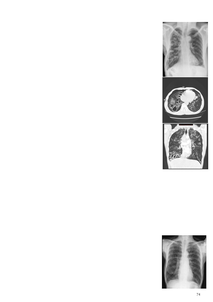
3- Bronchiectasis:
Refers to abnormal dilatation of the
considered irreversible.
CT is the most accurate modality for diagnosis (so every
bronchiectasis should go CT to determine the affected segment),
also can use bronchogram in the diagnosis.
It is largely considered irreversible
Causes:
o Post-infective (most common) necrotising bacterial
pneumonia (Staph. Aureus), granulomatous disease (TB),
bronchopulmonary aspergillosis (ABPA).
o Congenital congenital cystic bronchiectasis, cystic fibrosis
(CF), ciliary dysfunction syndromes (Kartagener syndrome).
o Bronchial obstruction bronchogenic carcinoma, inhaled
foreign bodies, chronic aspiration lung changes.
Plain radiograph:
o
seen in cylindrical bronchiectasis.
o Multiple Air-fluid levels seen in cystic bronchiectasis.
o
Honey comb shadow
.
o Bronchi seen end on may appear as ring shadows.
o Increase in bronchovascular markings.
o Pulmonary vasculature appears ill-defined (represent peri-
bronchovascular fibrosis).
4- Pulmonary emphysema:
Defined as the abnormal permanent enlargement of the airspaces distal to the
terminal bronchioles accompanied by destruction of the alveolar wall and without
obvious fibrosis.
Types of emphysema septal, bullous, generalized.
CXR not image emphysema directly, but rather infers the diagnosis due to associated
features (Except in the case of very advanced disease with bulla formation).
Radiological features:
o Flattened hemidiaphragm (s): most reliable sign.
o Tenting of the diaphragm.
o Increased and irregular radiolucency of the lungs.
o Increased retrosternal airspace.
o Increased antero-posterior diameter of chest (hyperinflation).
o Widely spaced ribs.
o Sternal bowing.
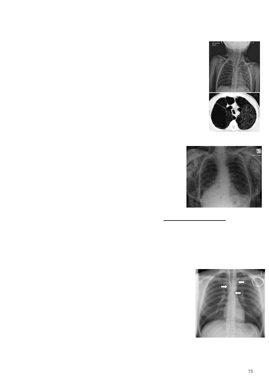
o Saber-sheath trachea.
o Absent pulmonary markings in outer 1/3 of the lung fields.
o Pulmonary arterial hypertension (pruning of peripheral vessels, increased calibre
of central arteries, right ventricular enlargement).
Unilateral obstructive emphysema:
o Causes elderly (tumor), child (TB).
o Radio-opaque foreign body be demonstrated.
o Aspirated foreign bodies have a predominance for the right
tracheo-bronchial tree.
Pulmonary bullae:
o Are focal regions of emphysema with no discenible wall.
o Measure more than 1cm in diameter.
5- Subcutaneous emphysema:
Refers to air in the subcutaneous tissues.
But the term is generally used to describe any soft tissue
emphysema of the body wall or limbs, since the air often
dissects into the deeper soft tissue and musculature along
fascial planes.
Radiological features if affecting the anterior chest wall:
o Subcutaneous emphysema can outline the pectoralis
major muscle, giving rise to the
o Dissecting air along tissue fat planes appears as multiple lines of lucency.
6- Pneumomediastinum:
It is the presence of extra-luminal gas within the mediastinum.
Gas may originate from the lungs, trachea, central bronchi, oesophagus, and track
from the mediastinum to the neck or abdomen.
Radiographic features:
o Small amounts of air appear as
linear or curvilinear
lucencies
outlining mediastinal contours such as
subcutaneous emphysema.
o
air anterior to pericardium.
o
air around pulmonary artery
and main branches.
o
air outlining major aortic branches.
o
air outlining bronchial wall.
o
Continuous diaphragm sign
due to air trapped posterior to pericardium.
o
Extrapleural sign
air between parietal pleura and diaphragm.
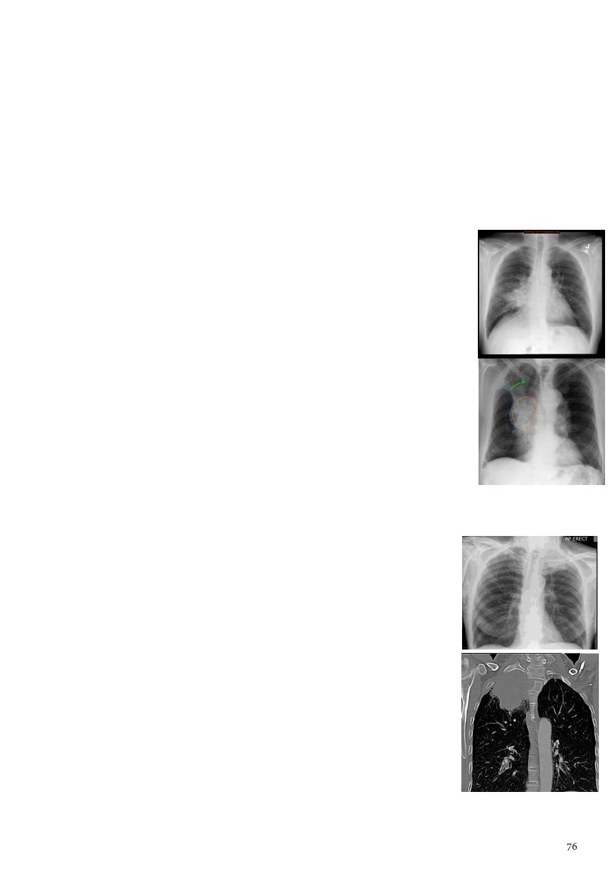
7- Bronchogenic carcinoma (BGCA):
It is the most common cause of cancer in men, and the 6th most frequent cancer in
women worldwide.
It is the leading cause of cancer mortality worldwide in both men and women (20% of
all cancer deaths).
Subtype
"different radiographic appearance, demographic, prognosis"
:
"most common primary lung malignancy to cause paraneoplastic
syndromes and SVC obstruction".
o Adenocarcinoma.
o Large cell carcinoma.
o Small cell carcinoma.
o Other malignant pulmonary neoplasms include lymphoma.
Radiological features:
o Central BGCA (mostly squamous cell carcinoma) bulky hilum
representing the tumor & local nodal, the lesion is homogenous
irregular outline with
spiky or sun ray speculation
.
o Peripheral BGCA (mostly adenocarcinoma) rounded or
speculated mass, cavitation, A/F level (in Large cell CA).
o Direct signs rounded or oval mass, 3 cm or more, irregular or
speculated margin, may show necrosis that lead to cavitation.
o Indirect signs pleural effusion, lobar collapse,
Golden S sign
,
destruction of the adjacent rib, evidence of soft tissue growing,
elevated hemi-diaphragm due to phrenic nerve palsy.
8- Pancoast tumor (superior sulcus tumor):
Refers to a relatively uncommon situation where a primary
bronchogenic carcinoma arises in the lung apex and invades the
surrounding soft tissues.
Adeno CA being the most frequent type.
Radiological features:
o Soft tissue opacity at the apex of the lung.
o Rib involvement.
o Extension into the supraclavicular fossa.
o Surrounded bony destruction.
o Lordotic views may be helpful.
Most important complication involvement of the sympathetic
chain (Ptosis, Meiosis, unhydrosis).
It is not TB because it has shape of mass and the ribs are
destructed minimally.
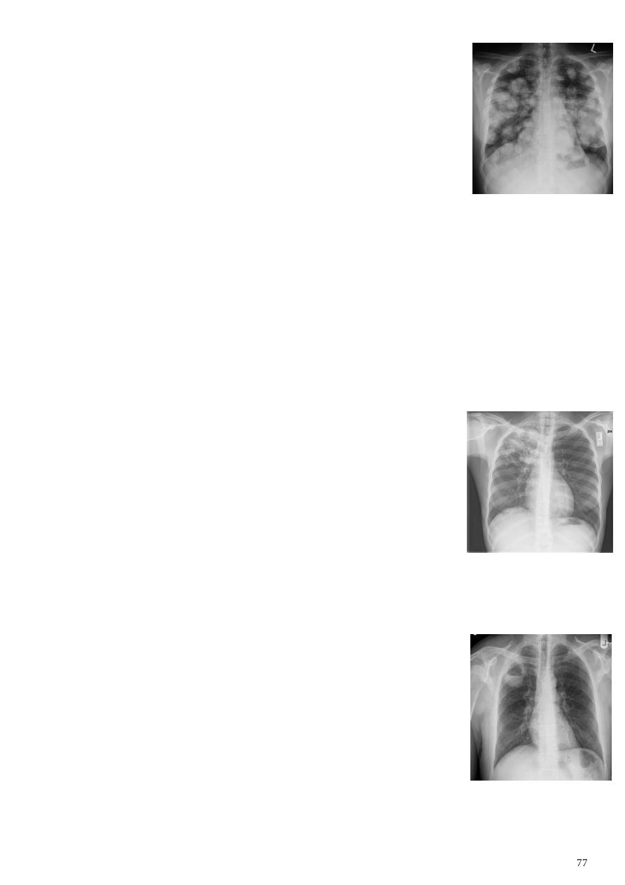
9- Secondary lung tumor:
Cannonball metastases:
o Large (1-3 cm), well circumscribed, round, multiple opacities
like cannonballs "
cannon balls appearance
".
o DDx is multiple hydatid cyst, so use CT scan.
Lymphangitis carcinomatosis:
o Tumor spread through the lymphatics of the lung.
o Most commonly seen secondary to adenocarcinoma.
o Up to a quarter of patients have normal chest x-rays.
o The most common findings reticulonodular pattern, thickening of the
interlobular septae which may resemble Kerley B lines, + /- pleural effusion.
Innumerable small metastases (miliary pattern).
o DDx is military TB (smaller + same size and density).
10- TB of the lung:
The lungs are the most common site of primary infection by tuberculosis and are a
major source of spread of the disease.
Primary pulmonary TB:
o The initial focus of infection can be located anywhere within
the lung.
o Ranging from too small to be detectable, to patchy areas or
consolidation or lobar consolidation in RT upper or RT
middle lobe.
o
Ghon lesion
+/- ipsilateral hilar or paratracheal
lymphadenopathy usually right sided.
o
Primary complex
= Ghon focus + LAP.
o
= calcified node + calcified Ghon lesion.
o Pleural effusions (more frequent in adults).
Post primary TB:
o Also known as reactivation tuberculosis or secondary
tuberculosis.
o Occurs years later, due to decreased immune status.
o Develops in either posterior segments of the upper lobes or
superior segments of the lower lobes.
o Alternating lesion patchy consolidation or poorly defined
linear and nodular opacities in both apices, upper zone in
one lung & lower zone in other lung.
o Cavitate, multiple abscess formation, A/F level in the
posterior segments of the upper lobes.
TB cavity
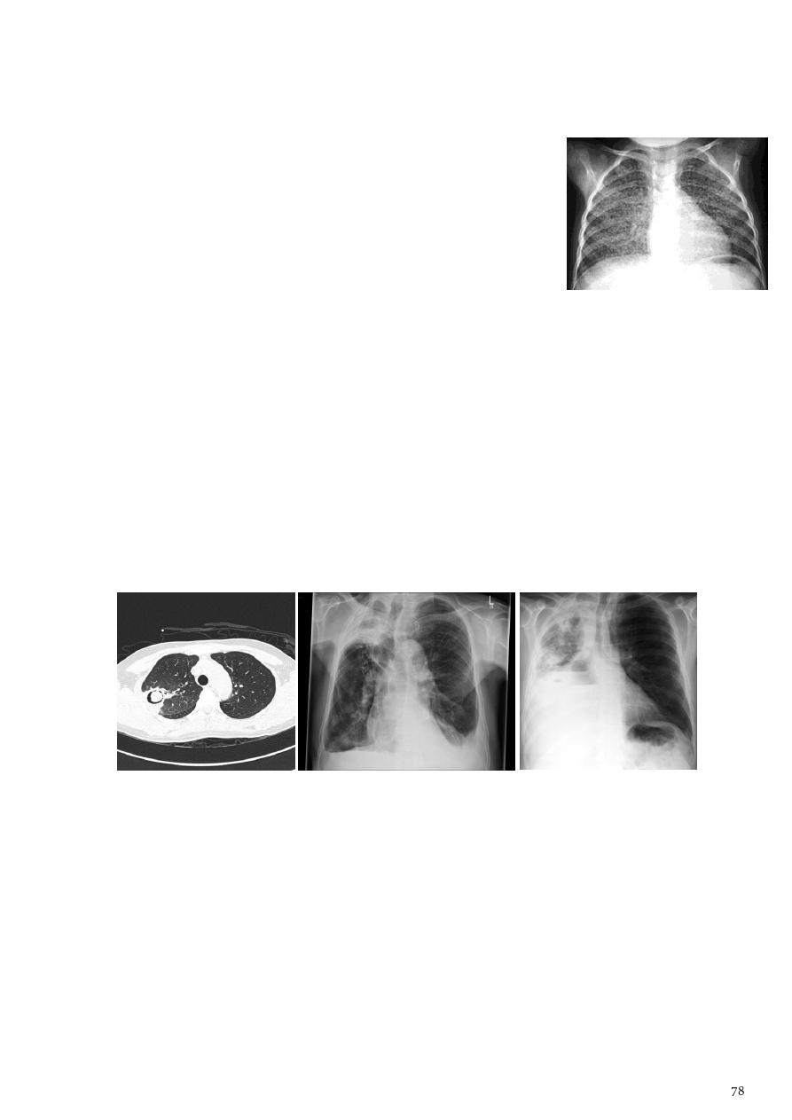
o Tuberculomas well defined rounded mass in the upper lobes.
o Post primary TB bronchopneumonia
cotton wool sign
.
Miliary tuberculosis:
o Is uncommon but carries a poor prognosis.
o It represents hematogenous dissemination of an
uncontrolled tuberculous infection.
o It is seen both in primary and post-primary tuberculosis.
o The lungs are usually the easiest location to the image.
o Description multiple well defined very fine
homogenous opacification 1-3 mm and they are uniform
in size and disrupted uniformly in both lung felids
Complications:
Mobile ball inside cavity so appear as cavity with crescent lucency.
Could lead to bilateral diffuse bronchiectasis.
o Bronchopleural fistula (occur after surgery).
o Bronchiectasis.
o Arterial pseudoaneurysms.
o Bronchial artery pseudo aneurysm.
o Pulmonary artery pseudo aneurysm / Rasmussen aneurysm.
o Empyema.
o Ribrothorax.
11- Lung abscess:
It is a circumscribed collection of pus within the lung, is are potentially life
threatening.
Divided into acute (< 6 weeks) and chronic (> 6 weeks).
Primary abscess:
o Develop as a result of primary infection of the lung.
o They most commonly arise from aspiration, necrotising pneumonia
(Staphylococcus aureus, Klebsiella pneumonia, Pseudomonas), chronic pneumonia
(pulmonary tuberculosis).
Aspergilloma or myctoma or
hematoma or ruptured hydatid cyst
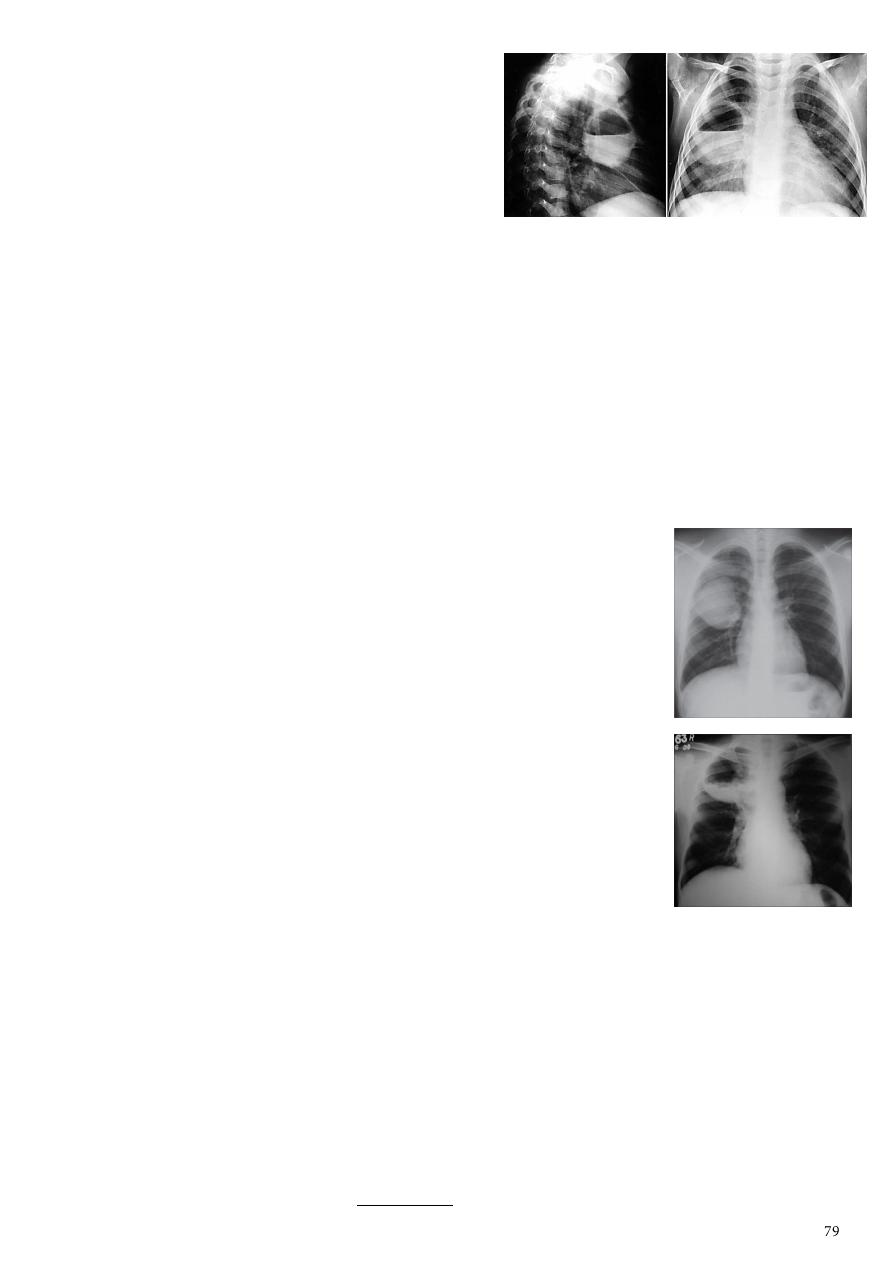
Radiological features:
o Cavity containing an air-fluid level.
o Abscesses are round in shape, and
appear similar in both frontal and lateral
projections.
12- Hydatid cysts:
General information:
o Result from infection by the Echinococcus.
o Can result in cyst formation anywhere in the body.
o Humans are accidental host.
o The infection occurs by ingesting food contaminated with Echinococcus eggs.
o The lung is the second most common site of involvement in adult, and the most
common site in children.
o The coexistence of liver and lung disease is present in only 6% of patients.
Chest XR features:
o Simple hydatid cyst:
Multiple or solitary rounded opacity.
Diameter of 1-20 cm.
Unilateral or bilateral.
Predominantly found in the lower lobes.
o Complicated cysts:
Meniscus sign
or
Combo sign
endocyst and pericyst has the appearance of an onion.
is seen in hydatid infections when there is
detachment of the endocyst membrane which results in
floating membranes within the pericyst that mimic the
appearance of a water lily.
Note: Water lily is irregular A/F level or Dome shape A/F level or
Double line A/F level.
Ruptured cyst consolidation adjacent to the cyst.
13- Pleural effusion:
Can be divided into exudates and transudates depending on the biochemical analysis
of aspirated pleural fluid.
Chest radiographs are the most commonly used examination to assess for presence of
a pleural effusion.
Routine erect CXR (PA, PA) 250-600 ml of fluid is required to become evident.
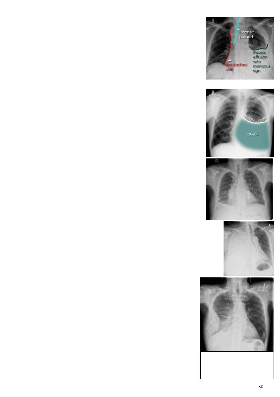
Lateral decubitus film most sensitive, able to identify even a
small amount of fluid.
Supine films can mask large quantities of fluid.
CXR (lateral decubitus):
o Obtained with the patient lying on their side, effusion side
down, with a cross table shoot through technique.
o Can visualize small amounts of fluid layering against the
dependent parietal pleura.
CXR (erect):
o
Blunting of the costophrenic angle
.
o
Blunting of the cardiophrenic angle
.
o Fluid within the horizontal or oblique fissures.
o
Meniscus
curved line seen in the upper aspect of
homogenous opacity.
Note – if there is A/F level it means empyema or hydro-pneumothorax.
Subpulmonic effusion (infrapulmonary effusion):
o May be seen when there is previously established
pulmonary disease.
o But can also be encountered in normal lungs.
o They are more common on the right, and usually unilateral.
Large volume effusions:
o Mediastinal shift occurs away from the effusion.
o Note: if coexistent collapse dominates then mediastinal shift may
occur towards the effusion.
14- Empyema:
Radiological features:
o Hyperinflation.
o Devoid of lung markings in the outer one third.
o Flattening of diaphragm.
Empyema VS pleural effusion:
o Shape and location.
o Empyema form an obtuse angle with the chest wall.
DDx: localized pleural effusion,
empyema, fibroma.
Can differentiated by CT scan.
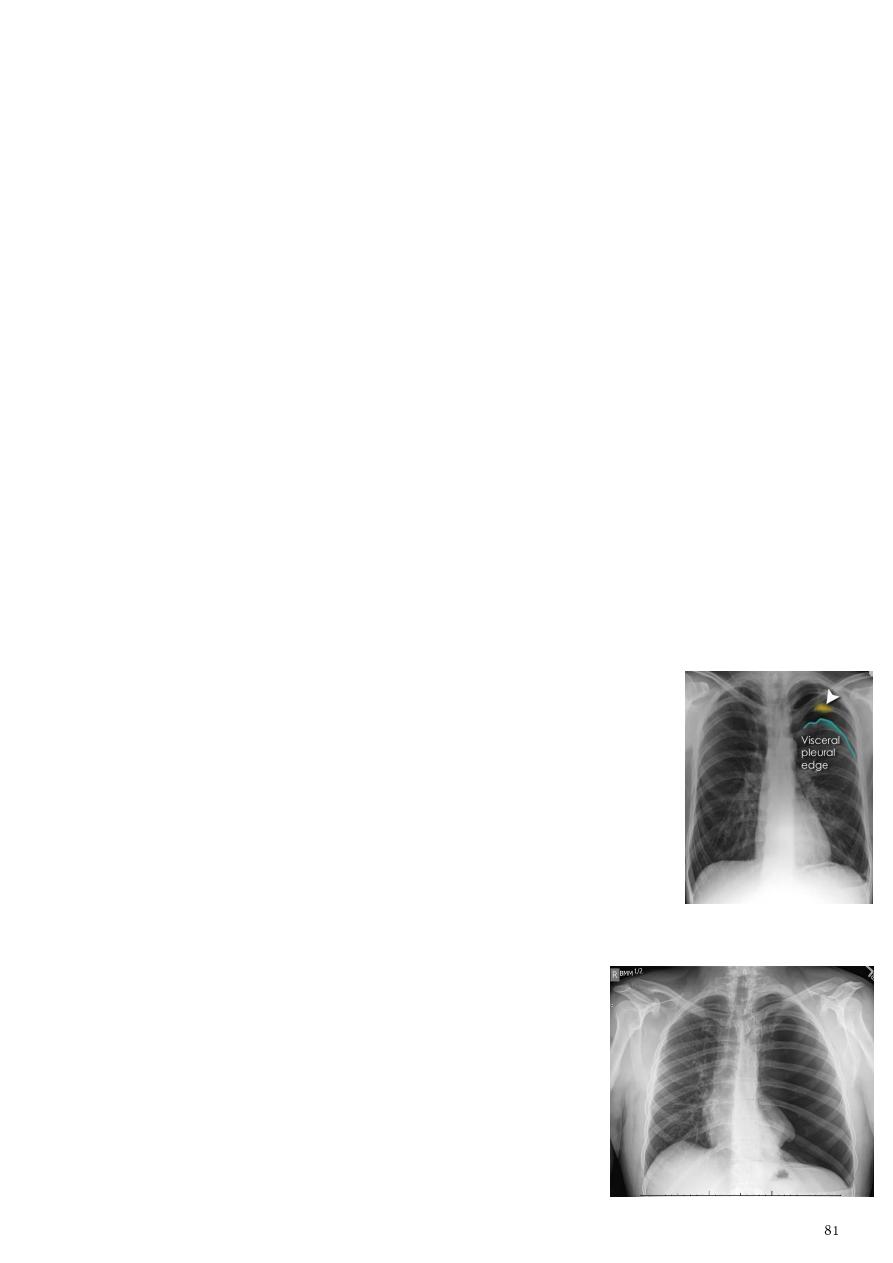
o Empyema is unilateral or markedly asymmetric / pleural effusion is usually
bilateral and similar in size.
o Empyema is lenticular in shape (bi-convex) / pleural effusion is crescentic in shape
(concave towards the lung).
Empyema VS peripheral pulmonary abscess:
o Relationship to adjacent bronchi / vessels:
Abscess will abruptly interrupt bronchovascular structures.
Empyema will usually distort and compress adjacent lung.
o Angle with pleura:
Abscess usually have an acute angle (
).
Empyema have obtuse angles.
o
thickening and separation of visceral and parietal pleura is a
sign of empyema
o Abscess has thick irregular walls, empyema is usually smoother.
15- Pneumothorax:
Pneumothorax refers to the presence of gas (air) in the pleural space.
Categories of pneumothorax:
o Primary spontaneous no underlying lung disease, marfan syndrome, Elher
danus syndrome, alpha-1 antitrypsin deficiency.
o Secondary spontaneous underlying lung disease is present.
o Iatrogenic/traumatic.
Radiographic features:
o Visible visceral pleural edge (very thin, sharp white line).
o No lung markings are seen peripheral to this line.
o Radiolucent peripheral space compared to adjacent lung.
o The lung may completely collapse.
o The mediastinum should not shift away from the
pneumothorax unless a tension pneumothorax is present.
16- Tension pneumothorax:
Occur when intrapleural air accumulates progressively in
such a way as to exert positive pressure on mediastinal
and intrathoracic structures.
It is a life threatening occurrence requiring rapid
recognition and treatment.
Radiographic features:
o Has the same features as a run-of-the-mill
pneumothorax.
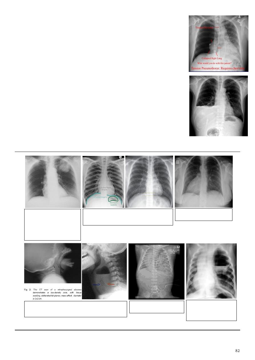
o Additional signs indicate over expansion of the hemithorax:
Ipsilateral increased intercostal spaces.
Shift of the mediastinum to the contralateral side.
Depression of the hemidiaphragm.
17- Hydropneumothorax:
It is concurrent presence of a pneumothorax as well as
a hydrothorax (air and fluid) in the pleural space.
Erect CXR easy to diagnose, show air-fluid level.
Supine CXR more challenging, show sharp pleural line is
bordered by increased opacity lateral to it within the pleural
space.
Lucency in the upper margin and opacity in the lower margin.
No cavity wall, if there is wall it indicate abscess.
Above 5 cm hydatid
cyst.
Below 5 cm it is coin
lesion with many DDx.
Look carefully on both diaphragmatic cruse
costo & cardio phrenic angles.
Dextrocardia
Retropharyngeal abscess = widening of
retropharyngeal space.
Diaphragmatic hernia
congenital cystic
adenomatous
malformation

CXR signs ...
Sign
Disease
Air Bronchogram Sign
Atelectasis, Pneumonia, Consolidation,
Tumor, Lymphoma.
Air Crescent Sign
Pulmonary aspergillosis, Tumor,
Hematoma, Wegener granulomatosis,
Hydatid cyst, TB, Nocardiosis, Bacterial
abscess.
Monod’s sign
Mycetoma, Aspergelloma.
Continuous Diaphragm Sign
Pneumomediastinum.
Deep Sulcus Sign
Pneumothorax.
Ring Around Artery Sign
Pneumomediastinum.
Flat Waist Sign
Left lower lobe collapse.
Finger-in-Glove Sign
Indicates mucoid impaction within an
obstructed bronchus.
Golden S Sign
RUL collapse, bronchial carcinoma, primary
mediastinal tumor, metastasis, enlarged
lymph nodes.
Luftsichel Sign
Left upper lobe collapse.
Double Density Sign
Left atrial enlargement.
Doughnut Sign
Mediastinal adenomegaly.
Cervicothoracic Sign
Retro sternal goiter.
Garland triad or 1-2-3 sign
Sarcoidosis.
Epicardial Fat Pad Sign
Pericardial effusion.
Westermark Sign
Dilatation pulmonary arteries proximal
embolus + collapse of distal vasculature.
Fleischner Sign
Massive pulmonary embolism.
Fallen Lung Sign
Tracheo-bronchial ruptures.
CT Angiogram Sign
Lobar Broncho-alveolar carcinoma.
Reverse Halo Sign
Crypto-genic organizing pneumonia (COP).
Split Pleura Sign
Empyema, bacterial pneumonia, cancer,
viral infection, PE.
Tree-in-Bud Sign
Tuberculosis.
Comet Tail Sign
Asbestosis, CHF, Dressler, infarct, TB,
parapneumonic effusions, histoplasmosis.
Signet Ring Sign
Bronchiectasis.
For more information visit
www.muhadharaty.com/lecture/8052

Part5
: Radiology in women
Obstetric Ultrasound
Types of ultrasonography:
Trans-abdominal ultrasonography (TAS).
Trans-vaginal ultrasonography (TVS).
Doppler ultrasound.
Tissue harmonic imaging (THI).
Three-dimensional ultrasound (3D USG).
Characteristics:
Safe, non-invasive, accurate, cost-effective.
Ultrasound useful in:
Establish the presence of a living embryo or fetus.
Estimate the age of the pregnancy.
Diagnose congenital anomalies of the fetus.
Evaluate the position of the fetus.
Evaluate the placenta (position, level, separation).
Detect multiple pregnancies.
Determine the amount of amniotic fluid.
Check for opening or shortening of the cervix.
Assess fetal (sex, age, stage of development, heart of fetus, weight, well-being).
Note:
In early pregnancy US full bladder is necessary.
In Transvaginal US empty bladder is necessary.
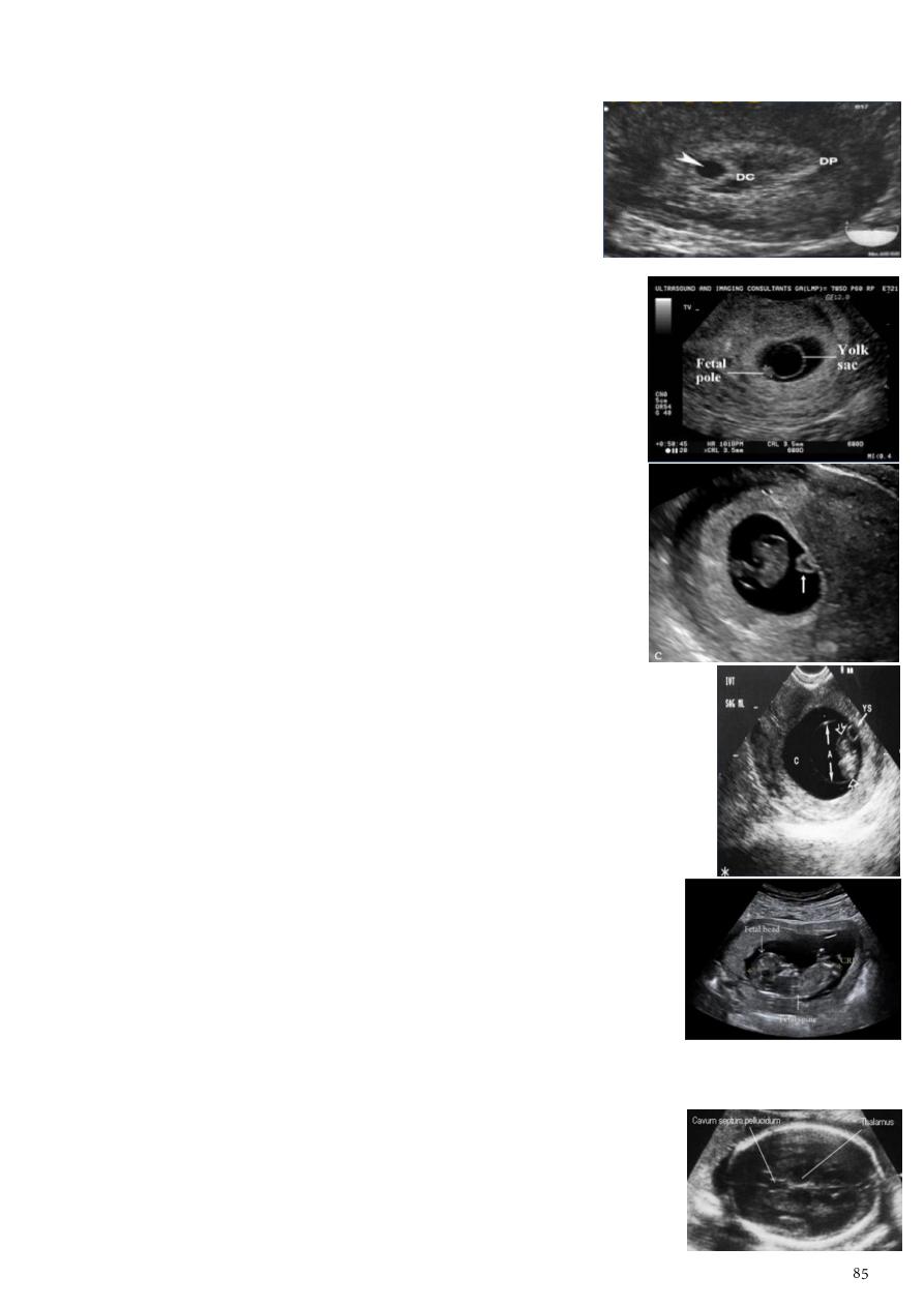
1- Age of the pregnancy:
Gestational sac:
o Earliest sign of pregnancy (4-4.5 weeks).
o It is intradecidual surrounded by decidual reaction.
o Can be used for gestational dating.
o Normal gestational sac grows by 1 mm per day.
Trimesters:
o First T 13 weeks (3 months).
o Second T 28 weeks (6 months).
o Third T 40 weeks (9 months).
Time in pregnancy:
o Women getting pregnant (pregnancy appear in US after
at least one week missing of the cycle) 7 days there is
thickening of endometrium and 10 day (sac appear)
sac of early pregnancy increased 1 ml of each day
this sac appear as black dot surrounded by white color.
o Sac = 5 mm pregnancy =< 4 weeks.
o Sac = 6-7 mm pregnancy = 5 weeks, with small (3-5
mm) thin wall ring within the sac this is yolk sac.
o Sac = 8-10 mm pregnancy (5-6 weeks) + fetal node.
o Sac = 10-12 mm pregnancy 7 weeks (fetal cardiac
pulsation appear at this time).
First trimester:
o Gestational sac = 4 to 5 weeks.
o Yolk sac = 5 to 6 weeks.
o Fetal pole = 6 to 7 weeks.
o Cardiac activity = 6 to 7 weeks.
o Mean sac size (MSS) = 4 to 5 weeks.
o Crown rump length (CRL) = 7 to 13 weeks.
Measure from top of head to rump.
Always measure in neutral position.
Very accurate (dating with the CRL can be within 3-4
days in LMP).
It should not be changed by a subsequent scan.
Second and third trimesters:
o Record placental localization.
o Biometry (BPD, FL, AC, HC) to estimate fetal weight,
gestational age, EDD.
o Bi-parietal diameter (BPD) = 13 weeks to full term.
Arrow shape Landmarks = midline flax cerebri, cavum
septum pellucidum, thalami, lateral ventricle.
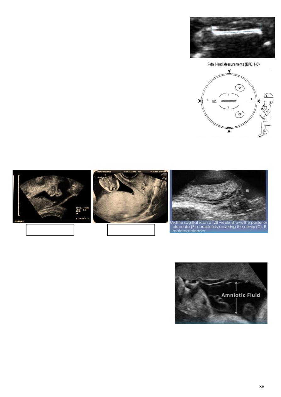
o Femur length (FL) = 14 weeks to full term.
US beam should be perpendicular to the bone.
Measurement is made along the femur diaohysis.
Exclude the distal femoral diaphysis.
o Abdominal circumference (AC).
o Head circumference (HC).
2- Placenta:
Placental location:
o Anterior/Posterior/Lateral/Fundal.
o Placental previa.
o Low-lying.
Placenta previa:
o The placenta overriding the cervix.
o Types: marginal, partial, complete.
3- Liquor:
Measure the dimensions of the largest vertical
pocket of amniotic fluid.
Pocket of fluid:
o <1cm = oligohydramnios.
o 1-2cm = decrease fluid.
o 2-8cm = normal.
o >8cm = polyhydramnios.
Amount of amniotic fluid normally is 2-8 cm (6-6.5 cm very typical).
This wide range (2-8 cm) is this because it depend on age of the fetus.
Causes of polyhydramnios D.M, congenital neural defect, esophageal atresia.
Causes of oligohydramnios Pott's syndrome (renal agenesis of fetus) indicate
severe oligohydramnios.
Low-lying placenta
Placenta previa
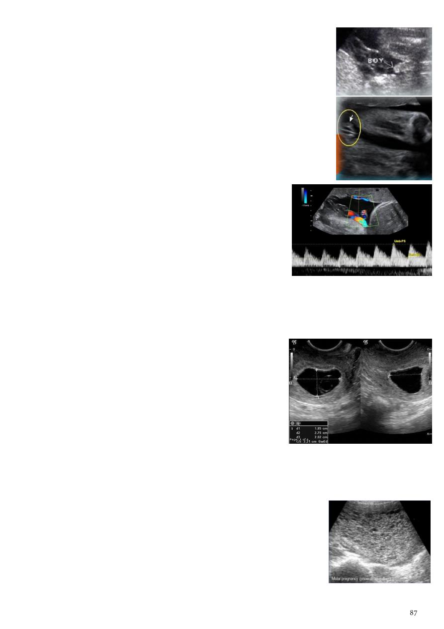
4- Fetal sex:
The earliest time for determining the sex of fetus is 12 weeks if in
right position.
Best done between 17-20 weeks.
Male: penis or scrotum.
Female: the 3-lines sign which denotes the labia.
5- Doppler ultrasonography:
Used to detect:
o Fetal cardiac pulsation.
o Pulsation in various fetal blood vessels.
Do umbilical artery Doppler is the following conditions:
o Low liquor.
o Low movement.
o Confusing measurements (parameters).
Doppler waveform give useful information about intra-
uterine growth retardation.
6- Blighted ovum:
Anembryonic pregnancy is a form of a failed early
pregnancy, where a gestational sac develops, but the
embryo does not form.
The term blighted ovum is synonymous with this, but
is falling out of favour and is best avoided.
By TVS, TAS (RCOG criteria) Blighted ovum = no
fetal pole + MSD ≥25 mm.
MSD = means sac diameter.
7- Molar pregnancy:
Gestational trophoblastic disease (GTD) results from abnormal
proliferation of trophoblastic tissue.
Including hydatidiform mole, complete mole, partial mole,
invasive mole, chorio-carcinoma (gestational choriocarcinoma)
Radiographic features by US:
o
Snowstorm appearance
solid collection of echoes with
numerous small (3-10 mm) anechoic spaces.
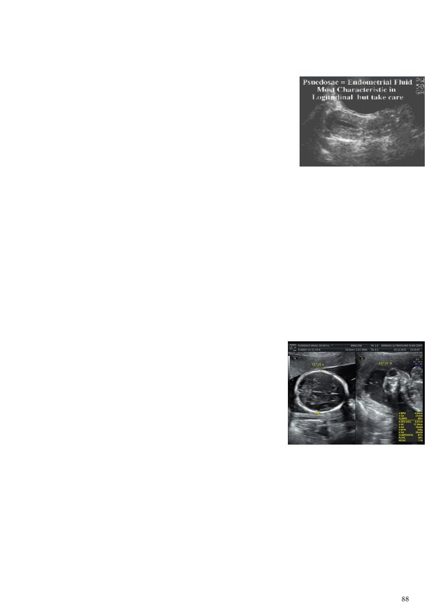
o
Punch of grapes sign
which represents hydropic swelling of trophoblastic villi.
o Enlarged uterus.
8- Ectopic pregnancy:
The ultrasound exam should be performed both
transabdominally and transvaginally.
The transabdominal provides a wider overview of the
abdomen.
The transvaginal is important for diagnostic sensitivity.
1-Empty uterine cavity (no evidence of intrauterine pregnancy).
2-
Pseudo gestational sac
or
decidual cyst
(10-20% of ectopic pregnancies).
3-Direct visualization of the sac at the adenxia.
9- Anencephaly:
General information:
o It is the most severe form of cranial neural tube defect (NTD).
o Characterized by absence of cortical tissue, absence of the cranial vault.
o Brainstem and cerebellum may be variably present.
o Associated with spina bifida, congenital heart defects, cleft lip/palate,
diaphragmatic hernia, spinal dysraphism, clubfeet, omphalocoele
Antenatal ultrasound:
o Anencephaly may be sonographically detectable as
early as 11 weeks.
o Ultrasound can be a non-invasive.
o
Frog sign appearance
.
o No parenchymal tissue is seen above the orbits.
o Calvarium is absent (parts of the occipital bone and
mid brain may be present).
o Urinary tract abnormalities (hydronephrosis most common).
10- Hydropis fetalis:
General information:
o Hydrops fetalis is defined as accumulation of fluid +/- edema involving at least two
fetal components.
o It is due to heart failure, volume overload, decreased oncotic pressure,
increased vascular permeability.
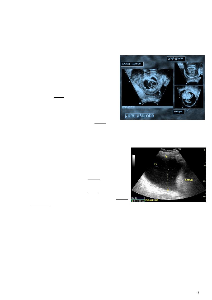
o It may manifest as fetal pleural effusion, fetal pericardial effusion, fetal ascites,
placental enlargement, Polyhydramnios, generalized body edema (fetal anasarca
/nuchal edema/cystic hygroma).
o One of the most common causes of hydrops fetalis is TORCH infection.
o Most common causes of increased placental thickness are hydrops fetalis & D.M.
Sonographic features:
o Can be similar for both immune and non-
immune hydrops.
o Increased amniotic fluid volumes.
o Larger placental size (placentomegaly).
o Increased placental thickness (placental
edema) maximum normal thickness of
placenta = 4 cm.
o Presence of a fetal pleural or pericardial
effusion.
o Generalized fetal body swelling (fetal
anasarca and skin thickening > 3 mm).
11- Polyhydramnios:
Polyhydramnios refers to a situation where the
amniotic fluid volume is more than expected for
gestational age.
It is generally defined as:
o Amniotic fluid index (AFI) > 25 cm.
o Largest fluid pocket depth (maximal vertical
pocket (MVP)) greater than 8 cm.
o Overall amniotic fluid volume larger than 1500-
2000 cc3.
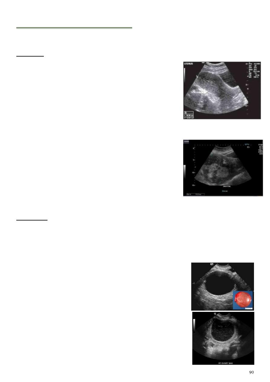
Gynecological Ultrasound
Uterus:
1- Normal uterus:
Shape of pear.
Length = fundus to cervix (7.5-8 cm).
Depth = anterio-poseterior (4.5-5 cm).
Width = coronal view (2.5-3 cm).
2- Adenomyosis.
3- Patient with abnormal bleeding.
4- Fibroid:
Show multiple hypoechoic and hyperechoic patchy areas.
Sometimes degenerative changes can take place in
fibroid with areas of necrosis and hemorrhage and result
in varying appearance from cystic to inhomogenous
appearance.
Ovaries:
1- Ovarian cyst:
Small cystic ovarian structures should be considered normal ovarian follicles unless
the patient is pre-pubertal, post-menopausal, pregnant, or the mean diameter is >3.0
cm.
Radiographic features:
o Anechoic.
o Intraovarian or exophytic.
o Has an imperceptible wall.
Important things to check in ovarian cyst:
o Thin or thick wall?
o Content?
o Soft tissue part?
o Doppler for vascularity?
Criteria of malignant cyst:
1- Thick wall 2- Soft tissue mass.
3- Vascular 4- Sepetation (more than 3 mm).
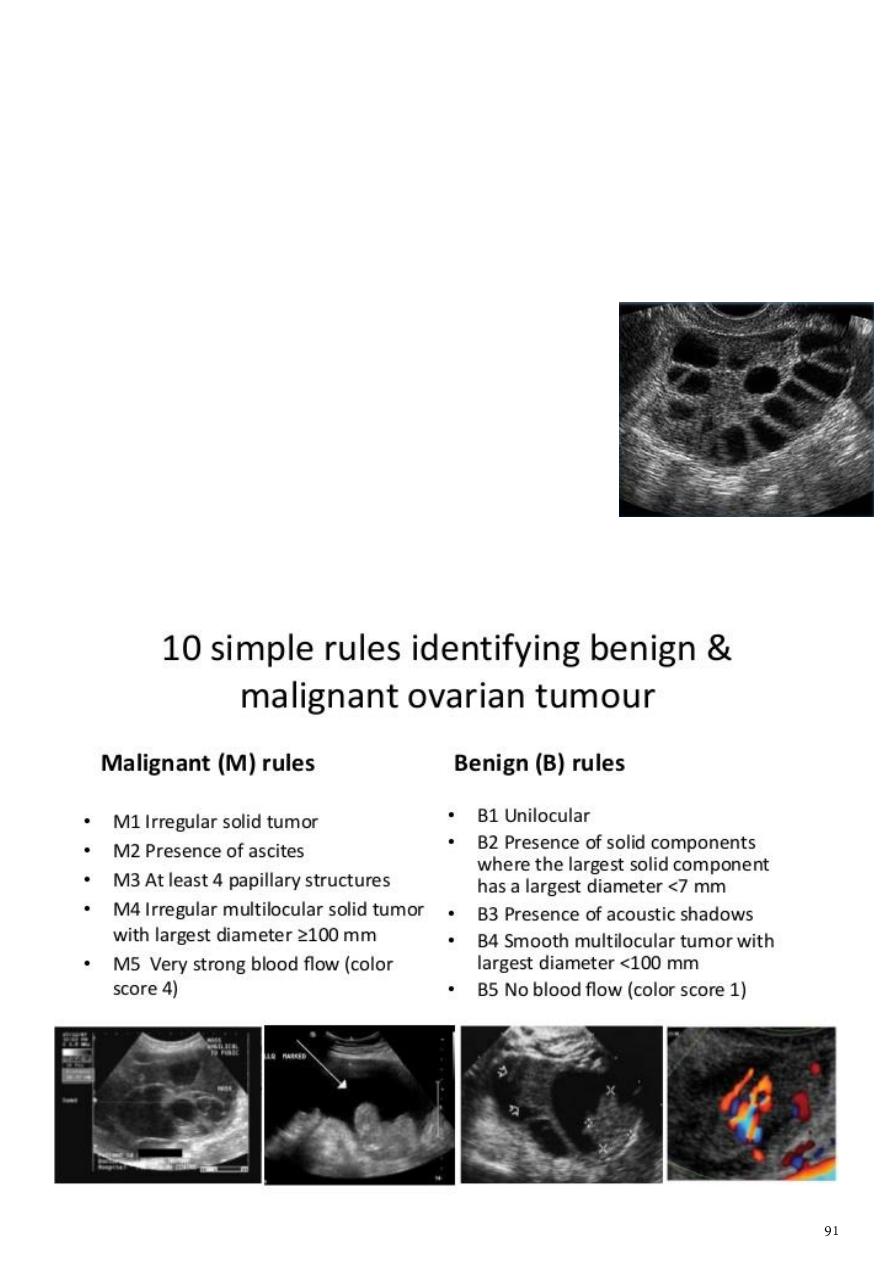
2- Endometriosis.
3- Teratoma.
4- Polycystic ovarian syndrome (PCOS):
Sonographic findings alone are not specific.
The diagnosis is made on the combined clinical, biochemical and sonographic grounds
The classic triad of PCOS oligomenorrhea, hirsutism, obesity.
Sonographic features of polycystic ovaries:
o Bilateral enlarged ovaries with multiple small follicles.
o Increased ovarian size (>10 cc).
o 12 or more follicles measuring 2-9 mm.
o Follicles of similar size.
o Peripheral location of follicles (which can give a
string
of pearl appearance
).
o Hyperechoic central stroma.
o The ovarian outline may be slightly irregular
5- Ovarian tumors:
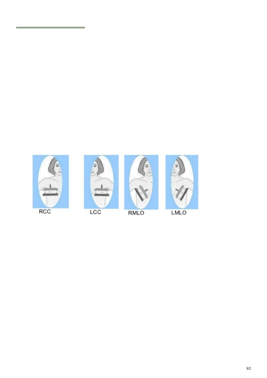
Mammography
Types of views in mammography:
Standard views:
Used for diagnosis and screening.
o The mediolateral oblique (MLO) view:
Also called Lundgren's view.
It is the most important projection as it allows to depict most breast tissue.
o The craniocaudal view (CC) view:
It must show the medial part as well the external lateral portion of the breast
as much as possible.
Supplementary views:
Used for diagnosis
.
o Lateromedial (from side towards center of chest).
o Mediolateral (from the center of the chest out).
o Spot compression view.
General information:
Mammography is done by the following breast is briefly compressed between 2
plates attached to the mammogram machine- an adjustable plastic plate on top and a
fixed plate on bottom which holds the x-ray film or the digital detector that makes the
image.
Mammography is screening test for any women above 35-40 years.
Any women with family history of breast cancer should make mammography every 3
months.
It can help reduce the number of deaths from breast cancer among women ages 40 to
70.
Mammograms are images of the breast tissue produced on X-ray film.
Mammograms are the most efficient screening method to detect early breast cancer.
A mammogram only takes a few seconds and may be mildly uncomfortable.
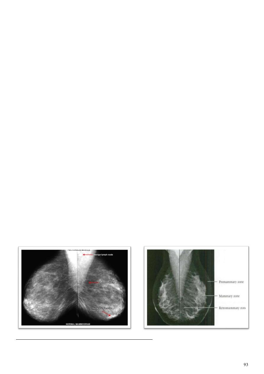
An abnormal mammogram does not necessarily mean that a cancer is present; other
tests, including biopsy, MRI, or ultrasound examination may be performed for further
clarification of an abnormal mammogram.
A normal mammogram does not exclude the presence of cancer.
False negative results occur in younger women with dense breast.
Overall, screening mammogram miss about 1 in 5 breast cancer.
Screening mammography is performed at regular intervals to detect abnormalities.
Benefits of mammography:
o Improves a physician's ability to detect small tumors.
o Increases the detection of ductal carcinoma in situ (DCIS).
o Useful for detecting all types of breast cancer, including invasive ductal and
invasive lobular cancer.
o No radiation remains in a patient's body after an x-ray examination.
o No side effects in the typical diagnostic range for this exam.
Risk of mammography:
o There is always a slight chance of cancer from excessive exposure to radiation.
Mammography in young women:
o Difficult to read because breast tissue is dense and it can hid the tumor.
o Women who are at high risk yearly MRI or mammograms are recommended at
age of 30 years.
o Risk factors: gene mutation, strong family history, other factors.
Breast density depend on how much fibrous and glandular tissue, how is the
distribution within breast tissue, how is breast made up of fatty tissue.
Dense breast are not abnormal but they are linked to higher risk of breast cancer.
Normal mammogram:
Abnormalities that can be detected by mammography mass, calcification, inflammatory
L.N, tubular density, architectural distortion, asymmetrical breast finding, other associated
findings.
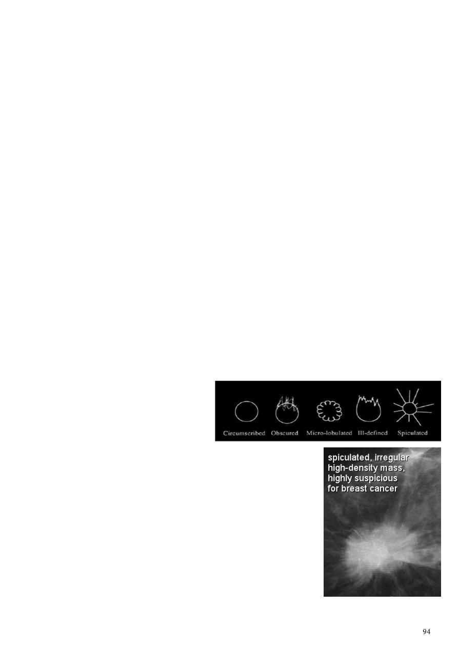
Breast lump:
Should be examined by self-exam, health care provider exam, mammography, US,
other imaging studies, fine needle aspiration.
The majority (about 80%) of breast lumps are not due to cancer they are cysts,
benign tumors (fibroadenoma), area of thicker or denser breast tissue, changes in
consistency due to the menstrual cycle, certain patterns of calcium deposits.
Abnormal mammogram does not mean there is cancer need other investigations.
Women with a breast lump need to have a mammogram of both breasts.
A mammogram is estimated to be able to detect about 90% of breast cancers.
Breast cancer:
There are two main types of breast cancer:
o Ductal carcinoma starts in the tubes (ducts) that carry milk from the breast to
the nipple, most breast cancers are of this type, and it is diagnosed earlies
because it lead to edema of the breast.
o Lobular carcinoma starts in lobules, which produce milk, it is difficult to
diagnose and need more time.
o In rare cases breast cancer can start in other areas of the breast.
If there is family history and +ve BRACA1 or 2 it means breast cancer.
Computer-aided detection (CAD) is a tool that helps the radiologist find areas of
concern that need further attention.
After taking the mammograph you should focus on the breast density and the margin
of the mass.
Mammographic findings:
o Rather well defined speculated irregular highly dense
mass breast cancer.
o Smooth outline of the mass cyst (echolucent by US)
or fibroadenoma (hyperechoic by US).
o Micro-calcifications underlying CA.
o Macro-calcifications (pop-corn) fibroadenoma.
o Linear calcification it is vascular calcifications (not
dangerous).
o Cyst with calcifications is rare.
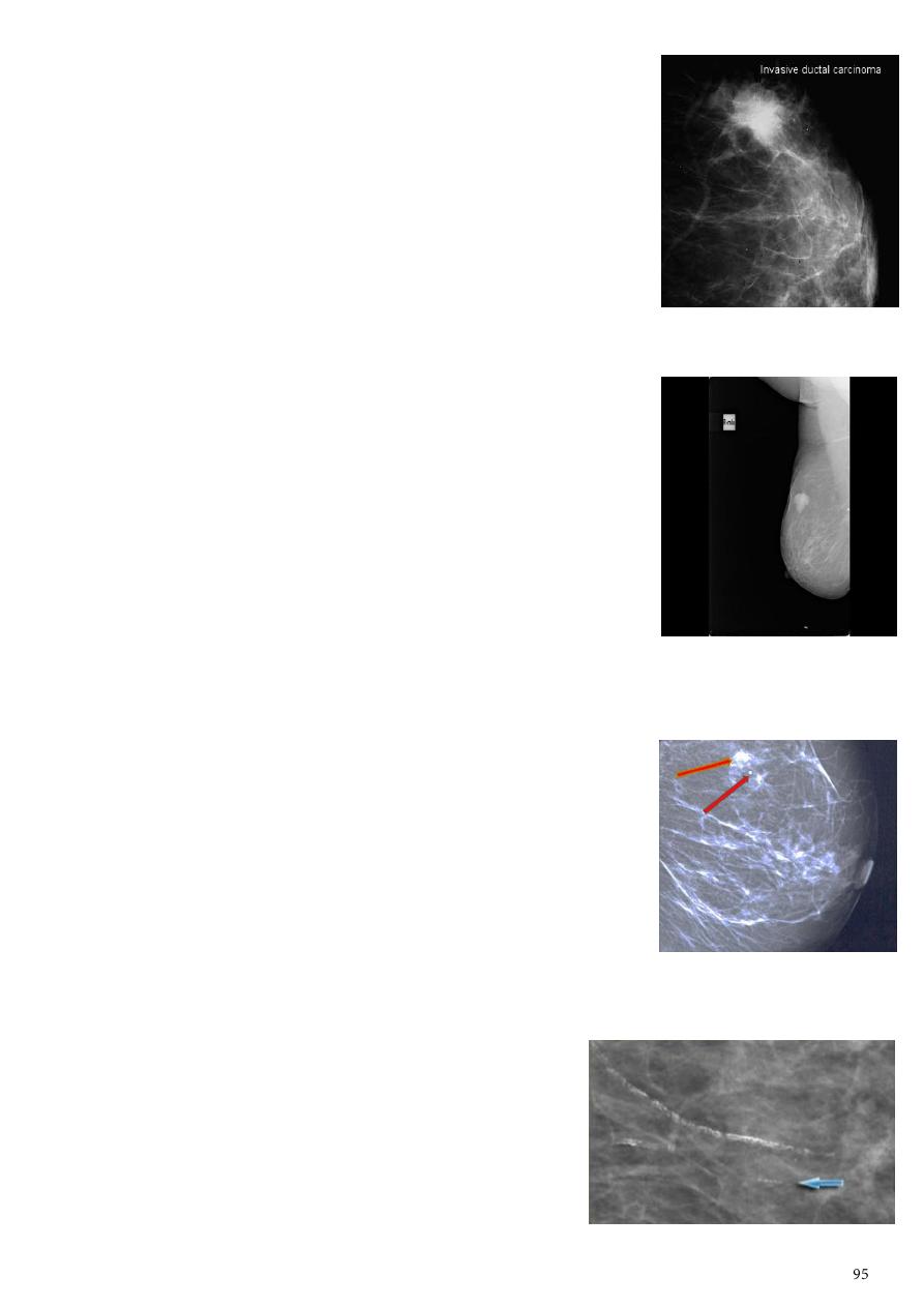
1- Invasive ductal carcinoma (IDC):
Is a subset of ductal carcinoma.
It is an infiltrating, malignant and abnormal proliferation of
neoplastic cells in the breast tissues.
It is the most frequently seen breast malignancy.
Mammogram spiculated hyperdense lesion,
oval/lobulated lesion, microcalicifications.
2- Infiltrating or invasive lobular carcinoma (ILC):
It is the second most common type of invasive breast cancer.
Multicentric and bilateral (10-15%).
Progressive shrinkage or enlargement or reduced
compressibility of the involved breast.
Mammography is 55-80% sensitivity.
Sonography and MRI used in evaluating clinically
suspicious findings.
ILC are more commonly seen on the craniocaudal (CC),
compared to the mediolateral oblique (MLO).
3- Microcalcifications:
Microcalcifications are tiny specks of calcium in the breast.
They do not always mean that cancer is present.
The shape and layout of microcalcifications help the
radiologist judge how likely it is that cancer is present.
The presence of microcalcifications does not mean a biopsy
is needed.
If the microcalcifications have a suspicious look and pattern,
a biopsy will be recommended.
A biopsy is the only way to tell if cancer is really present.
4- Vascular calcifications:
Linear or parallel tracks.
Associated with blood vessels.
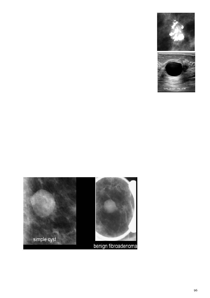
5- Popcorn calcifications:
It indicate fibroadenoma.
Not indicate ductal carcinoma.
6- Cyst:
Simple cyst simple fluid-filled sacs, benign (not cancer), don’t
need to be biopsied.
Complex cystic and solid masses be partially solid, it is of
more concern and might need to be biopsied to be sure it isn’t
cancer.
7- Fibroadenoma:
It is solid benign non-cystic lesion.
Results from excess proliferation of connective tissue.
Characteristically contain both stromal and epithelial cells.
Mammography:
1- Well circumscribed discrete oval mass hypo- or isodense to the breast glandular
tissue.
2- Mass with macrolobulation or partially obscured margin.
3- Involuting fibroadenomas in older, typically postmenopausal patients may contain
calcification, often producing the classic, coarse popcorn calcification appearance.

Part6
: Computerized tomography (CT scan)
Basic information:
CT scan is a form of XR examination.
It is the technique of rotating a radiation detector around the patient so that the
image obtained gives additional three- dimensional information.
CT scan can produce an image of slice through the body at a particular level.
When the number of the detector increase, the clearance of the image is increase,
and the radiation dose also increase.
The usual number of detectors is used about 400.
The XR can penetrate any organ & when penetrate it, it will face many different
density tissues like bone, muscle, fat, fluid or CSF.
The higher the thickness, the higher the Molecular weight, the higher the
concentration of material the more the absorption of the XR & more attenuation,
examples:
o Bone white color, because it absorb much XR so it is highly attenuated
material.
o Soft tissues gray color, because it have less attenuation.
o Air black color, because it has low absorption & low attenuation.
o Air / water / fat all appear black in color.
CT terms:
o Iso-dense = same density to organ examine.
o Hyper-dense = higher than the organ examined.
o Hypo-dense = lower than the organ examined.
Densities:
o All soft tissue organs lie within a range 10-90 HU, the highest is thyroid 70 HU.
o Water density 0 HU.
o Fluid 10-15 HU depending on their density.
o Hemorrhage 40-80 HU.
o Air -1000 HU.
o Fat -100 - -50 HU.
o Bone & calcification 100 & above HU.
o Ossification 1000 & even more HU depending on the density.
Pregnancy is absolute contraindication of CT scan.
CT scan give high radiation dose to the patient.
CT scan can differentiate between structures because it give the density.
CT scan take the axial view then the software will give sagittal and coronal views.
Side effects of CT Lead to cataract and thyroid problems in pediatrics, Lead to
fibrosis of genitalia in children, It is teratogenic.
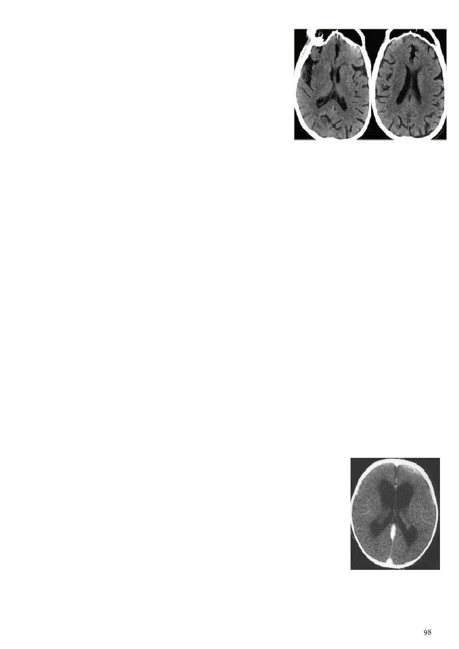
CT scan of the brain:
CT of the brain start from the vertex to the base of
skull except the eye region.
CT will show cerebral hemispheres, frontal lobe,
parietal lobe (banana shape), temporal lobe (no
bannan shape, appearance of frontal horn), occipital
lobe, posterior fossae, cerebellum, brain stem,
lateral ventricles.
CSF circulation lateral ventricles foramen of morno third ventricle
cerebral aqueduct forth ventricle foramen subarachnoid space arachnoid
villi venous drainage.
Pituitary fossa:
o Anterior cliniod process.
o The region of the sella tursica.
o Posterior cliniod process.
o Channel like area black in color (cerebello pontine angle).
o Lesser wing of the sphenoid bone & below it immediately the greater wing of
sphenoid.
1- Encephalitis:
General information:
o Age young & adolescence mostly.
o Clinical picture headache, vomiting, blurring of vision.
o Usually the disease preceded by flue or vaccination, herpes simplex virus.
o Complications Brain destruction, Hydrocephalous, Atrophy of the brain tissues
with subsequent loss of volume & dilatation of the ventricular system.
o Note: any inflammation in the body lead to edema which change the gray scale to
black color and lead to expansion and then compression of nearby structures.
CT findings:
o Edema (Low attenuation of white matter).
o Compressing the ventricular system (due to edema) specially
the frontal & the body of the lateral ventricle become slit like.
o Brain necrosis (destruction) lead to stretching.
o Effacement of sulci.
Herpes encephalitis:
o Same as usual encephalitis.
o There will be low attenuation or hypodensity area in the tempo frontal or tempo
priato frontal
o Evidence of patchy whitish area due to the clotted blood (hemorrhage).
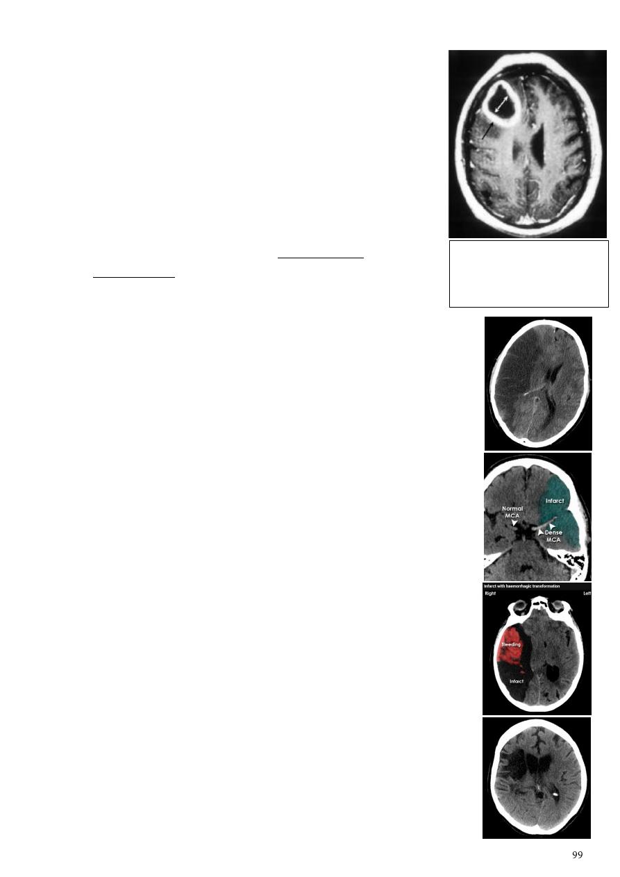
2- Brain abscess:
General information:
o Clinical picture high fever for long duration, focus of
infection, toxic, epilepsy (focal).
o DDx Secondary metastasis (history of primary
malignancy, no fever), primary necrotic tumor (no history
of primary malignancy, no fever).
CT finding:
o Hypodense area surrounded by large area of peri-focal
edema (cause shifting of the midline).
o After injection the lesion show ring pattern of
enhancement so the peripheral aspect of the lesion
become clearer.
4- Infarction:
General information:
o 90% of patient referred is due to infarction.
o Localize the site to detect the affected artery.
Acute infarction:
o History: sudden onset of hemiplegia, with slurred speech.
o CT features:
o Wedge shape area of hypodensity involve the tributaries of
anterior, middle or posterior cerebral arteries (priato frontal &
priato occipital area).
o Shift of the midline to the other side.
o Compression of the ipsilateral aspect of the regional ventricle by
the edema.
o
Dense middle artery sign
.
o Hemorrhagic infarction wedge shape area of hypodensity
contain area of hyperdensity inside it.
Chronic infarction:
o The necrotic area will be liquefy & change to the source of fluid
(por encephalon).
o CT features:
o Hypodensity area as a result of blockage of anterior, middle or
posterior cerebral arteries.
o Dilated ventricle to compensate for the loosed brain tissue.
o The area of subacute (chronic) infarction will convert to fatty
necrosis which lead to regional dilatation or stretching.
Axial CT section of the brain
showing well defined rounded
dense ring enhancement
lesion in the right hemisphere.
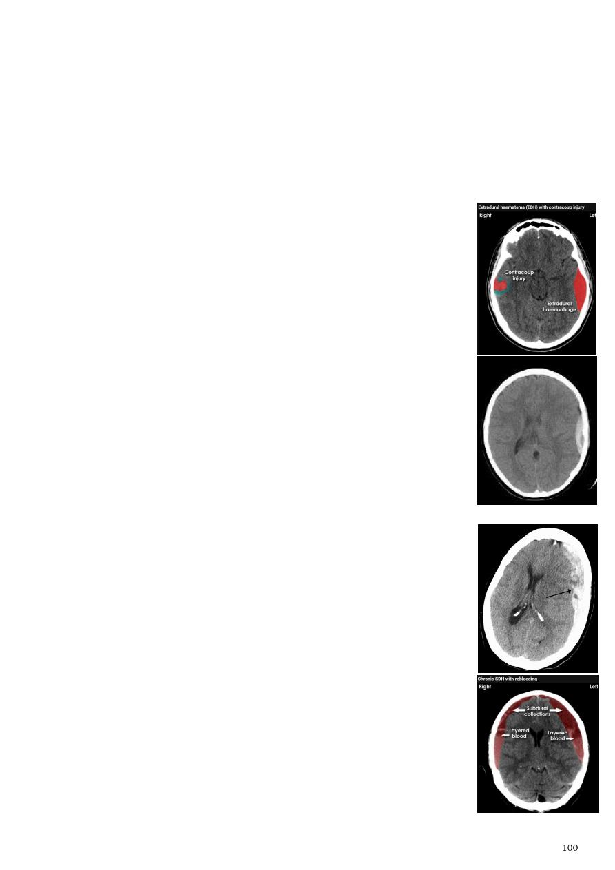
4- Hemorrhage:
Classification according to the site:
o Epidural EDH
o Subdural SDH
o Sub arachnoids SAH
o Intra cerebral ICH
o Intra ventricular IVH
Classification according to the onset of bleeding:
o Acute.
o Chronic.
- Acute Epidural hemorrhage:
Hyperdensity area 50-70 HU due to the clotted blood.
Elliptical (biconvex) in shape.
> 90 % associated with skull fracture.
May cause shifting of the midline or compression of the ipsilateral
ventricle.
No edema around the lesion.
Source of bleeding is middle meningeal artery.
Middle age group.
- Acute subdural hemorrhage:
Hyperdensity area their inner medial margin is irregular shaggy.
Crescent or oval shape.
Not associated with skull fracture.
Shifting of midline.
Source of bleeding is venous (disruption of subdural vein).
Extreme age group:
o In children shock baby syndrome (wide subarachnoid space
and wide cistern)
o In old age sudden movement or concussion (brain atrophied
wide subarachnoid space)
Note: hematocrit layer (layered blood) is characteristic for SDH.
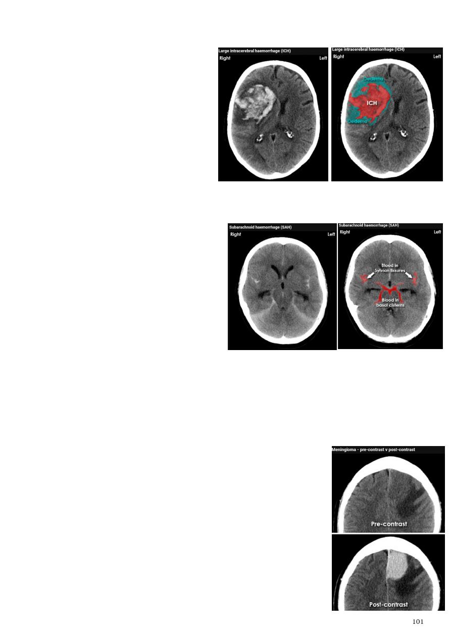
- Acute Intra cerebral hematoma:
Hyper dense area.
Surrounded by edema.
Anywhere within the brain
parenchyma.
Shifting of the midline.
Compression of the ipsilateral
ventricle.
Associated with infarction, fracture,
trauma concussion, tumor.
- Subarachnoid hemorrhage:
Hyperdensity is seen within the
subarachnoid space.
Hyperdense sulci, being filled with
clotted blood.
Opacified inter hemispheric fissure
(become white & more dense).
Opacification of the falx cerebri.
Causes os SAH ruptured aneurysm
(90 % of cases especially at the circle of
Willis), ruptured AV malformation, trauma.
5- Meningioma:
General information:
o Benign tumor arise from the arachnid cells of the
meningeal covering of the brain.
o Most common primary intracranial neoplasm.
o Usually present in middle age female.
o It is well-defined, rounded or sessile, plaque like, extra-
axial, located mainly at the convexity of the skull periphery.
o Note: any benign tumor in the brain could convert to
malignant.
CT features:
o Isodense area or slightly hyperdensity area.
o Surrounded by crescent area of hypodensity "
CSF cap
".
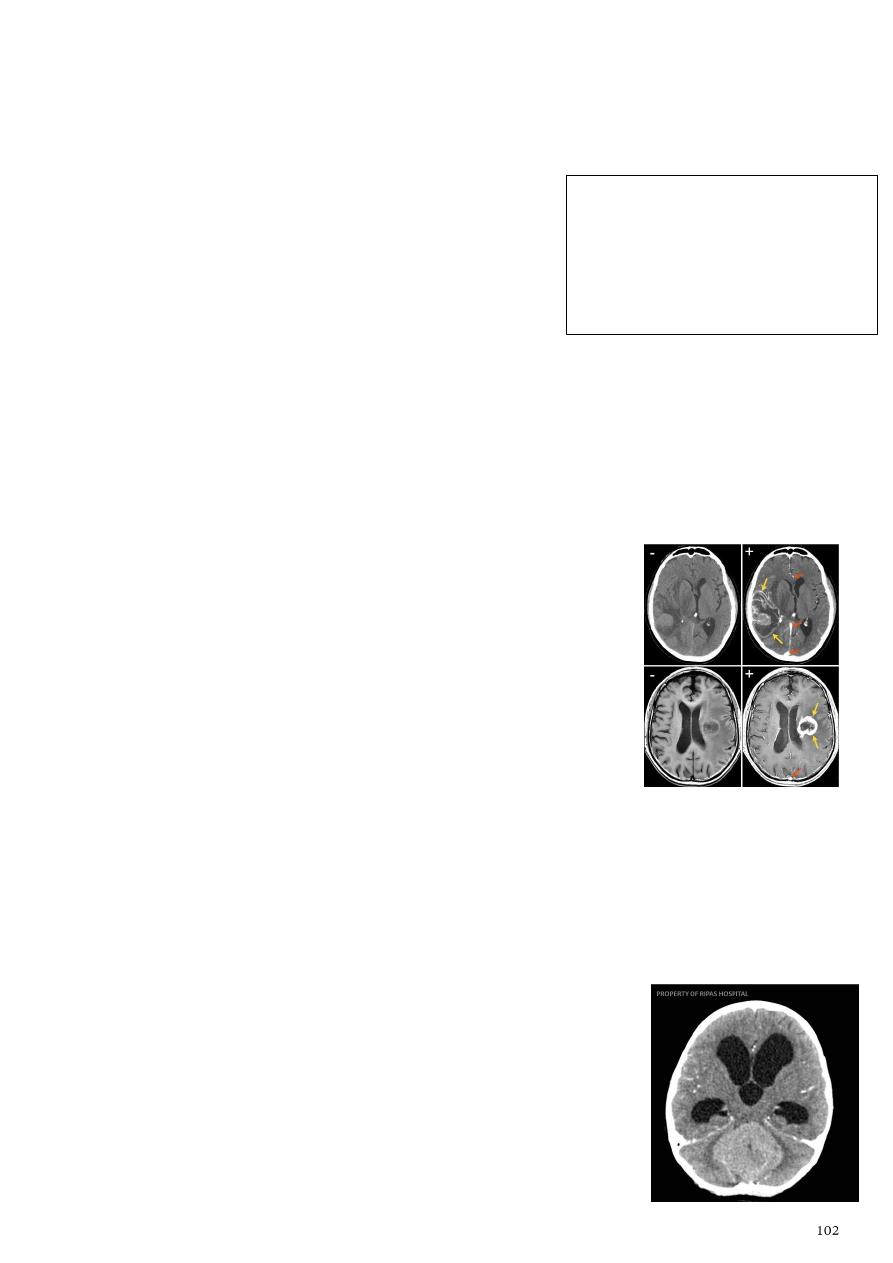
o Post contrast injection the lesion enhance homogeneously with
enhancing Dural
tail
.
o 20 % show calcification.
o Hyperostosis & thickening of the nearby bony part of the skull & diplioc space.
o Associated with little or no peri focal edema.
o If the lesion associated with central necrosis
with large perifocal edema meningiosarcoma
should be excluded.
6- Glioma:
Comments primary interracial tumor, vary greatly in malignancy.
Have many names depending on the histological type:
o Astrocytoma and Oligodendroglioma both of them are well differentiated
slowly growing tumor.
o Gliobtastoma multiforme (butterfly glioma) G IV highly malignant tumor, arise
from the anterior or posterior aspect of the corpus callosum, extend and spread
to both cerebral hemispheric sides.
Grading of malignant glioma depending on the following:
o Well defined or irregularity of the lesion.
o Surrounding edema or not.
o Associated shifting of midline & crossed midline lesion.
o Contrast enhancement.
o Associated hemorrhage, necrosis, cystic formation.
o Seeding via CSF & dissemination.
Grades:
o Low grade G. I well or ill-defined lesion isodence, not associated with edema,
no enhancement, no associated hemorrhage, necrosis.
o G II, III various previous finding.
o G IV (glioblastoma multiforme) highly malignant have all previous mentioned
features.
7- Posterior fosse tumor:
Medulloblastoma:
o Age: between 5-15 years.
o Represent about 35-40 % of posterior fossa tumors.
o Arise from the midline mainly within or from the roof of
the 4
th
ventricle.
o Can be seen in the distal part of the spinal canal.
Note: any lesion in the brain should give
contrast and if the lesion take the contrast it is
malignant cancer of any grade, no
enhancement is benign (except meningioma
which is benign and take the contrast).
Meningioma before enchantment is isodense
and after enhancement is hyperdense.
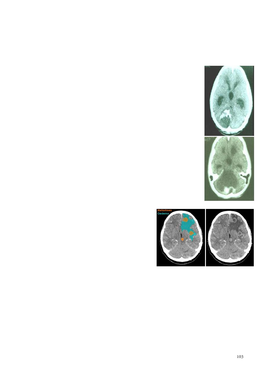
o CT finding well circumscribed, heterogeneous in density, have solid and cystic
part, the solid part is enhance, with scattered calcification, little surrounded
edema.
o 90 % present with obstructive hydrocephalous, with dilatation of the lateral
ventricle and third ventricle.
o 40 % have secondary metastasis.
Pilocytic astrocytoma:
o Age between 5-15 years.
o Arise within the vermis and cerebral hemisphere.
o CT finding well circumscribed lesion, hypo or hyper dense,
growing mainly with expansion, usually large lesion solid or cystic
or both of them, with enhancing mural nodule, 20% show
calcification of the nodule, may associated with edema.
Haemangioblastoma:
o Age between 30 -65 years.
o Represent 10 % of posterior fossa tumors.
o Arise from per vascular pericyte.
o GII to III in their malignancy.
o Intraaxial tumor arise from the cerebellum, brain stem, spinal
cord.
o CT finding smooth walled cystic lesion with enhancing mural
nodule rarely calcified.
Secondary metastasis:
o Age: above 50 years.
o Any lesion within the cerebellar hemisphere
it is secondary metastasis unless proven
otherwise.
o Female from breast cancer, male from
bronchogenic carcinoma.
o CT finding nodular single or multiple
lesion, hypo dense or hyper dense,
surrounded by per focal edema, enhanced as
solid or ring pattern of enhancement.
8- Supra seller tumor (cranio-phyrengioma):
Tumor situated above the sella tursica.
Arise from the remnant of the rathekes pouch.
The tumor might invade sella tursica, then go anterior, posterior, upward, sometime
reach the third ventricle.
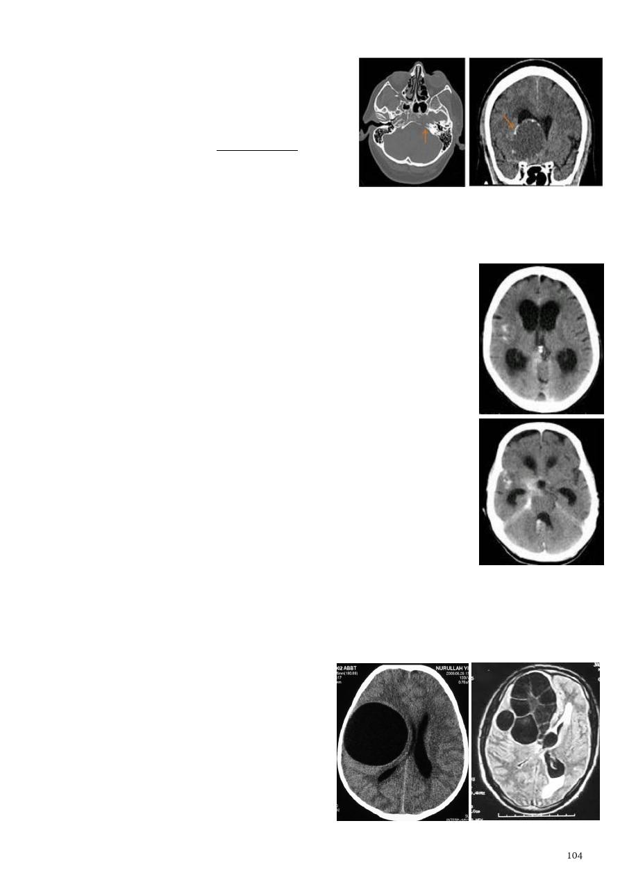
CT finding:
o Deformity of the supra sellar cistern.
o Have solid and cystic component.
o Post contrast solid part will get
enhancement, flecks of calcification seen
around the lesion as multiple rings.
The lesion might obstruct the third ventricle
and present with obstructive hydrocephalous.
9- Hydrocephalous:
Definition dilatation of ventricular system.
Obstructive (non-communicating):
o Forth ventricle is slit point.
o Causes focal lesion, secondary metastasis, infarction,
tumors (craniophyrengioma, medulloblastoma, ependymoma).
o Aquiduct stenosis, comments cause of obstruction being
congenital in nature.
Non-obstructive (communicating):
o Forth ventricle is dilated.
o No obstruction of the ventricular pathway.
o The absorption of the CSF at the level of arachnoids
granulation is occluded secondary to lodge by blood clot or
inflammatory cell or infection post meningitis.
o Most commonly to occur post SAH.
Most common cause of pediatric with obstructive hydrocephalus
and slit point forth ventricle is Toxoplasmosis due to aqueduct
stenosis, and treated by shunt.
We cannot determine if it is obstructive or non-obstructive hydrocephalus unless we
see the posterior fossa.
10- Hydatid cyst of the brain:
Axial CT section of the brain showing well-
defined rounded hypodense lesion.
Unilocular or multilocular.
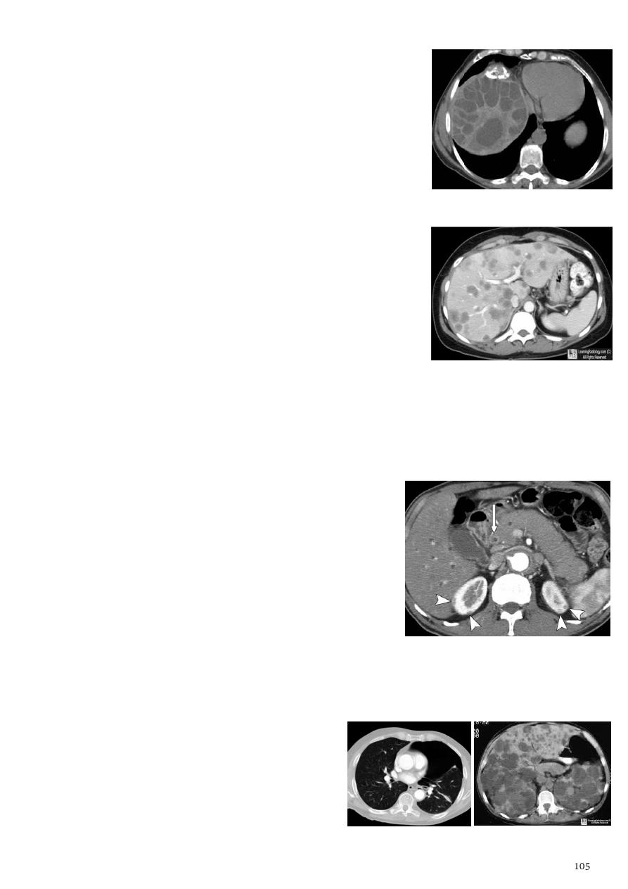
11- Hydatid cyst of the abdomen:
Appear as large oval hypodense area.
Density of fluid with well-defined margin.
Multiple flecks of calcification seen at their periphery.
Hydatid cyst with daughter cyst appear as multiple
hypo-densities rounded area within the main loculi with
multiple rim of calcification.
12- Secondary metastasis in the liver:
Multiple rounded hypo density areas of different density,
shape, size.
Pattern of enhancement is either uniform, target, bulls
eye pattern.
Hepatomegaly.
13- Pancreatitis:
Clinical features abdominal pain, vomiting, with or without jaundice, increase
amylase level.
CT finding:
o Focal or generalized increase in the size of
pancreas.
o Focal or generalized hypodensity within the
pancreas (due to the edema).
o Peri pancreatic fluid collection and edema around
the pancreas.
o The fluid around the pancreas if persist more than
6 w become encysted leading to the pancreatic
pseudo cyst.
o Edema of the wall of the stomach.
Normal pancreas = hypodense, slit like.
Pancreatitis = increase is size, per pancreatic collection.
Hemorrhagic pancreatitis = increase is size,
increase in density.
Pneumothorax Adult polycystic kidney
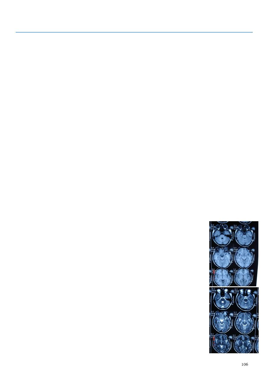
Part7
: Magnetic resonance imaging (MRI)
Why we use MRI when we have CT scan?
Higher tissue contrast resolution so it is more sensitive to detect brain lesion.
Beam hardening artifact is not present so visualization of posterior fossa is better on
MRI than on CT scan.
Direct coronal and sagittal section are taken.
Lack of ionizing radiation.
Basic information:
MRI employs radiowaves and magnetic fields.
The patient is placed in a magnet and radiowave sent in, the transmitters is turned off
and the patient re-emits radiowaves, which are received and used for reconstruction
of the image.
Use the nuclei of hydrogen atoms in water and fat which absorb and emit the
radiofrequency energy.
The magnetic field strength has a set value between 0.5-2.0 tesla (T), depending on
the machine.
1T = 20 000 times greater than the earth's magnetic field.
The MR image (T1, T2) of tissue controlled by two parameters set by the operator
TE (time to echo) & TR (time to repeat).
T1 weighted image:
o Longitudinal relaxation time.
o Short TR (300-600ms).
o Short TE (30ms).
o The shorter T1, the stronger the signal and the brighter the pixel.
o Fat (fatty bone marrow) is bright.
o White matter and CSF are dark.
T2 weighted image:
o Transverse relaxation time.
o Longer TE (90-140ms).
o Long TR (1000-2000ms).
o The longer T2, the stronger the signal and the brighter the pixel.
o Water and CSF appear brighter than fat.
– T1 = all fluid black, fat white, hemorrhage white.
– T2= all fluid white, fat white, hemorrhage white.
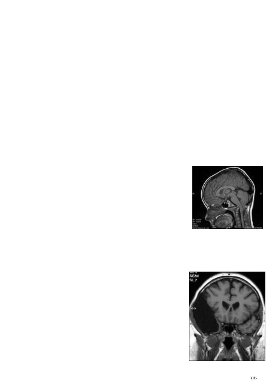
1- Congenital hydrocephalus:
Congenital lesion of brain are well seen by MRI & CT scan but MRI has the advantage
of absence of ionizing radiation so can be used safely in children.
Communicating congenital hydrocephalus:
o Free communication between ventricles and basal cisterns.
o Obstruction to flow of CSF is in the subarachnoid space or basal cisterns.
o This is due to meningeal irritation by hemorrhage, infection, trauma.
o All ventricles are enlarged and basal cisterns may be prominent.
Non-communicating congenital hydrocephalus:
o CSF flow is obstructed within the ventricular system at 3rd ventricle, aqueduct,
or 4th ventricular level.
o The ventricles are dilated above the level of obstruction.
o Most common cause aqueduct stenosis, it is congenital obstruction or stenosis
of the aqueduct which result in hydrocephalus involving the 3rd and lateral
ventricles.
2- Arnold Chiari malformation:
Downward herniation of cerebellar tonsil and medulla
oblongata beneath level of foramen magnum.
Elongation of 4th ventricle.
Associated with aqueduct stenosis, and myelomeningocele.
3- Arachnoid cyst:
Clear CSF filled cyst.
Occur due to congenital splitting of the arachnoid membrane.
They are intracranial but extracerebral.
Can be seen in middle, posterior cranial fossa, suprasellar
region, behind the 3rd ventricle.
They can be large enough to produce mass effect or
obstructive hydrocephalus.
There may be Hypoplasia of underlying cerebral tissue
such as the temporal pole.
Description coronal T1 weighted MRI image through a
brain lesion showing homogeneity of the lesion, lack of a
perceptible wall, lack of internal complexity, and CSF
signal intensity, there is associated remodeling of the
adjacent calvarium and brain displacement.
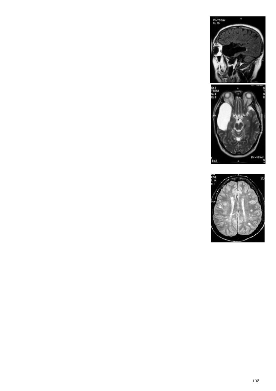
Description sagittal fluid-attenuated inversion recovery (FLAIR)
weighted image through a brain lesion), showing homogeneity of
the lesion, lack of a perceptible wall, lack of internal complexity,
and CSF signal intensity there is associated brain displacement.
Description Axial T2-weighted MRI image through the
midbrain, showing a right middle cranial fossa homogeneous
lesion) with CSF signal intensity and no perceptible wall or
internal complexity, there is associated remodeling of the
adjacent sphenoid bone and brain displacement.
4- White matter disease:
Have been classified as:
o Dysmyelinating there is abnormal formation of myelin
(leukodystrophies, metabolic disorders).
o Demylinating there is destruction of normally formed
myelin (multiple sclerosis).
Multiple sclerosis:
o Characterized by disseminated plaques of demyelination and
gliosis throughout the neuraxis.
o The sites of election are periventricular, optic pathways,
brainstem, spinal cord, cerebellar white matter and peduncles.
o Young adults are primarily affected.
o MRI is more sensitive than CT in the demonstration of MS plaques.
o MRI can even image lesions in the spinal cord, brainstem, cerebellum.
o Multiple cerebral lesions are seen in over half the patient presenting with a single
episode of optic neuritis.
o The most characteristic appearance peri ventricular nodular Hyper intense
lesions on T2 weighted images.
o The plaques are also well seen at the gray- white matter interfaces.
o Some lesion may show a central area of greater signal intensity, resembling a
target.
o Contrast enhancement after giving gadolinium occurs in the acute phase
indicating activity and in the chronic phase doesn't enhance.
o DDx ischemic lesions, following radiotherapy, encephalomyelitis.
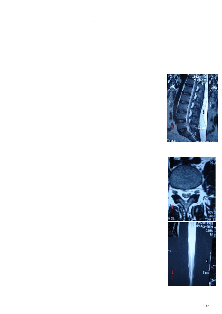
NEURORADIOLOGY OF SPINE
General information:
MRI indicated for precise localization of the level of a lesion which is difficult to be
done from clinical examination.
Advantage of MRI over CT MRI is a direct multiplanar acquisition (can be easily
applied in any plane, including optimal sagittal axis).
Annulus fibrosus / spinal ligaments / dura matter / cortical bone
of the vertebrae give low intensity signal.
Epidural / paraspinal fat give high intensity signal.
The gel of the normal nucleus pulposus of the normal
intervertebral discs gives high intensity signal on T2 weighted
image sequences.
In the normal adult disc a shelf of annulus causes a low signal
horizontal band resulting in a bilocular appearance.
With normal aging the intensity of the signal from the nuclei
decreases.
1- Normal anatomy:
The spinal canal is bounded anteriorly by vertebral bodies and
intervertebral discs, backed by the posterior longitudinal
ligament, posterolaterally by pedicles and laminae lined by
ligamenta flava.
The normal intervertebral foramens are oval or boot shaped and
symmetrical in the absence of scoliosis.
Disc content Center (neuclus pulposus), Annulus fibrosus.
Intervertebral foramen contain dorsal root ganglion, spinal
nerve root, epidural fat.
Potentially significant developmental narrowing If sagittal
diameter in the cervical and lumber regions below 12mm and 14
mm respectively.
The spinal cord descends from the medulla oblongata,
commencing at about the level of foramen magnum and
terminates at the conus medullaris, which lies between the lower
border of 12th thoracic and the upper border of the third lumber
vertebra.
MR MYELOGRAM
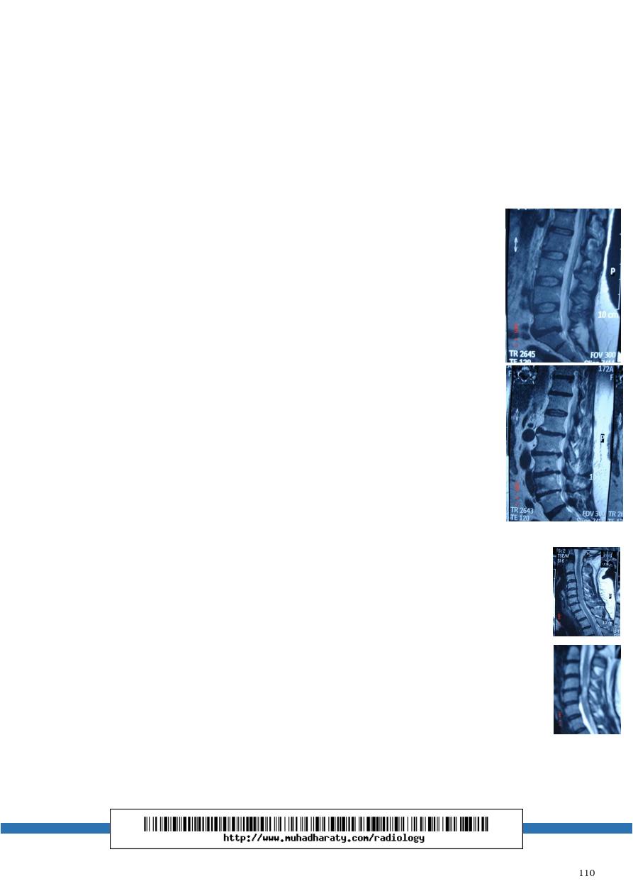
2- Spondylosis:
General information:
o This process involve intervertebral disc prolapse and degeneration.
o It is caused by wear and tear.
o It involve intervertebral discs, vertebral bodies, facet joints.
o It is the commonest cause of entrapment neuropathy and of neurological
disability due to spinal cord disease.
Disc prolapse:
o Extrusion of the softer material from within an intervertebral disc
into or through a posterior or posterolateral radial tear in the
annulus fibrosus.
o It take the form of focal broad based bulge in the margin of
annulus or a focal mass extending upwards or downwards in the
anterior epidural space.
o Far lateral protrusions or extruded fragments.
o Involve the intervertebral foramens, not the spinal canal.
o They are commonest in the lumber spine.
o Description no three layer sign in the disc, herniation of disc
material, compression on spine.
Degenerative changes:
o Loss of normal bright signal of the nucleus on T2 weighted image.
o Loss of the normal height of the disc.
o When involve articular surface leads to articular surface
irregularities & osteophyte formation.
o When involve ligaments leads to calcification or ossification which result in
diffuse thickening or a focal mass which may compress the neural tissue.
Posterior longitudinal ligament:
o Disc prolapse at c3-4, c4-5, c5-6, c6-7.
o Posterior longitudinal ligament thickening & ligamentum flavum
thickening.
Spinal cord compression:
o Spinal cord compression from disc prolapse & degeneration is damaging
cord substance.
o It is seen in the form of focal signal changes in the cord substance.
Significant spinal canal stenosis when:
o The stenosis is enough to eliminate CSF signal intensity on MR MYELOGRAPHY.
o Cauda equina entrapment.