Elbow joint exam
Practical rheumatology
Session-2-
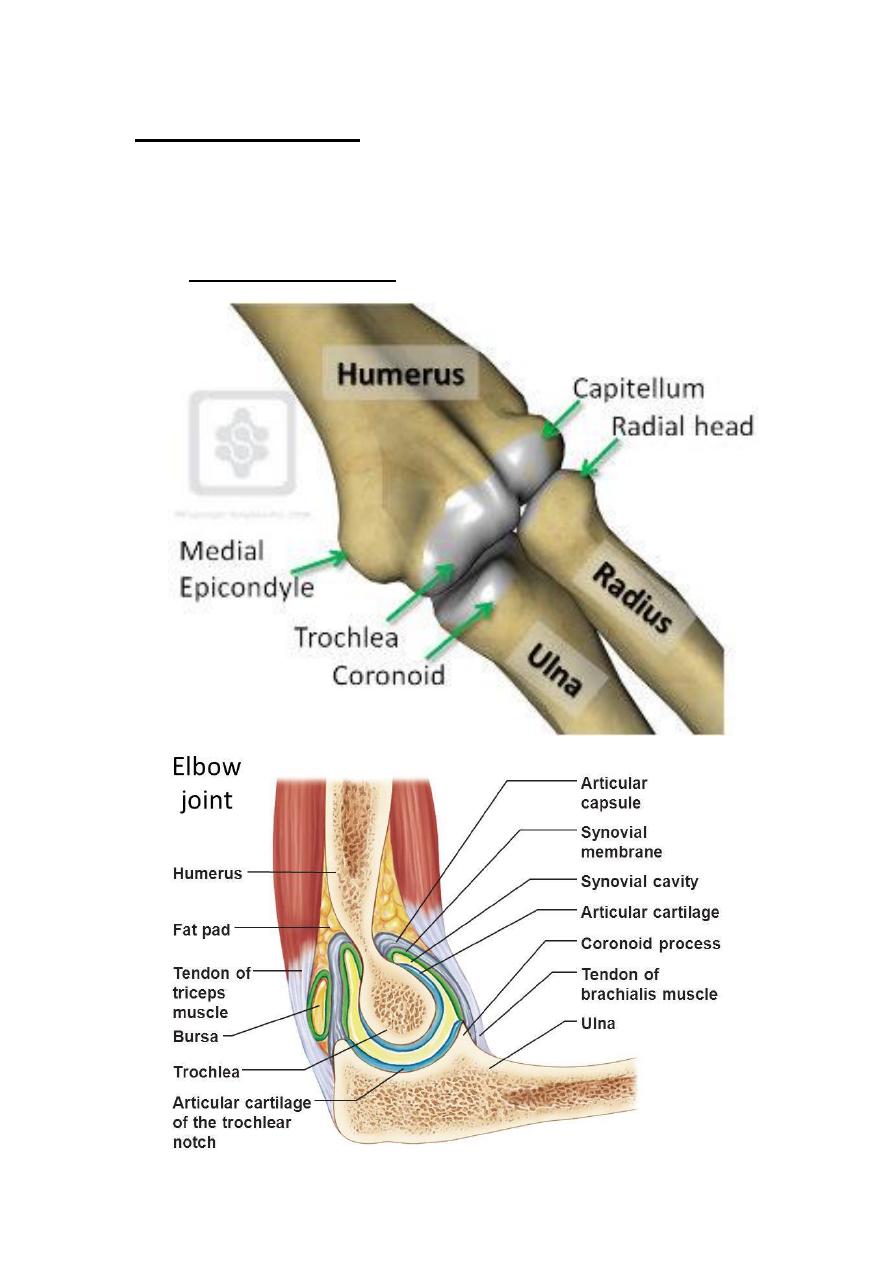
Elbow joint anatomy
Examination of elbow joint
Inspection :-
1-front
o At the overall alignment of the extended elbow
"CUBITUS VALGUS
o
o
injury / inflammatory arthritis / infection.
o
-13 degrees – females tend to have
more significant carrying angles than males.
2-Side
o Scars / Swelling / bruising / Erythema / rash / tophi.
o
o
is often most noticeable from this angle.
o Swelling of synovitis between the lateral epicondyle and
olecranon.
3-Back
*Rheumatoid nodules firm lumps on the elbow / olecranon –
indicate systemic rheumatoid disease.
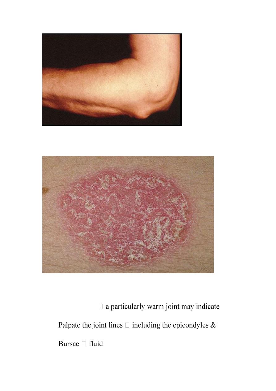
*Psoriatic plaques well defined pink / red elevated lesions
with silvery scale
2- Feel:
o Temperature
inflammatory arthritis or infection.
o
olecranon for any localised tenderness.
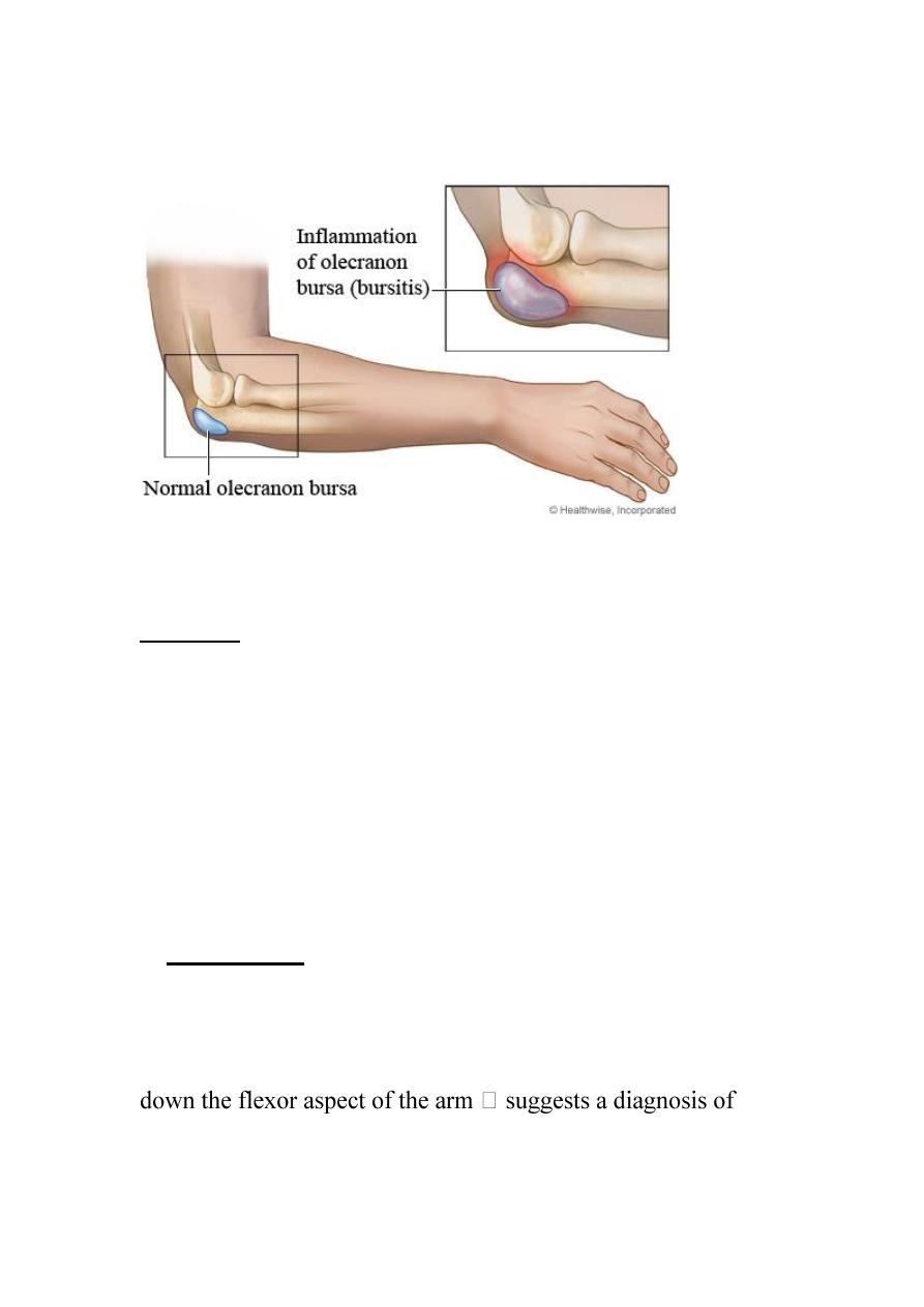
o
-filled sacs which are usually soft, but if
acutely inflamed or infected may be firm.
o Bony contours, sponginess, tenderness.
3-move :-
Assess each of the movements of the elbow joint actively &
passively.
-Elbow flexion - extension normal range is 0 – 145º
range less than 30–110° will cause functional
problems.
-Pronation 85º.
-Supination 90º.
-When moving the joint passively assess for crepitus.
4- Special tests:
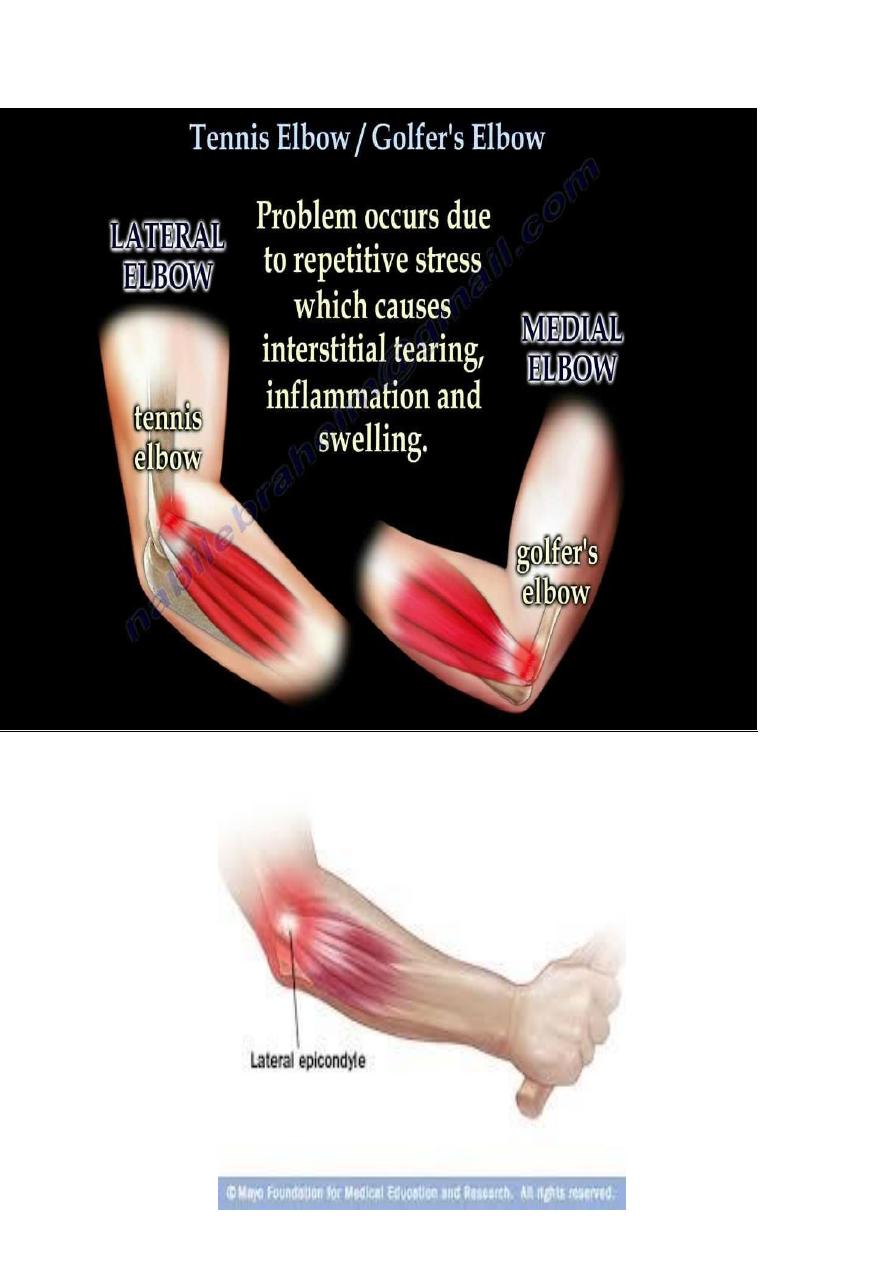
Medial epicondylitis – “Golfers Elbow”
*Ask the patient to actively flex the wrist whilst the elbow is
flexed.
*Localized pain over the medial epicondyle and may be referred
medial epicondylitis.
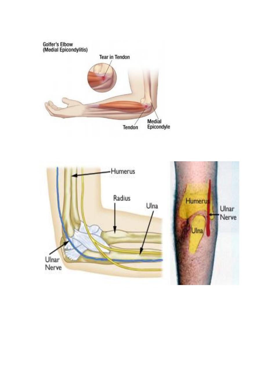
*palpate for ulnar nerve tenderness over its course.
--palpate for bicipital tendinitis
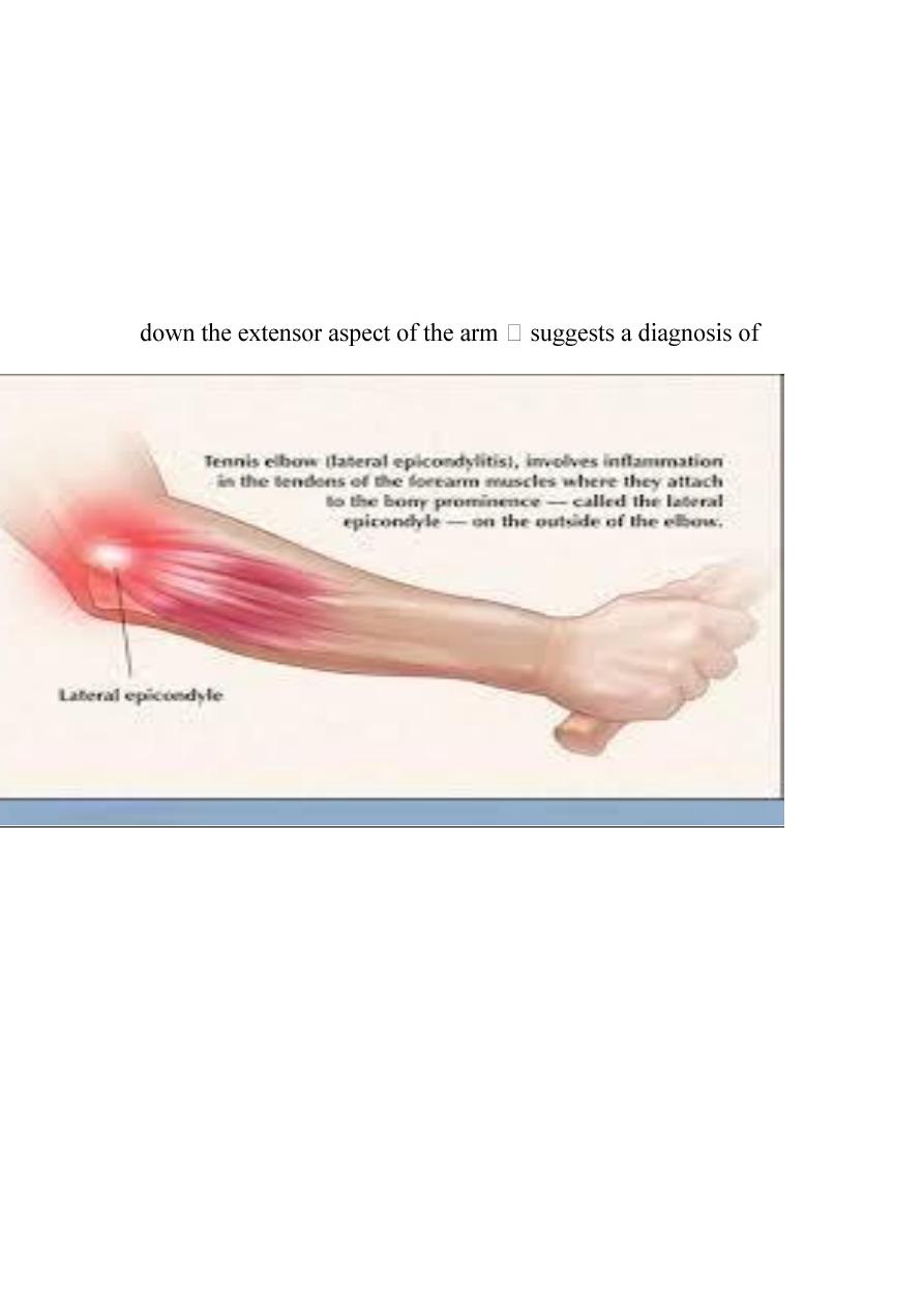
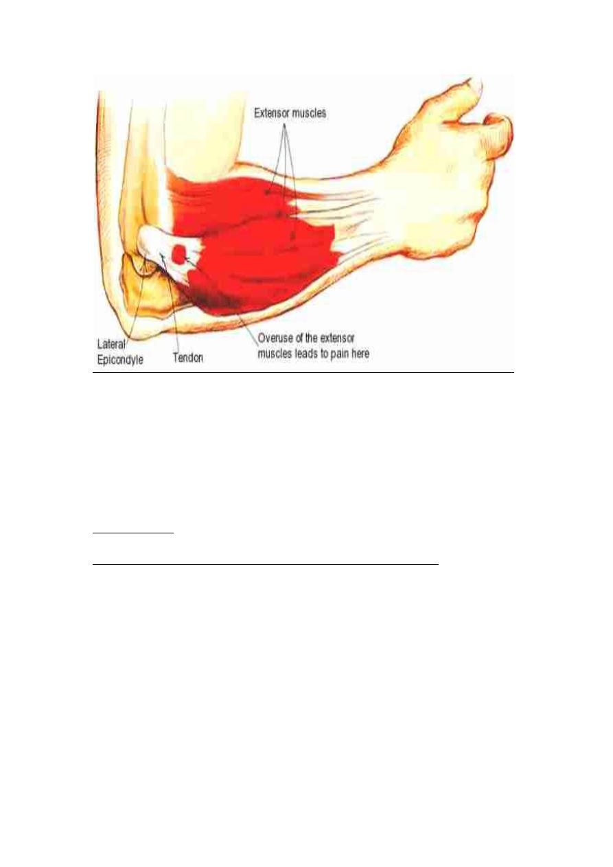
Lateral epicondylitis – “Tennis Elbow”
* Ask the patient to actively extend the wrist whilst the elbow is
flexed.
*Localized pain over the lateral epicondyle and may be referred
lateral epicondylitis.
WATCH
https://www.youtube.com/watch?v=2TALLv3ADz8
.