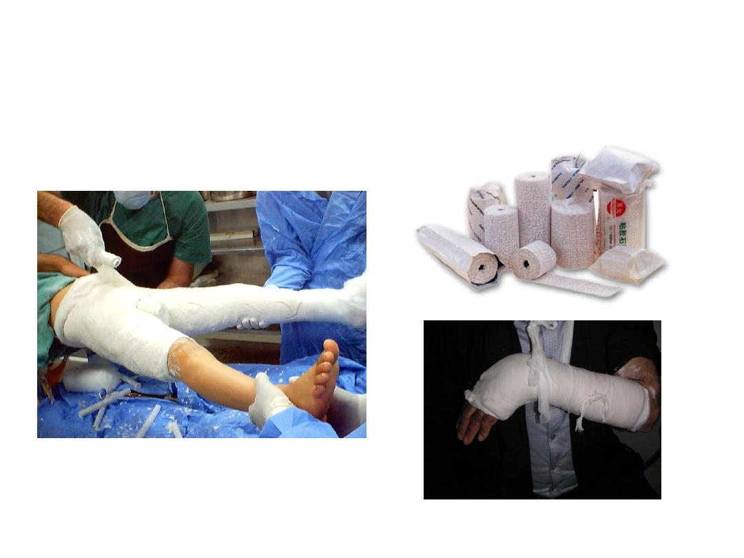
• What is this .
• Enumerate
other
methods
of
immobilizations.
1.What is this.
2.its complications.
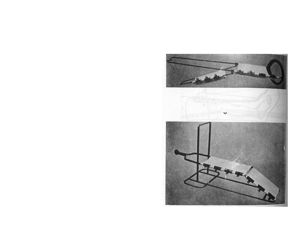
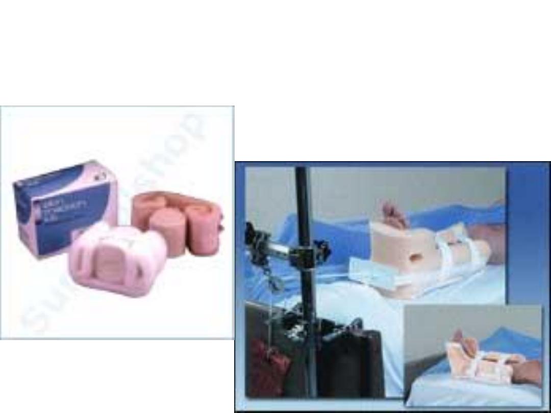
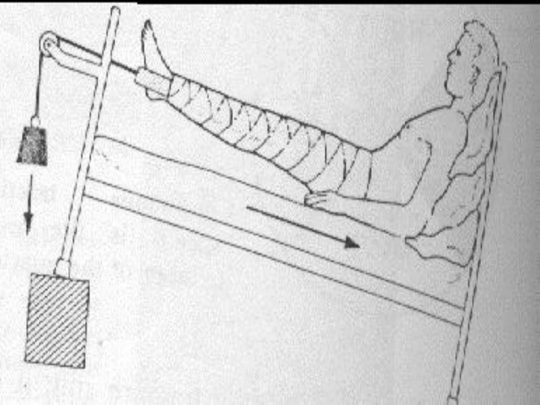
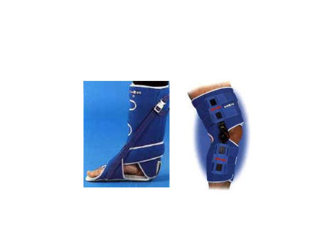
• What is this?
• When we are using it
• What is this.
• When we are using it.
When we use this jepsona?
What are the complications?
What is this?
What are the uses her?
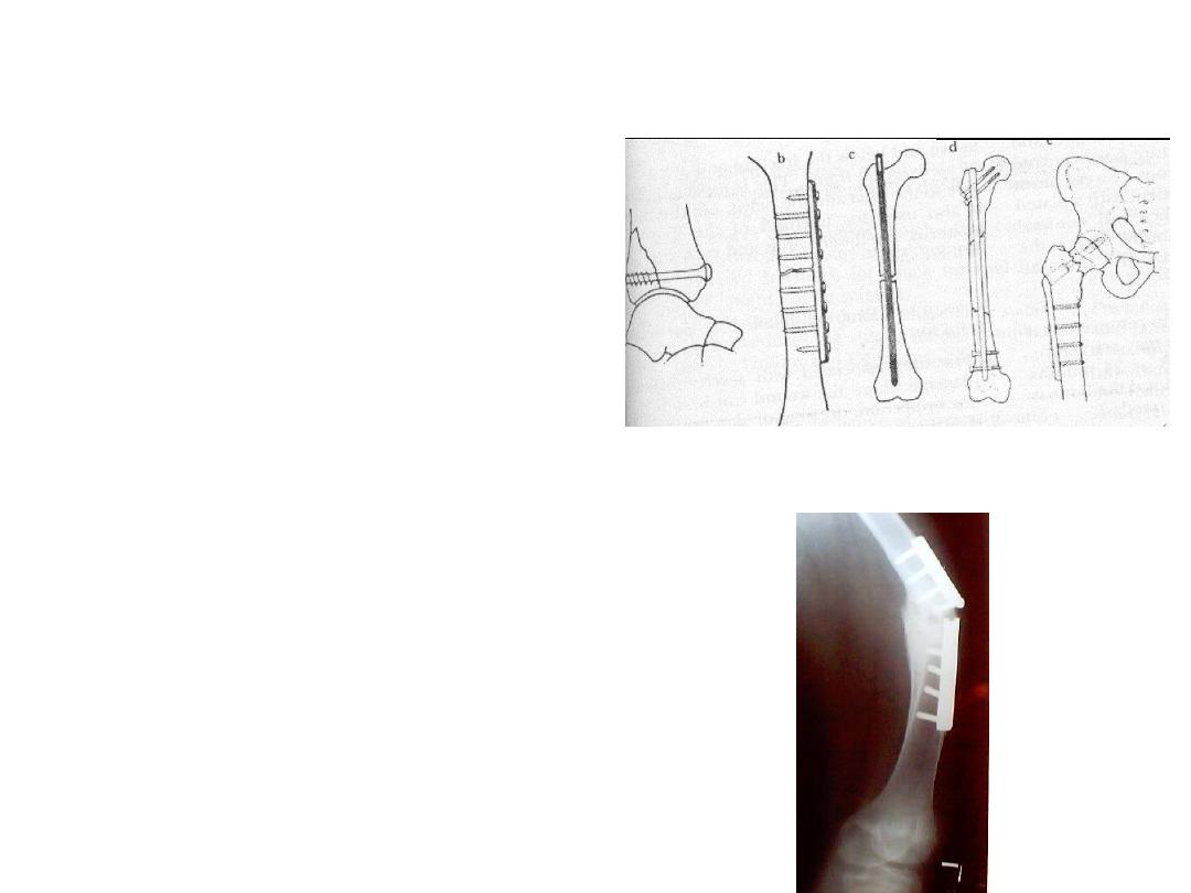
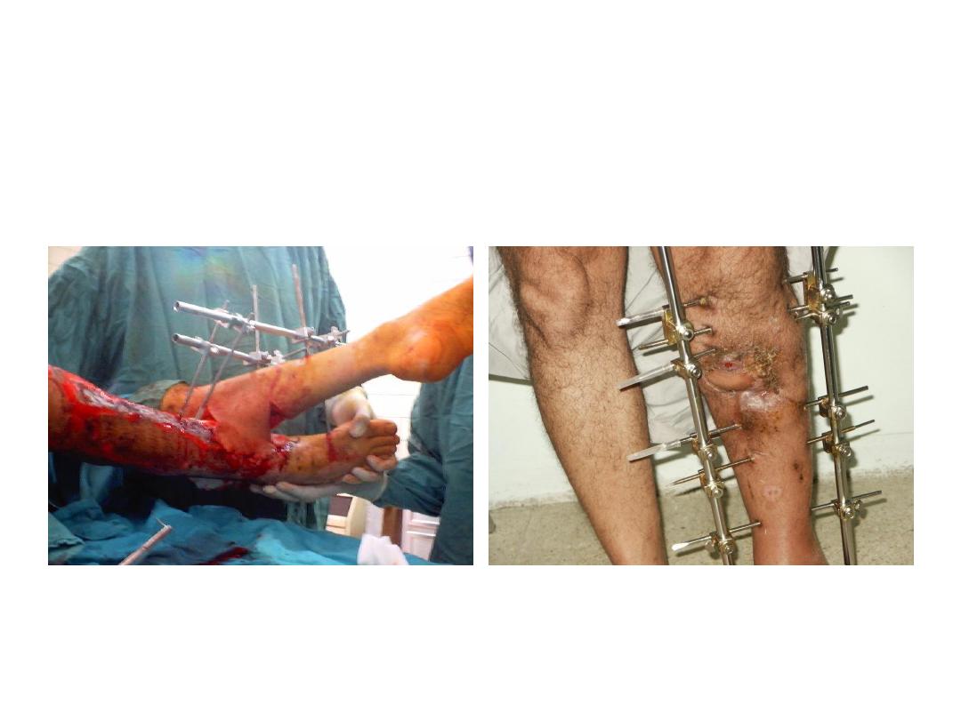
• Enumerate methods of
internal fixations you
see.
• Give 2 complications.
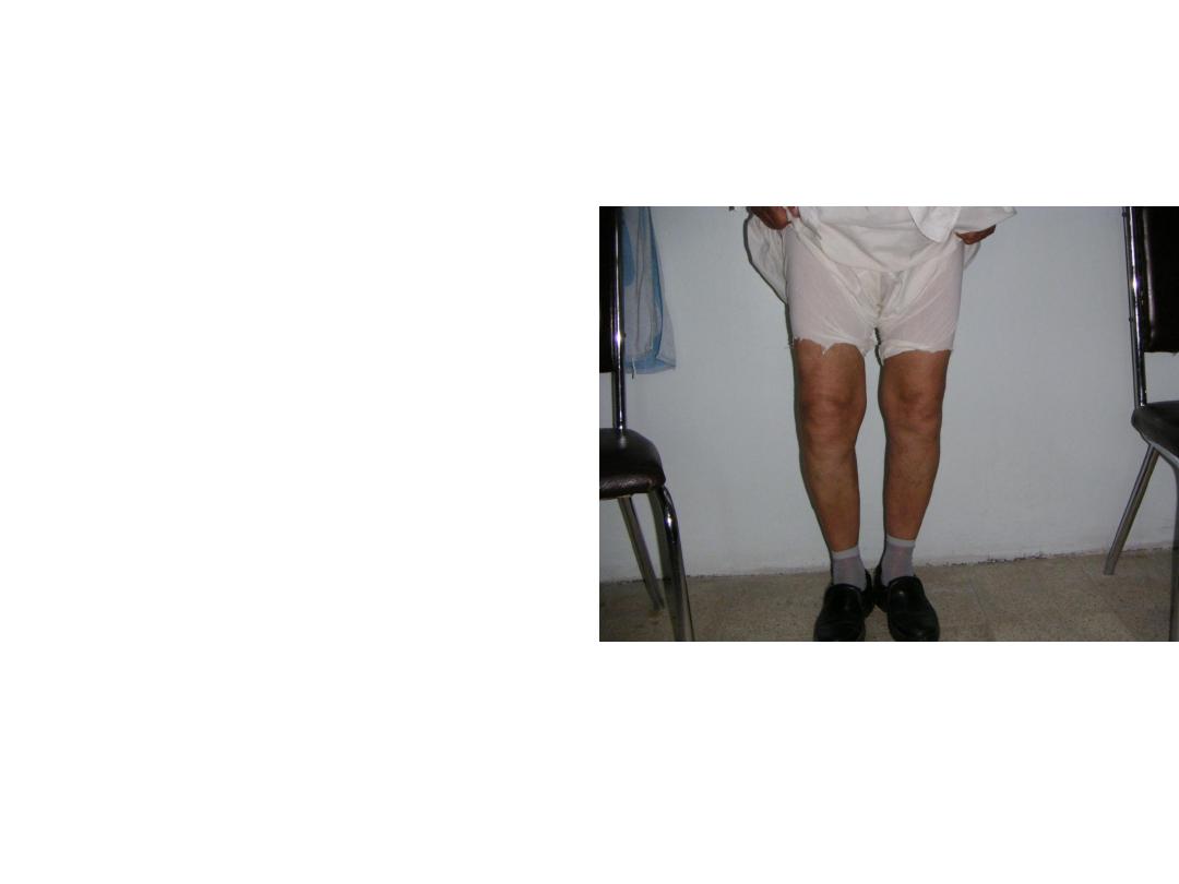
• Describe skin changes
you see.
• Enumerate
other
immediate soft tissue
complications
might
occurred in fracture.
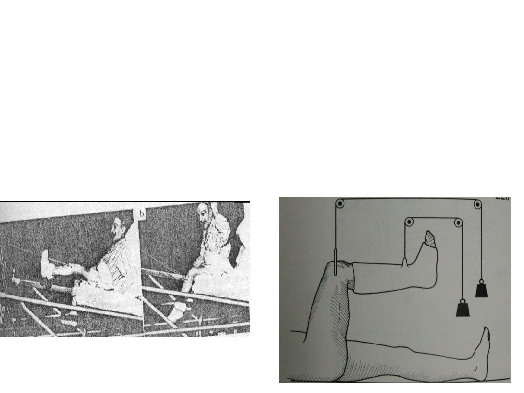
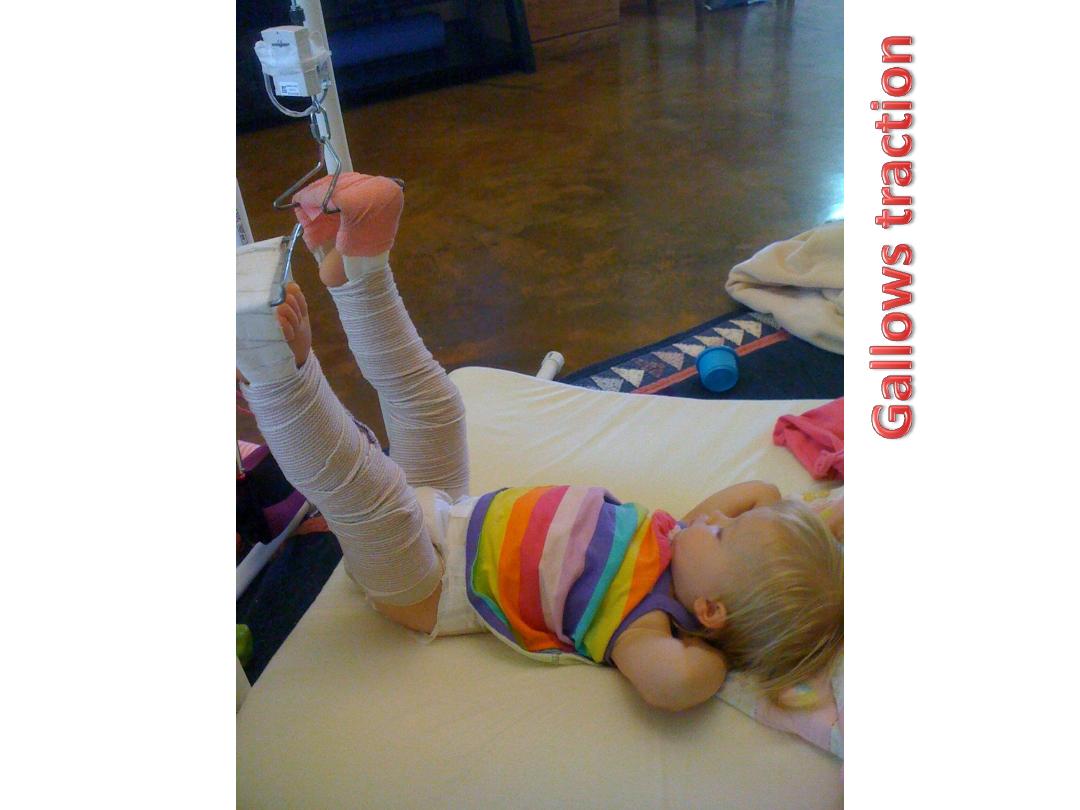
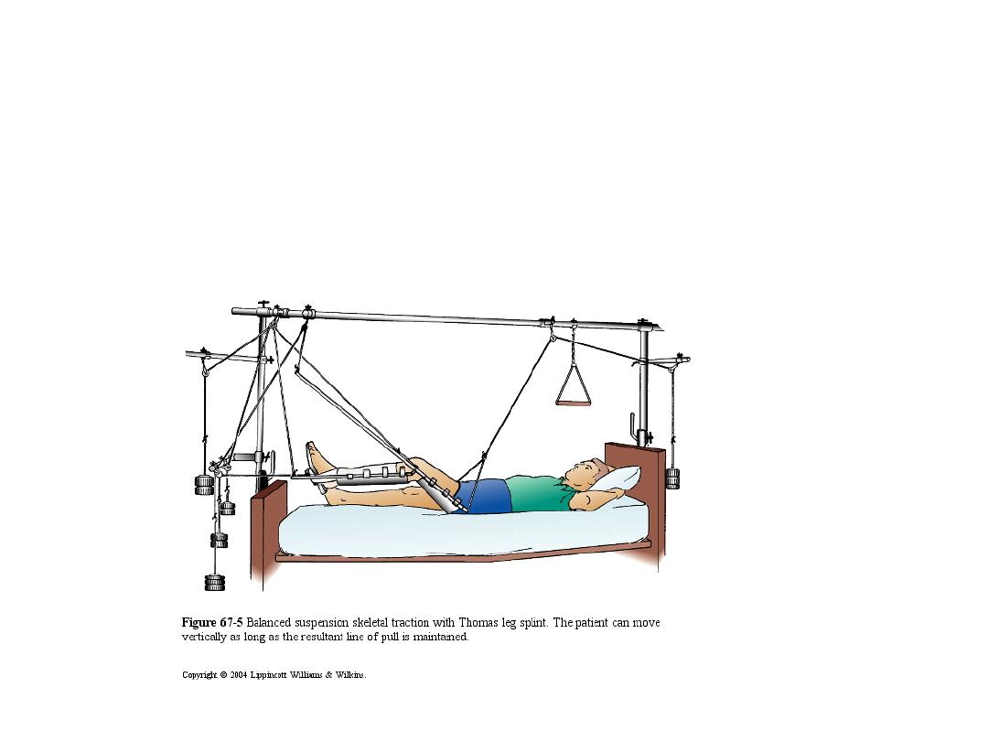
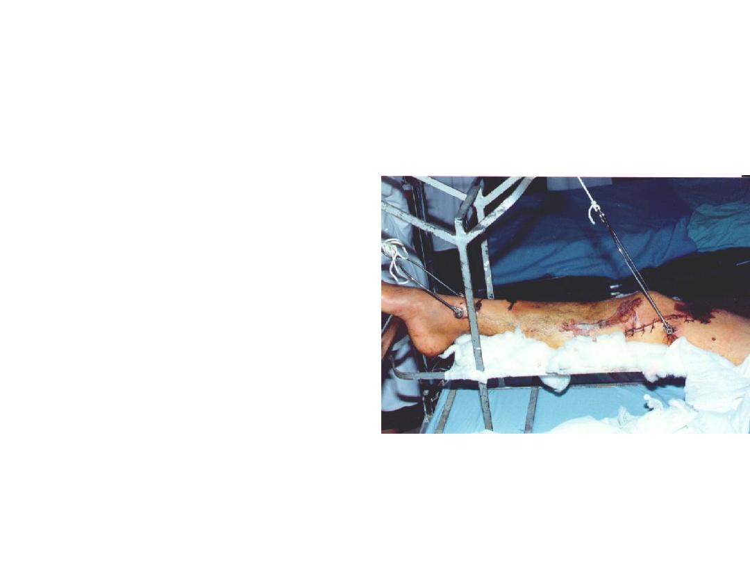
1.What is this.
2.How many weight you should apply.
What is this?
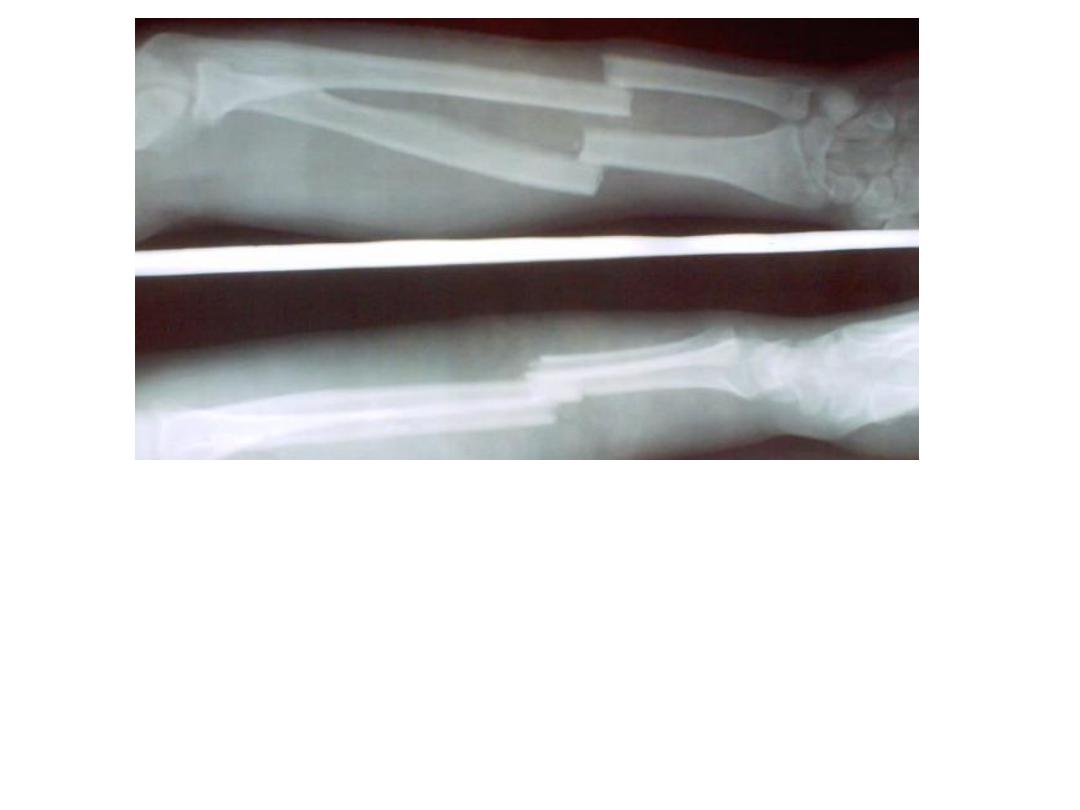
• Describe this film.
• How you can treat this patient
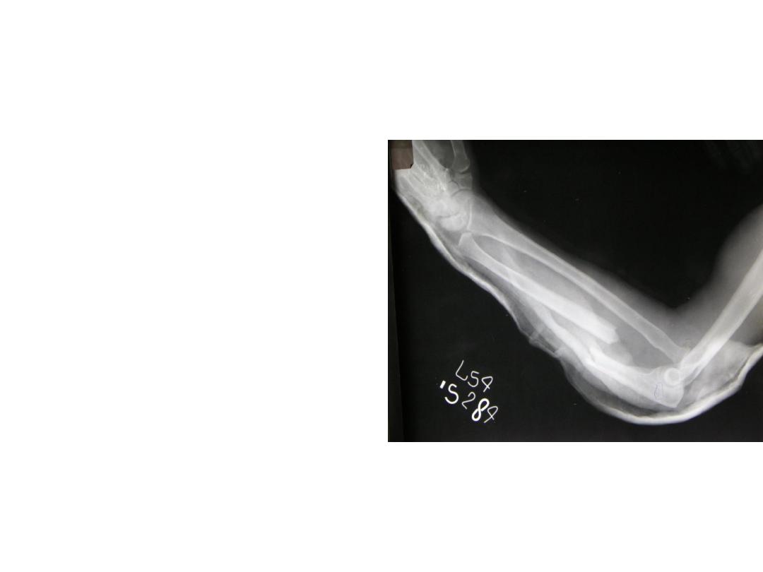
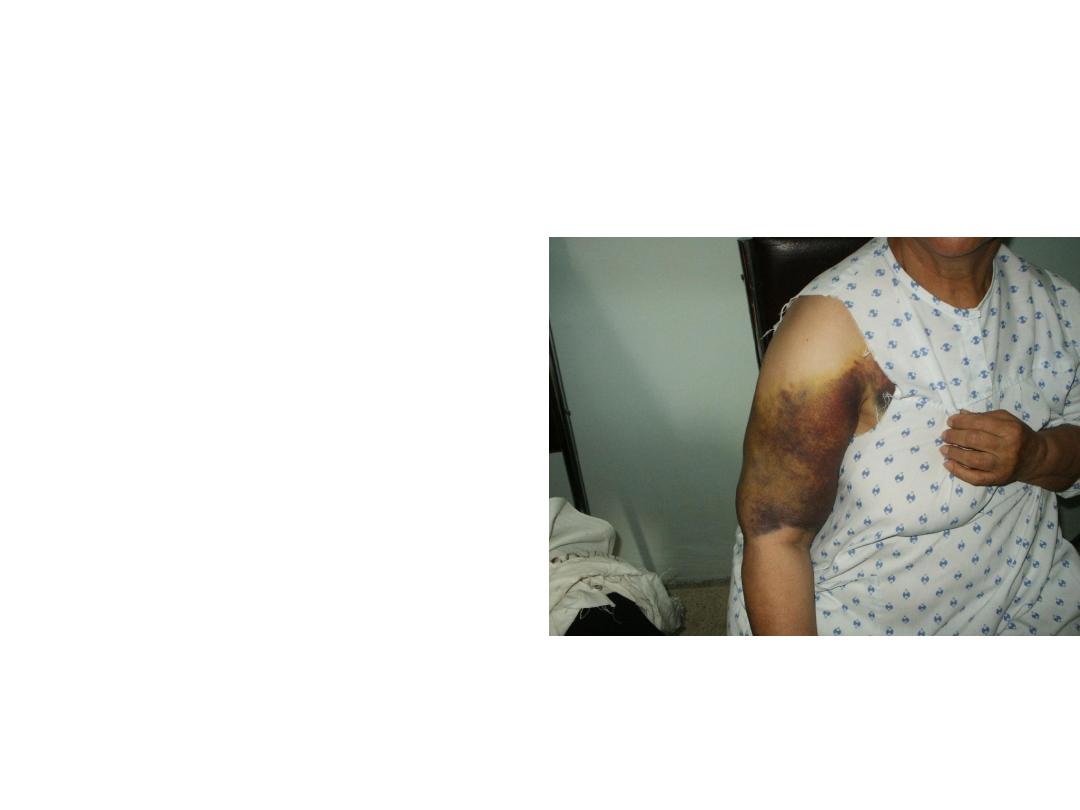
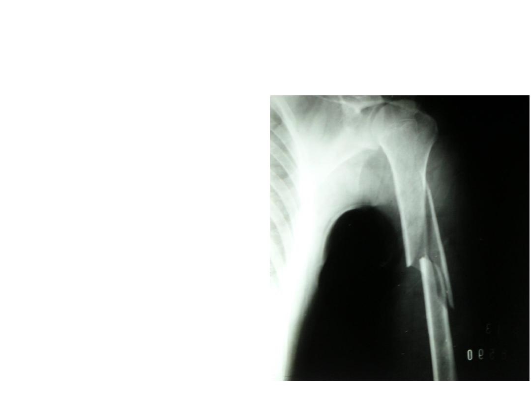
• Patient sustained trauma
to the upper limb.
• What is your diagnosis.
• Enumerate the principle of
treatment of this fracture.
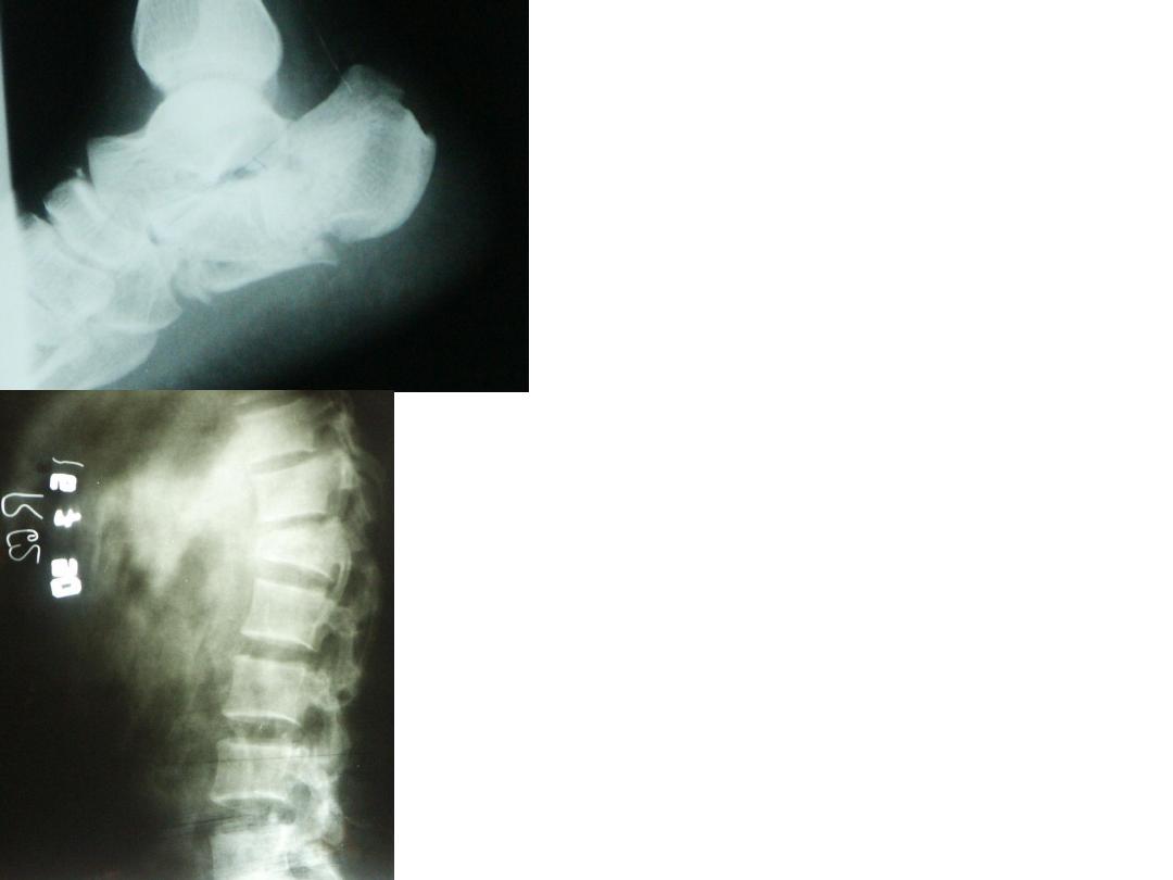
• This patient gave history
of fallen from height.
• Why we take 2 films.
• What is your diagnosis.
• How many views you
can see.
• Enumerate others with
there benefits.
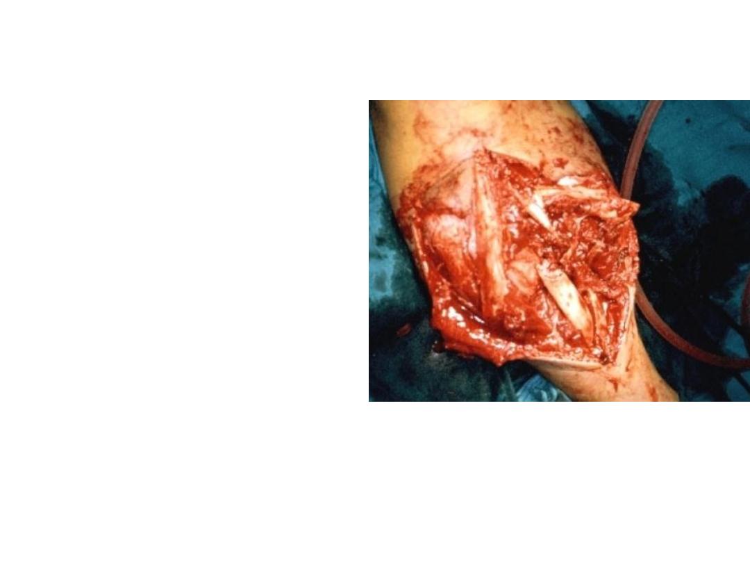
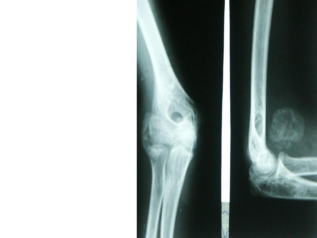
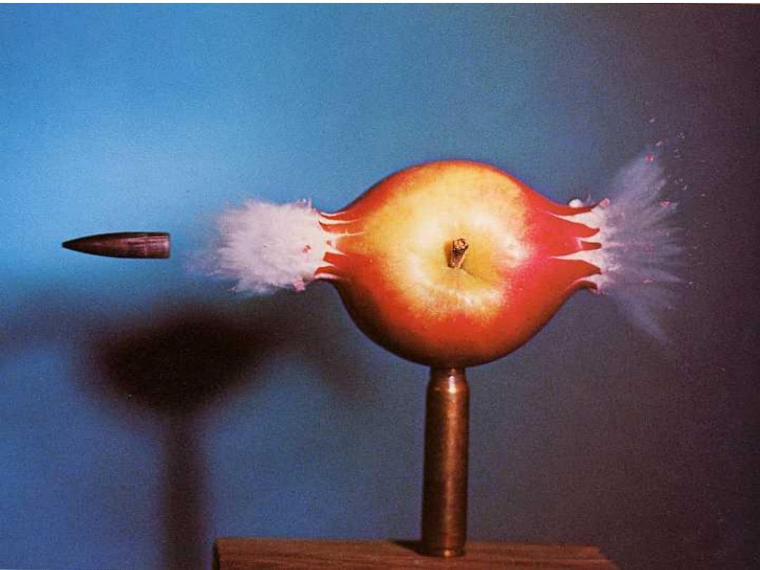
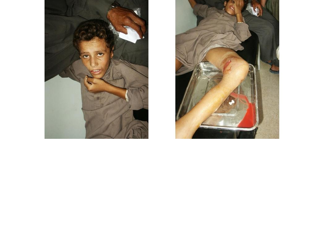
• What is this?
• What are the methods of
stoppage bleeding in such
patients.
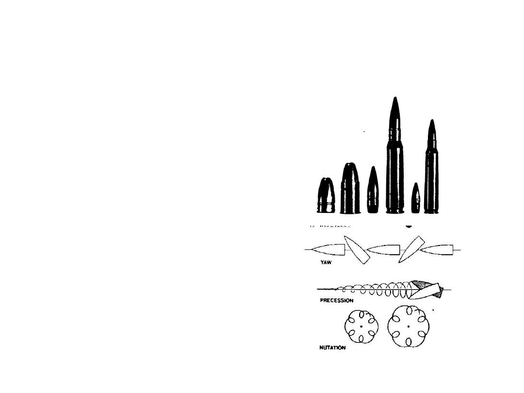
• How many types of
missile inquire you
know.
• What is the main
pathophysiologic
changes that happened.
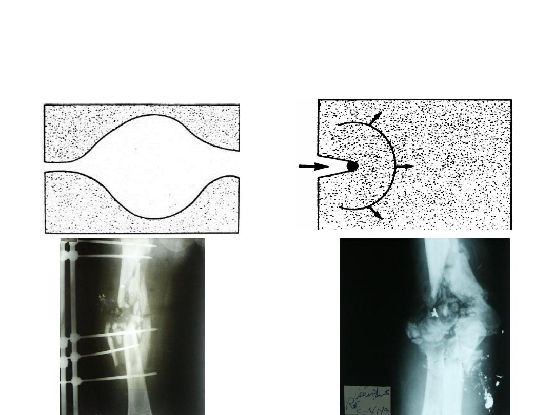
Describe this effect.
When it has been happened?
جـــــــــيه
• This female has been
fallen on the ground .
• What is your diagnosis?
• What are the main
clinical signs in this case
you might see?
• What is your diagnosis?
• What is the main
clinical sign in this
fracture you check for?
• How you can performed
it?
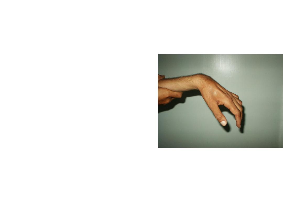
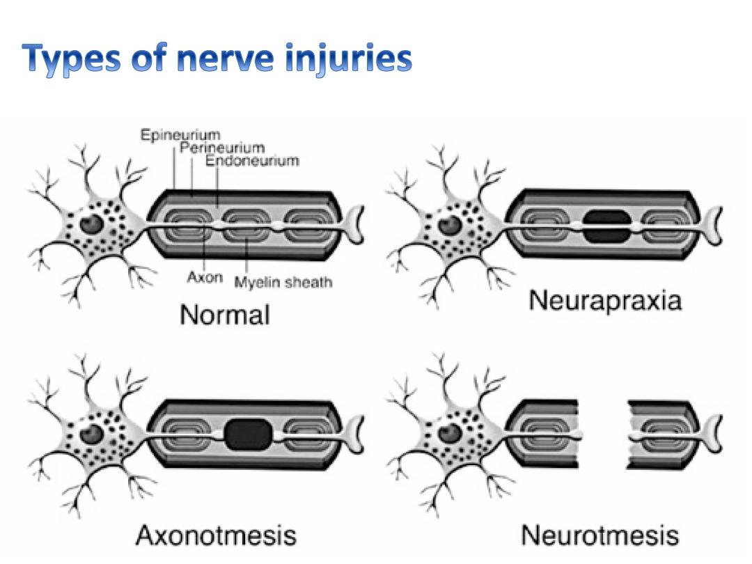
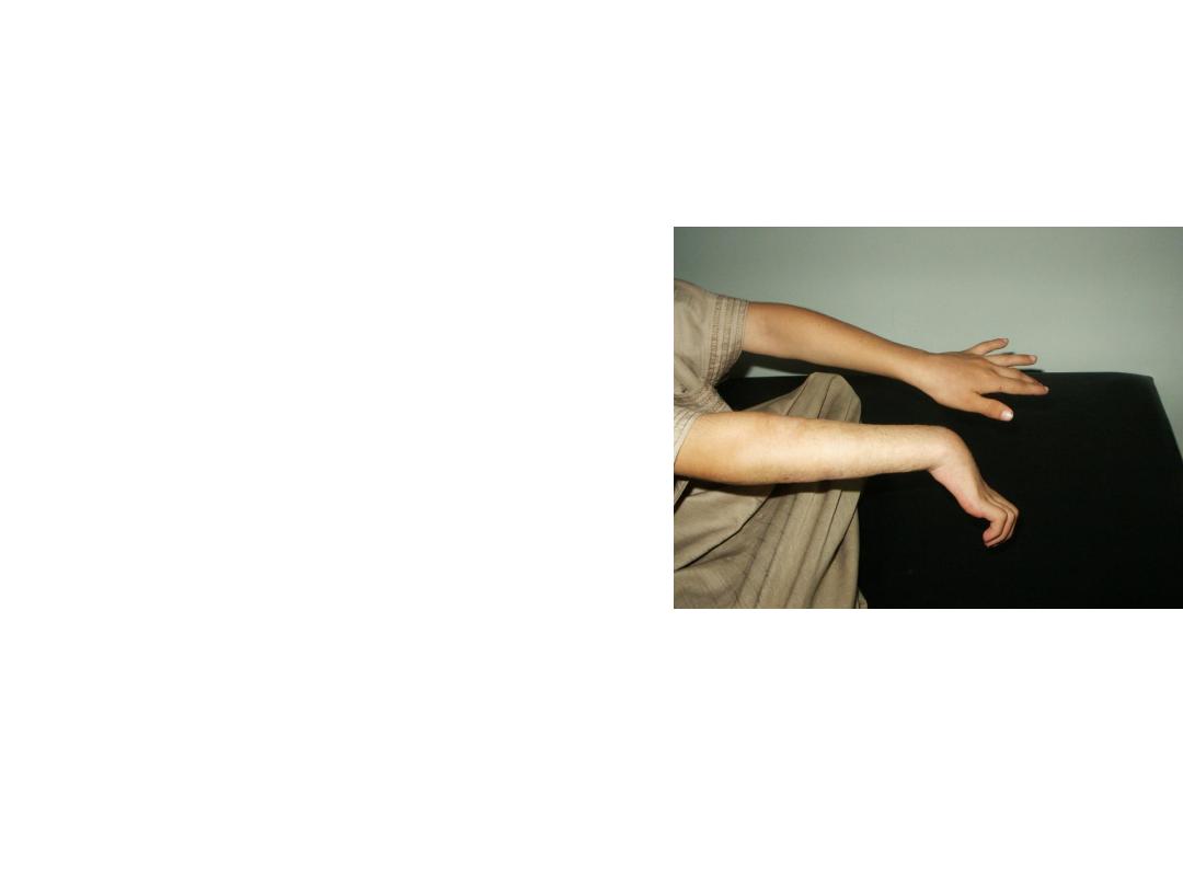
• This patient unable to
dorsiflex his wrist.
• What is your diagnosis?
• How many types of nerve
injure you know?
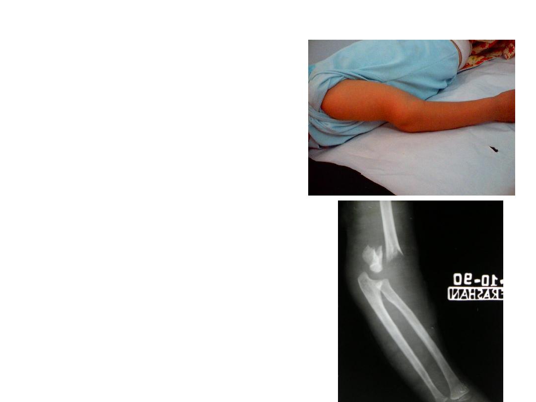
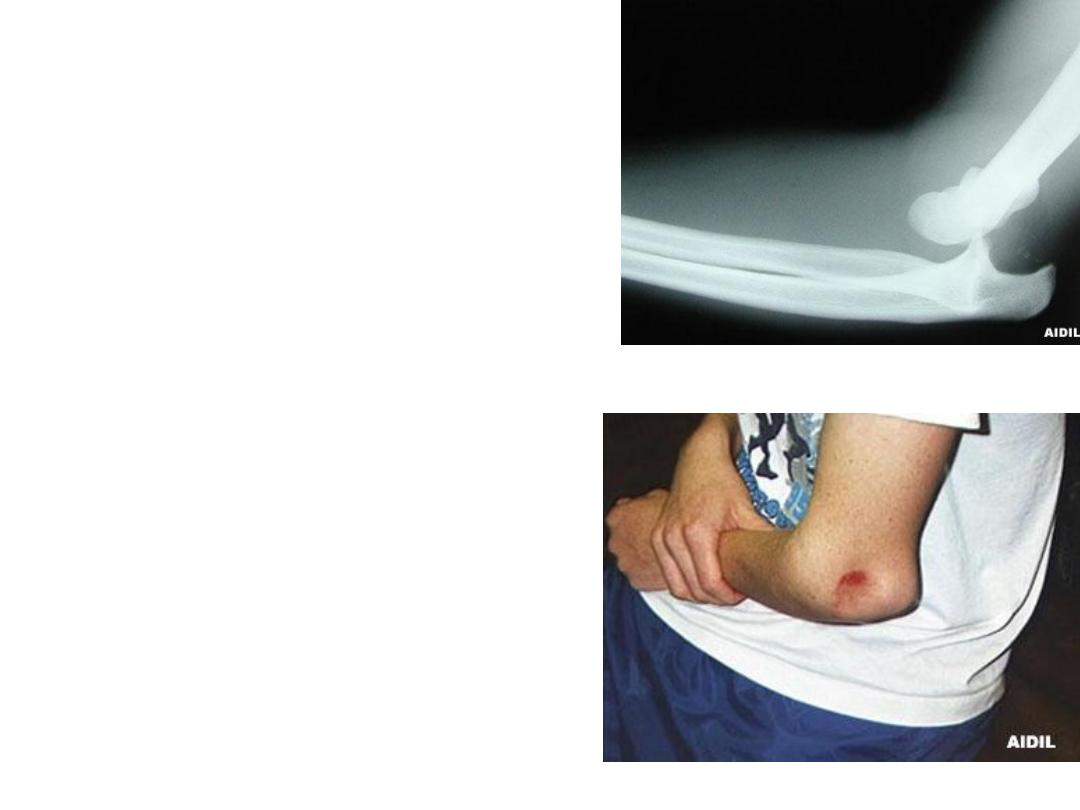
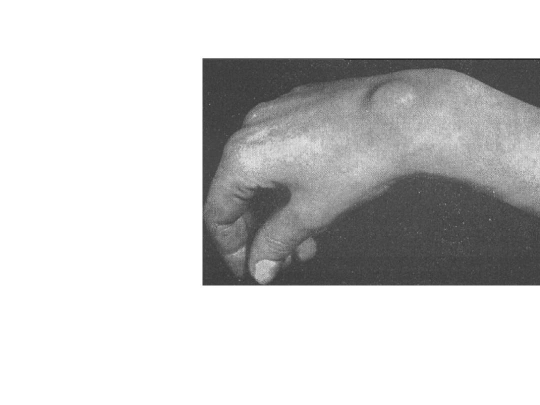
• This child fallen on the
ground. presented with
this deformity.
• Describe it?
• Mention main clinical
sign you check for?
• Describe
this
deformity?
• Mention
other
complication
might
occur in supracondylar
fracture
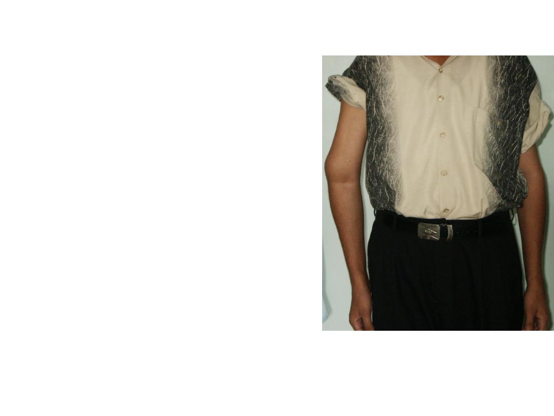
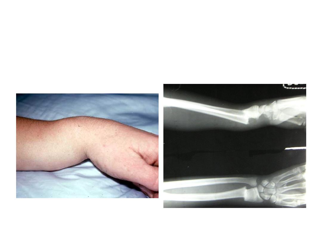
• This patient had fracture
radius and ulna. Treated by
bone setter. he developed
complication
from
bad
immobilization>
• What is the main complication
occurred?
• What is the cause?
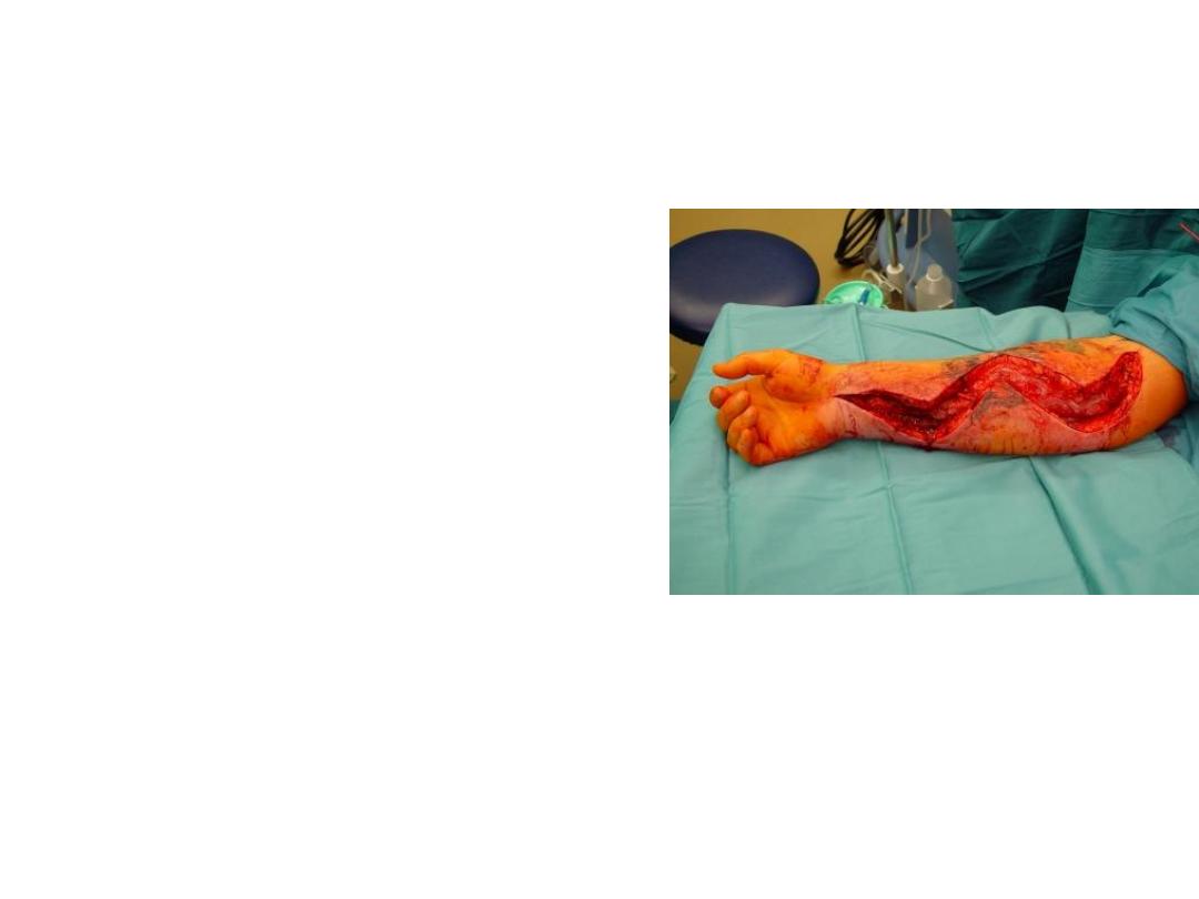
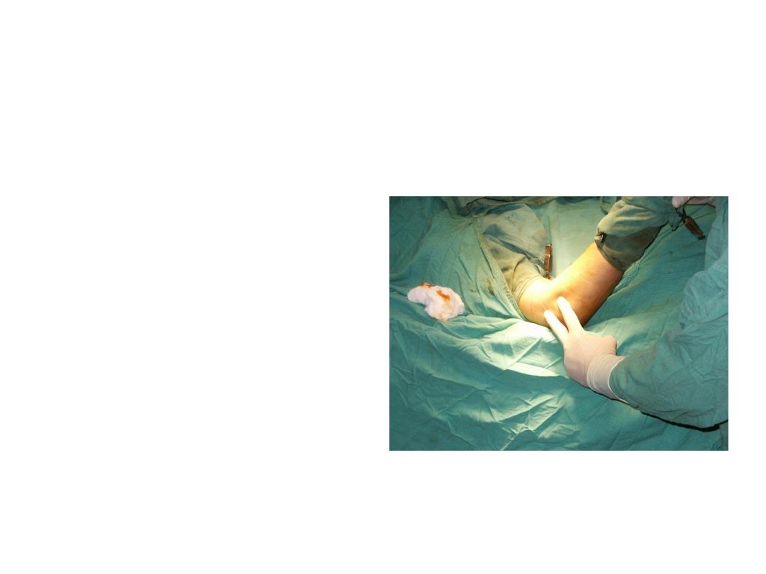
• This is fasciotomy has been
done for this patient?
• why we use it?
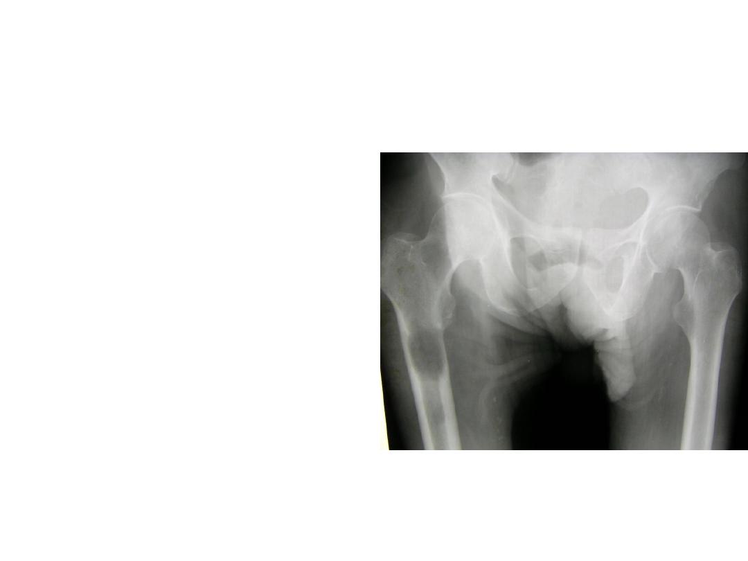
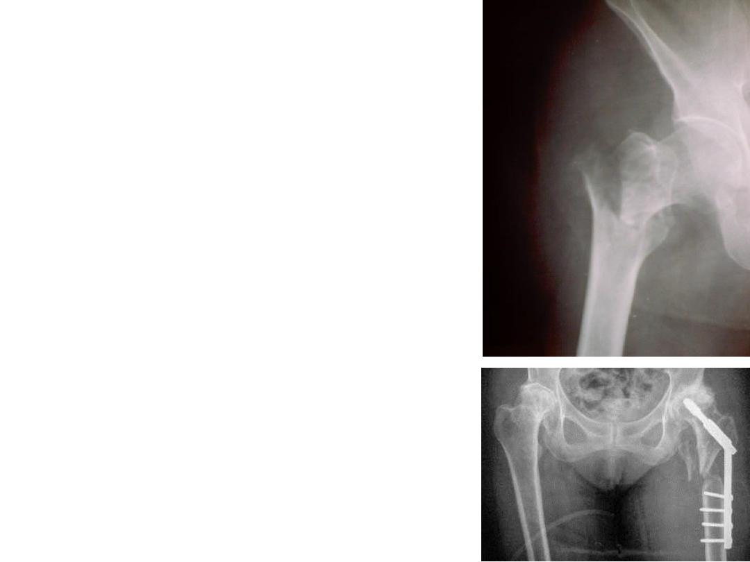
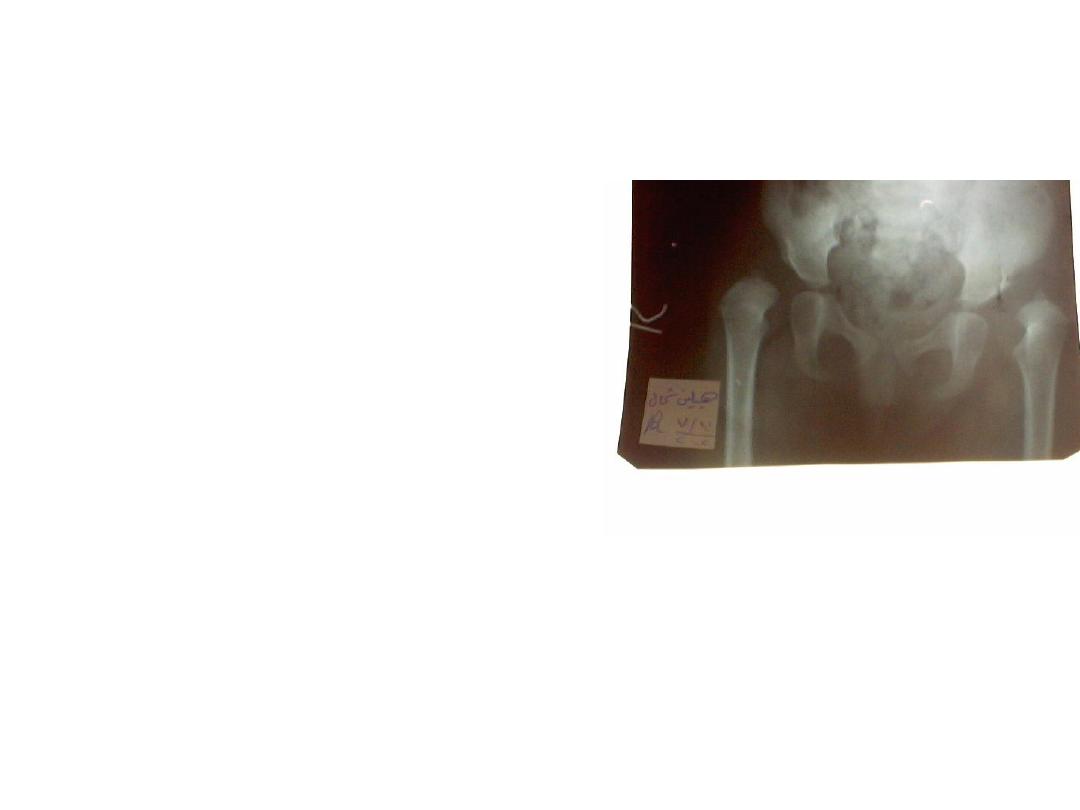
• This is an x ray for elderly
patient fallen on the
ground, With painful hip.
• What is your diagnosis?
• What is the type of
fracture here?
• This patient treated
conservatively
with
pop.
• What is your diagnosis?
• Do you agree about this
alignment?
• This patient gave a
history
of
acute
osteiomylites.
• What
is
this
late
complication?
• Give an example of
immediate complications
might happened?
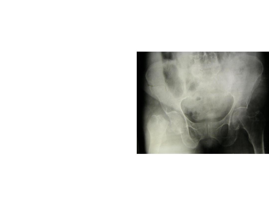
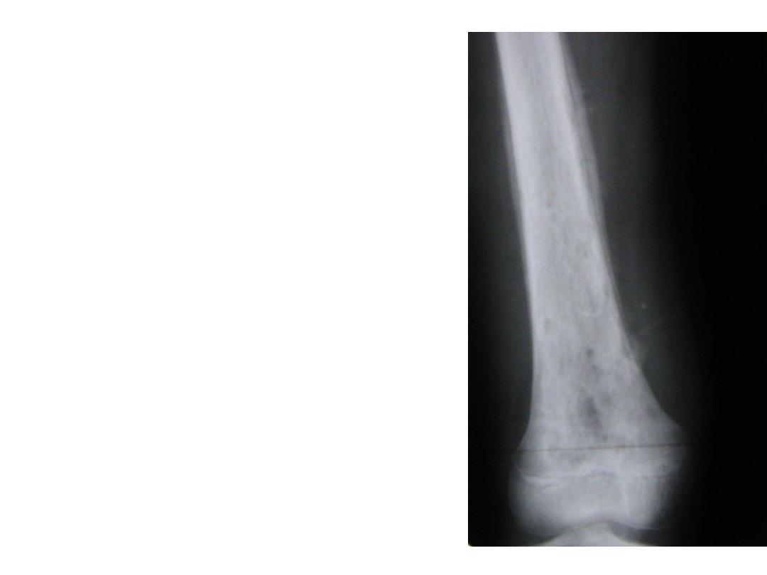
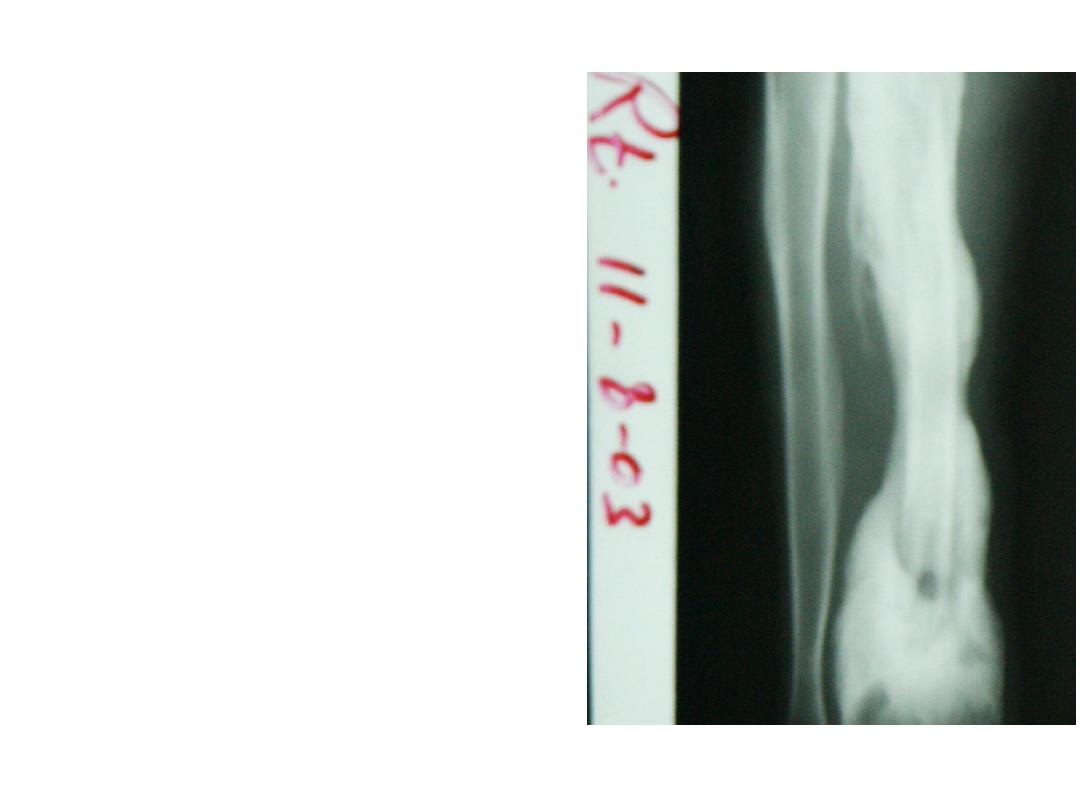
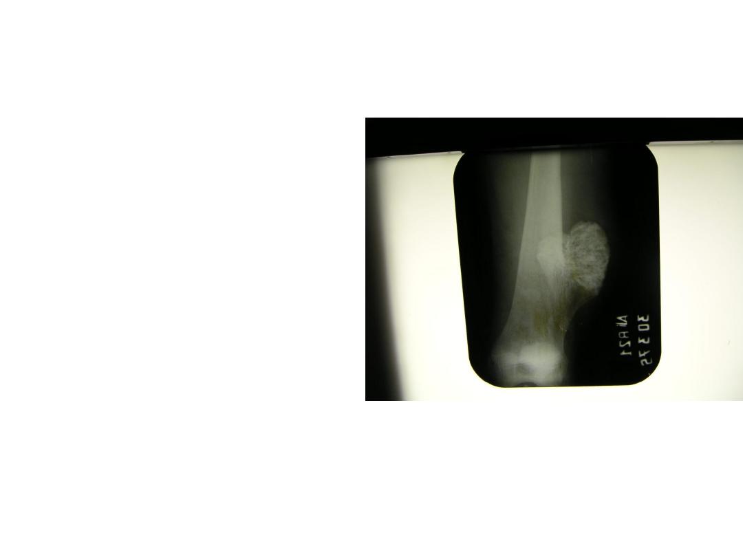
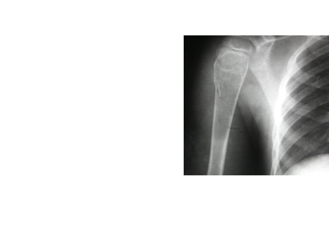
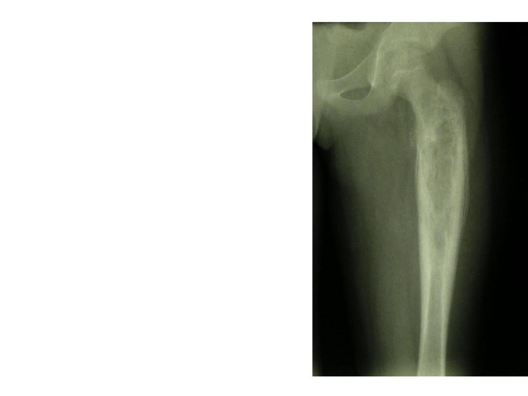
• This patient had history of
haemoptysis
.developed
pain in the thigh.
• Describe bony changes in
the femur.
• What is your diagnosis?
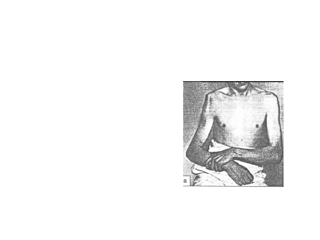
• This patient fallen on the
ground. developed sever pain
and inability to move his upper
limb.
• What are the clinical signs that
you find in this patient?
• What investigation needed to
confirm diagnosis?
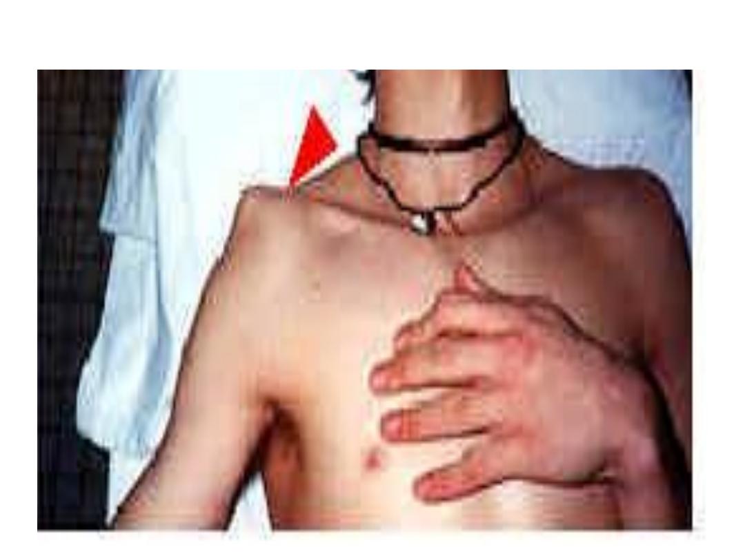
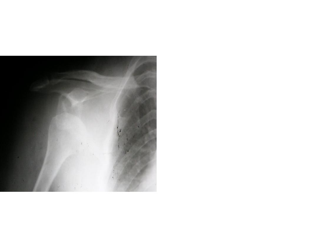
• Describe the radiological
changes in this x ray.
• What is the complications
associated
with
this
problem?
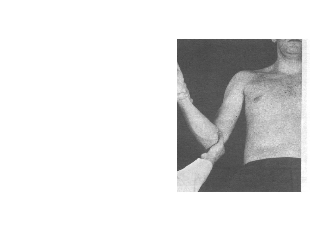
• Enumerate methods of
reduction for anterior
shoulder dislocation ?
• Describe
Kochers’
method as in this patient.
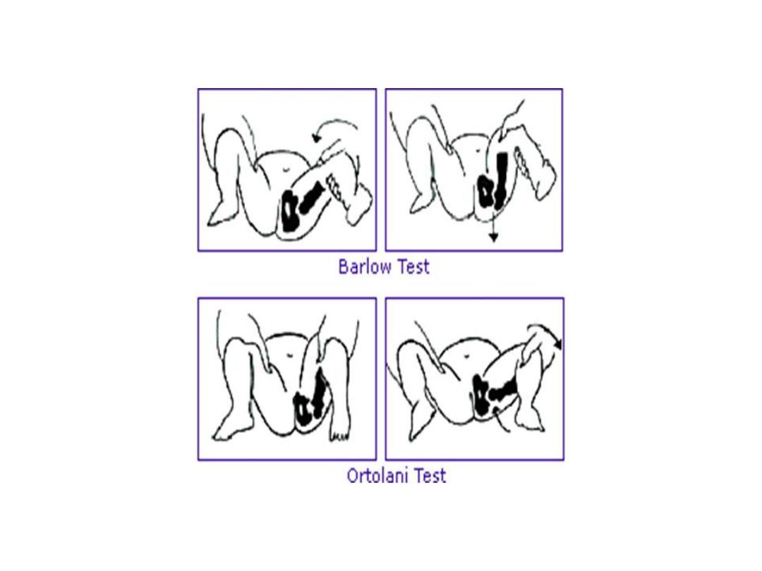
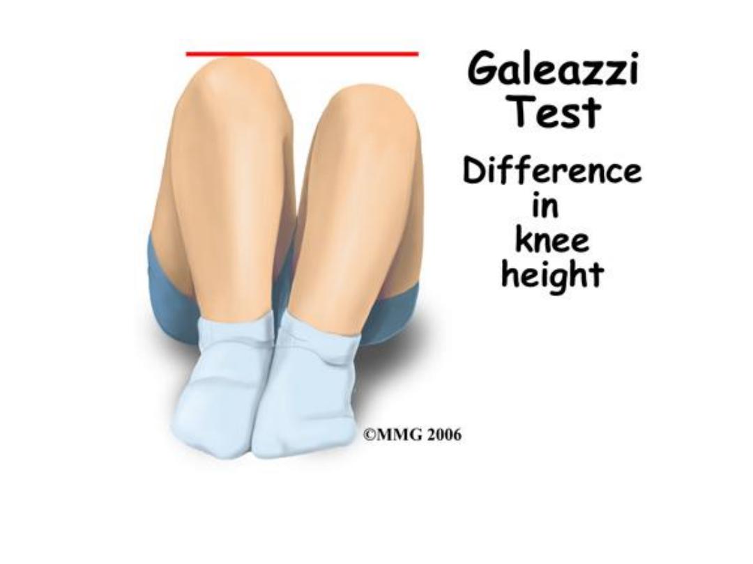
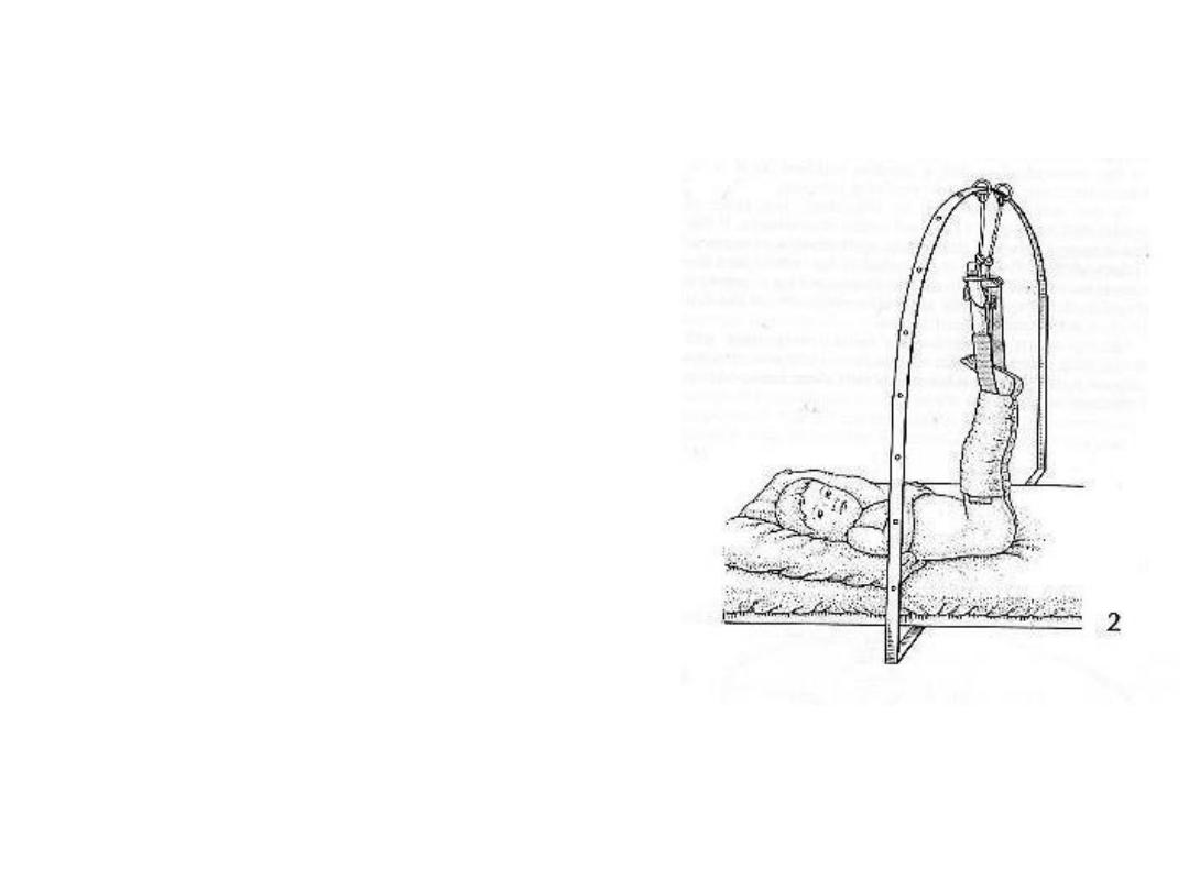
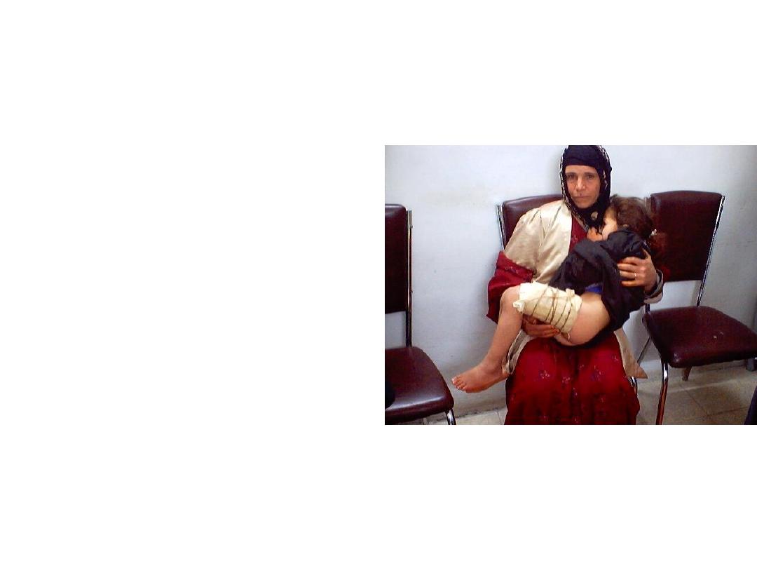
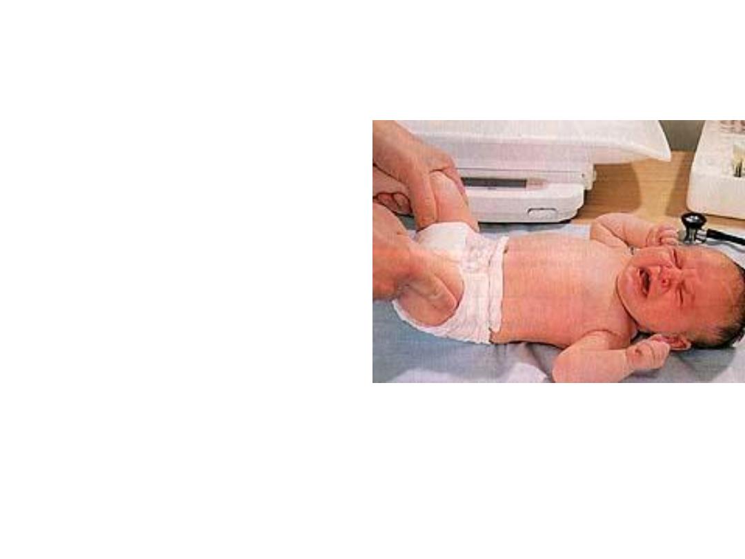
• Describe this method of
examination?
• When it was used?

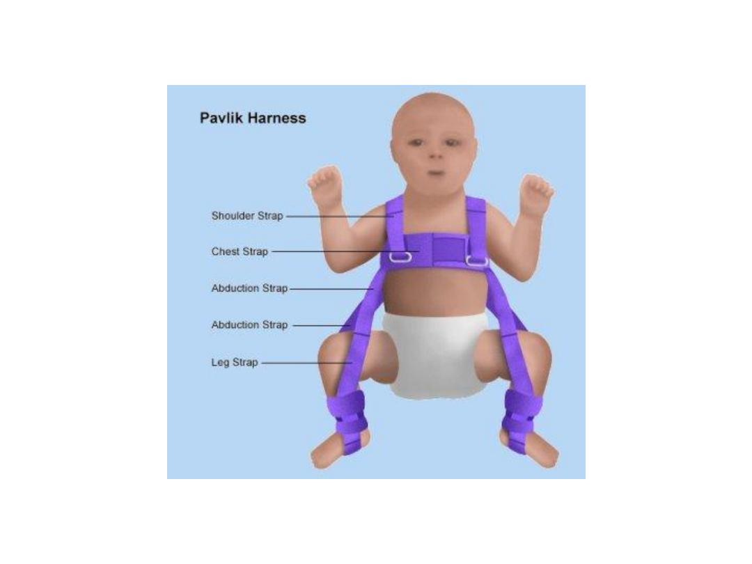
• What problem this way of
baby holding lead to?
• How you reduce it?
• Describe the clinical and
radiological change s
seen in this patient?
• Mention
its
complications?
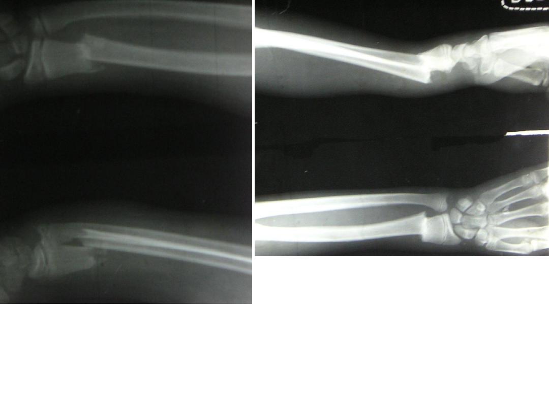
• This is an x-ray for a
patient
fallen
on
outstretched hand.
• Describe the direction
of
distal
piece
displacement?
• What is your diagnosis?
Describe this deformity?
Describe the radiological changes?
What is your diagnosis?
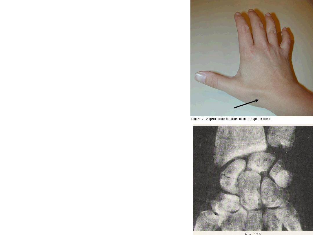
• This patient fallen on out
stretched
hand
with
painful snuffbox.
• Describe the radiological
changes?
• how you can hold this
fracture?
• What is this?
• How you treat this
patient?
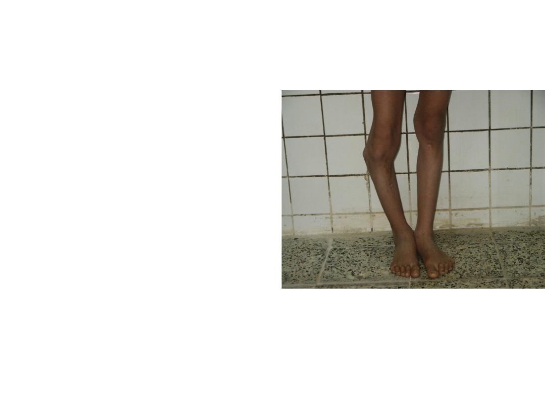
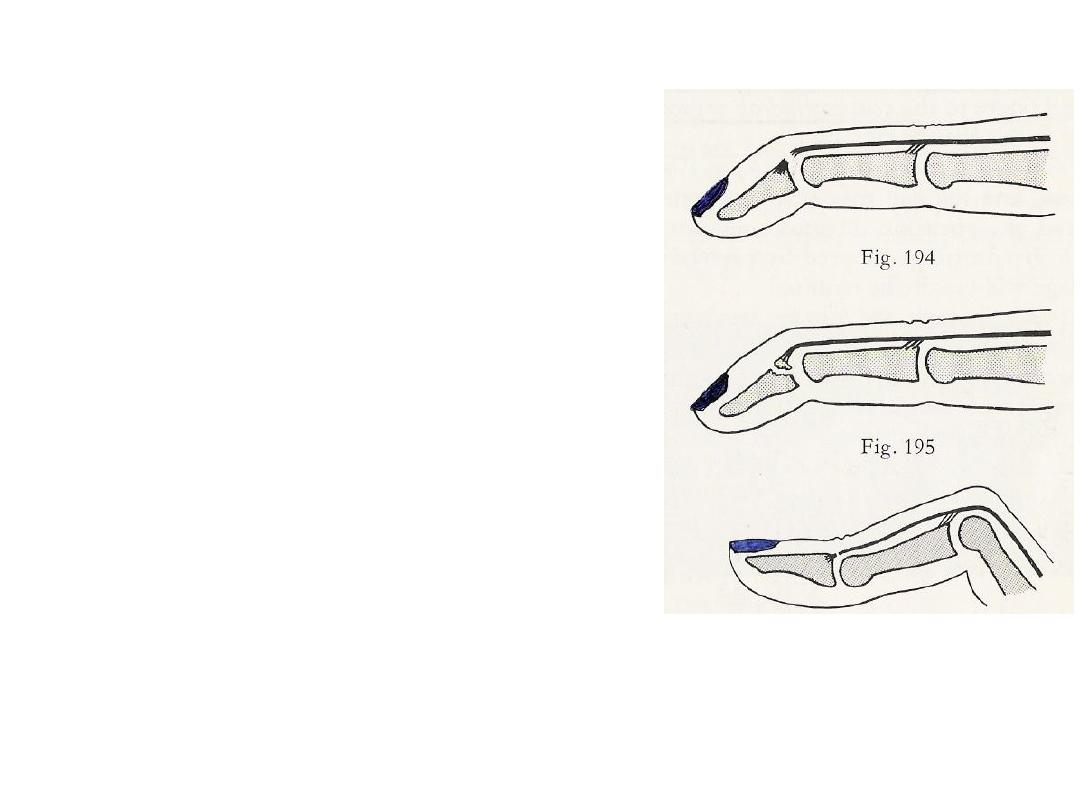
• Describe this
deformity?
• What is your diagnosis?
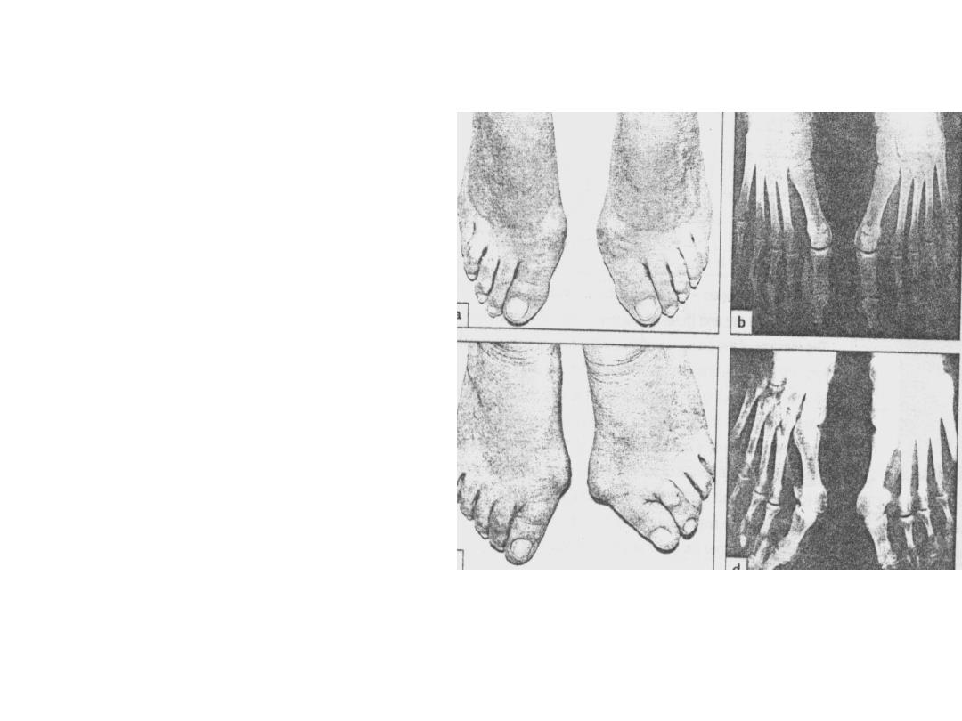
• How you describe this
clinical problem?
• What are the clinical
features
might
occurred?
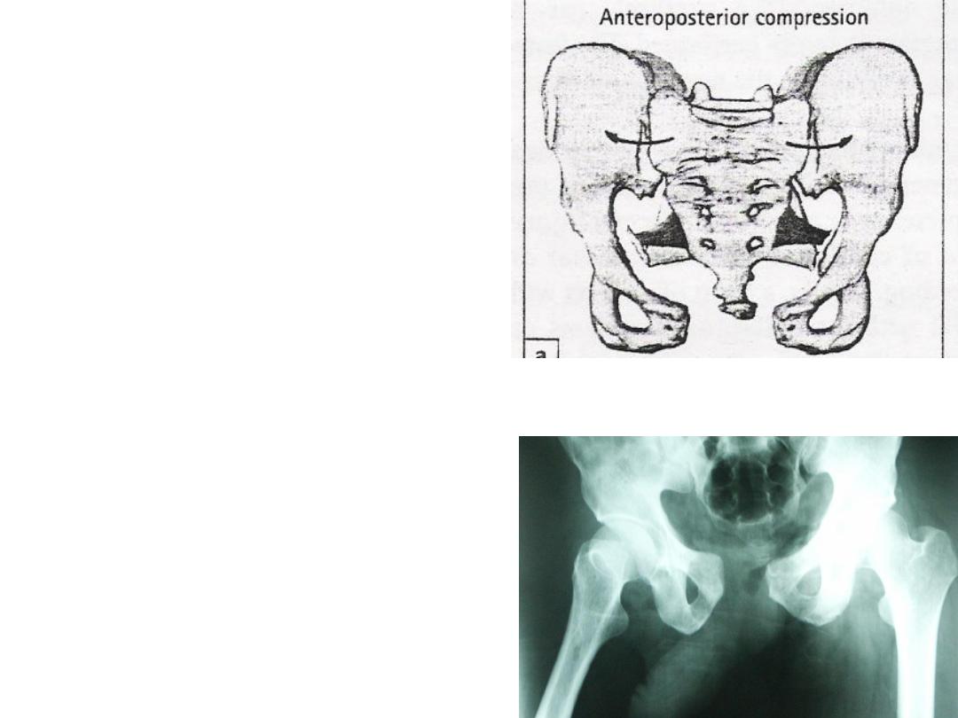
• How you describe this type
of fracture pelvis?
• What should you do in case
of possibility of urethral
injure?
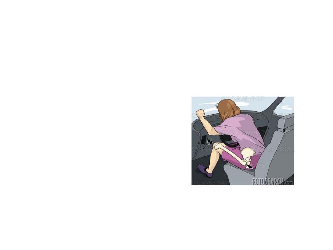
• This patient sustained
dashboard injure.
• What type of injure
might developed?
• Describe the clinical
findings?
• Describe the clinical
sings in this patient?
• How you reduce the
hip?
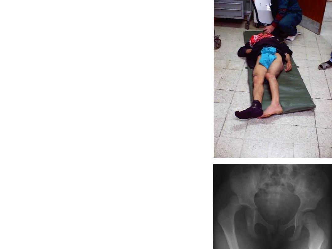
• What type of fracture might
lead this way of fallen on the
ground?
• Why we use this type of
fixation in this patient?
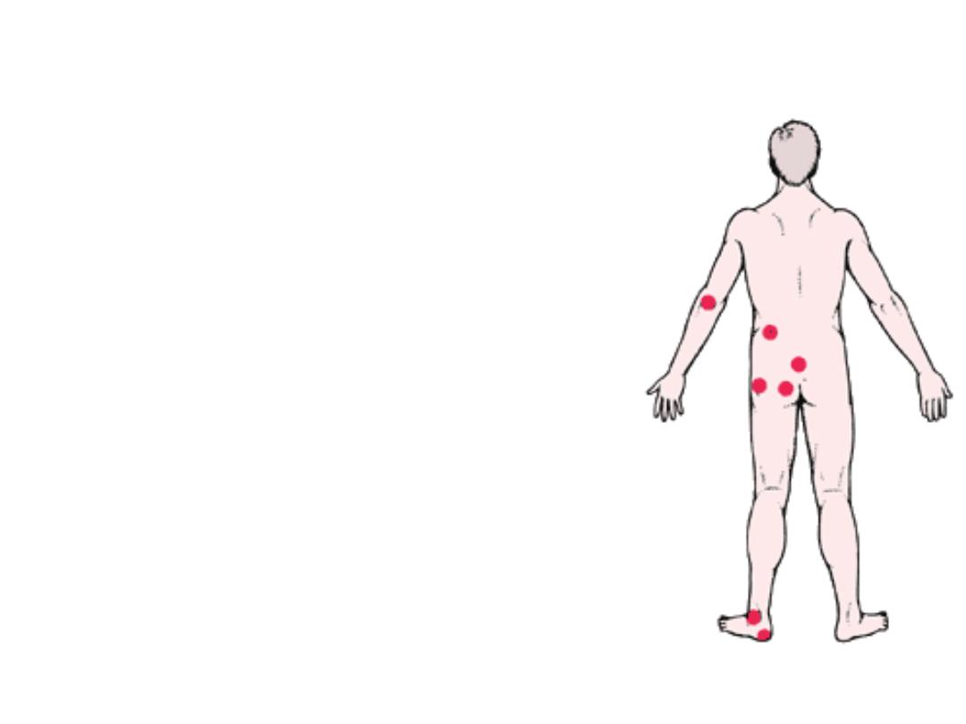
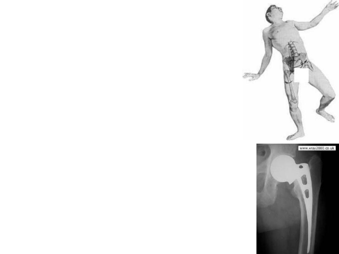
• Those are common site for
skin changes developed in bed
ridden elderly patient with
fracture neck femur.
• What you call this problem?
• Mention other problems might
developed in such patient؟
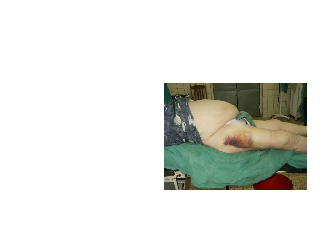
• This 60 years old female
fallen on the ground with
painful hip.
• Describe it ?
• What is your diagnosis?
• Describe
this
radiological findings?
• What you call this type
of fracture holding?
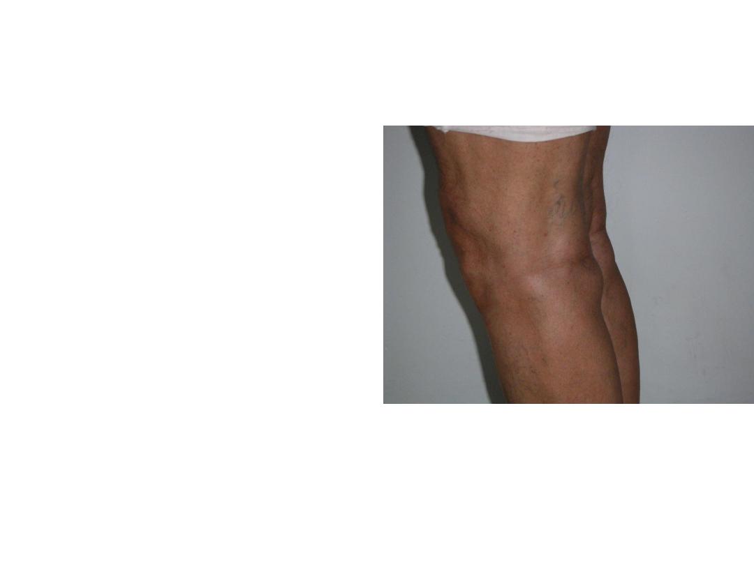
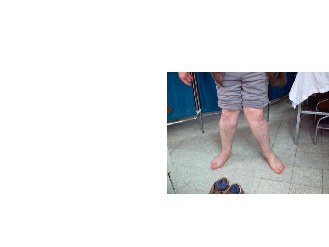
• Describe the knee changes
in this patients?
• Mention 3 causes of acute
knee joint swelling?
• This is posterior knee
joint swelling?
• What is your diagnosis?
• Mention other causes of
posterior swelling?
• Describe
this
pathology?
• What you advise for
managing this patient?
• This patient had fever
and swelling around the
knee.
• Describe the clinical
finding?
• What is your diagnosis?
• What is this ?
• Define it?
• Mention it causes?

• What is this ?
• Define it?
• Mention it causes?
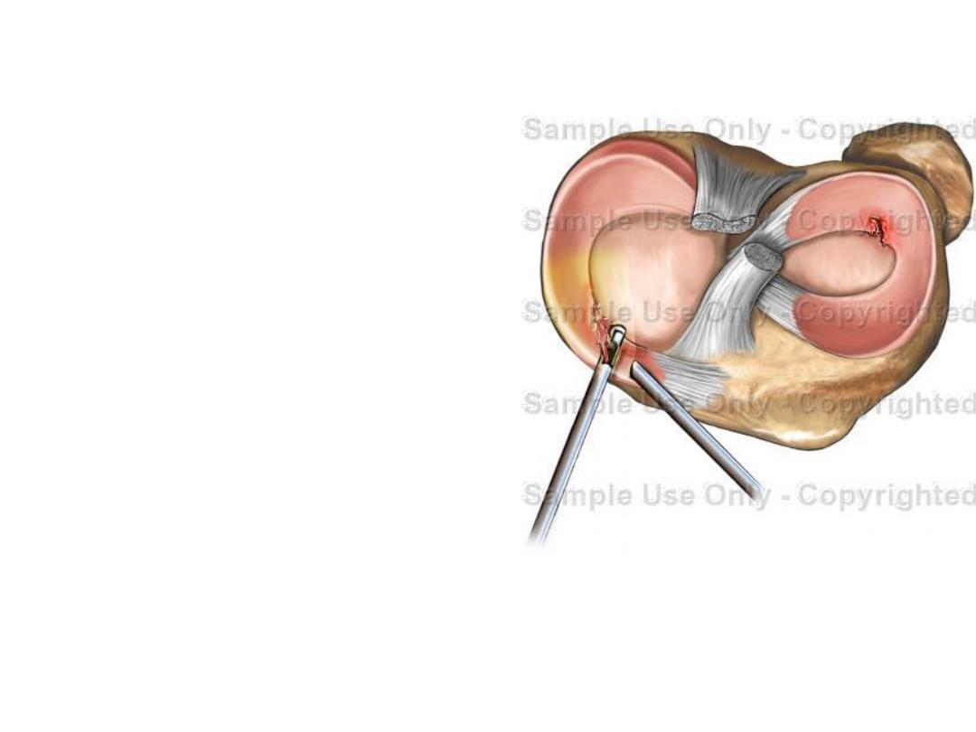
• This is superior view for the
exposed knee.
• Describe the pathology you
can find?
• Mention a test to confirm
your diagnosis
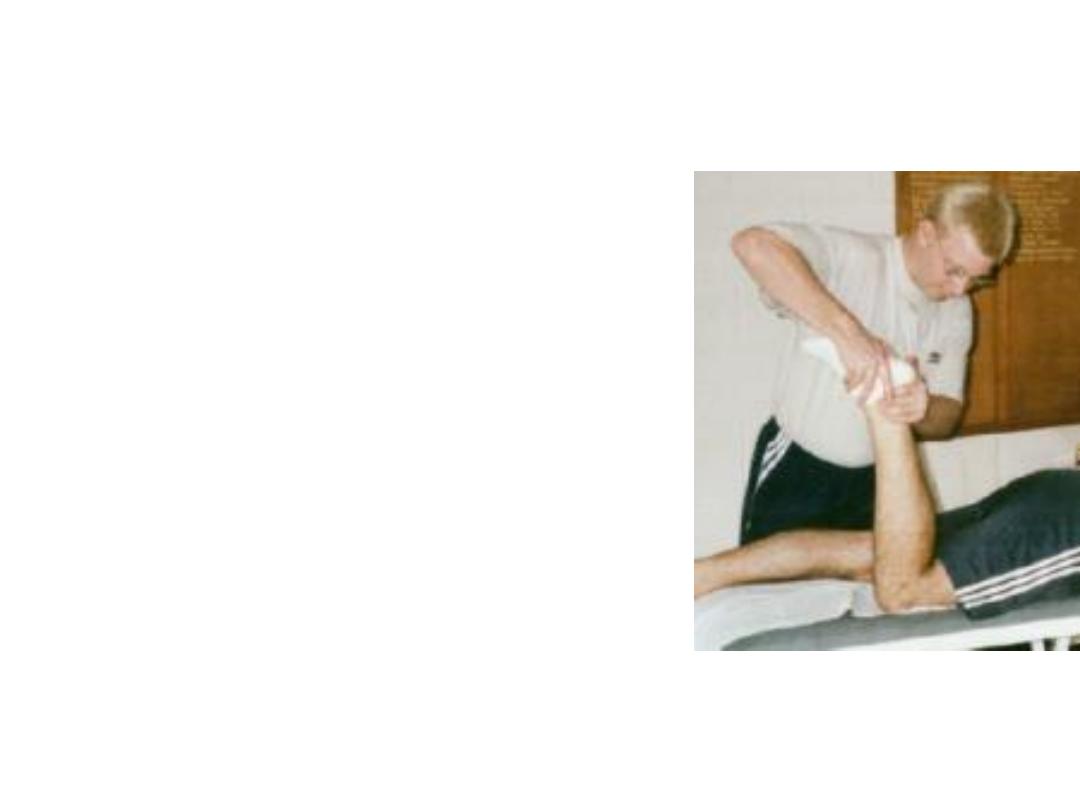
• How you call this
method
of
knee
examination?
• When you can used it?
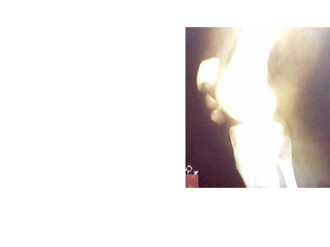
• Describe this radiological
finding?
• How you can confirm
clinically that this patient
had such a problem?
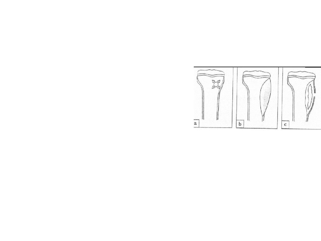
• Metaphyseal site of long bone
is the common site for
osteiomylites development.
• Mention the cause of this site
of predilection?
• What is this?
• Define it?
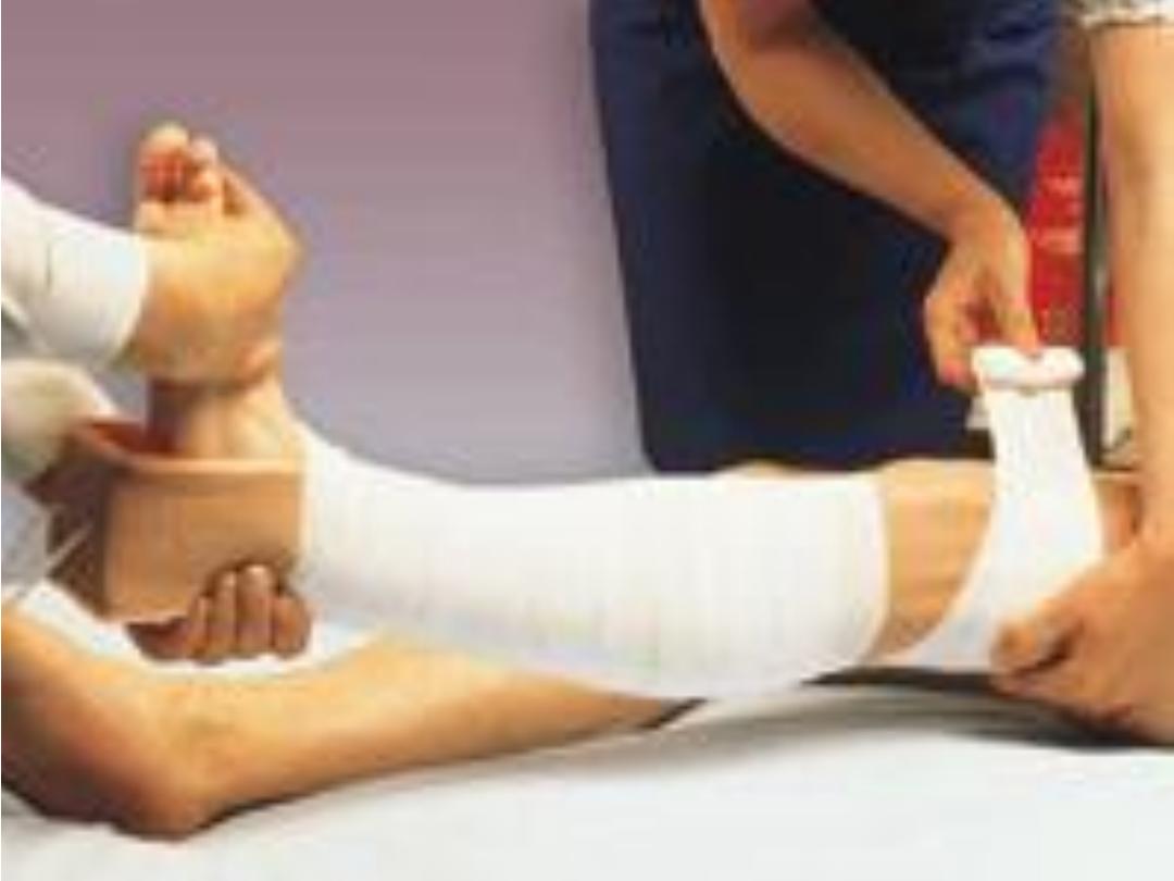
• This is primary way of
limb holding.
• Mention other methods
of limb holding in
fracture?
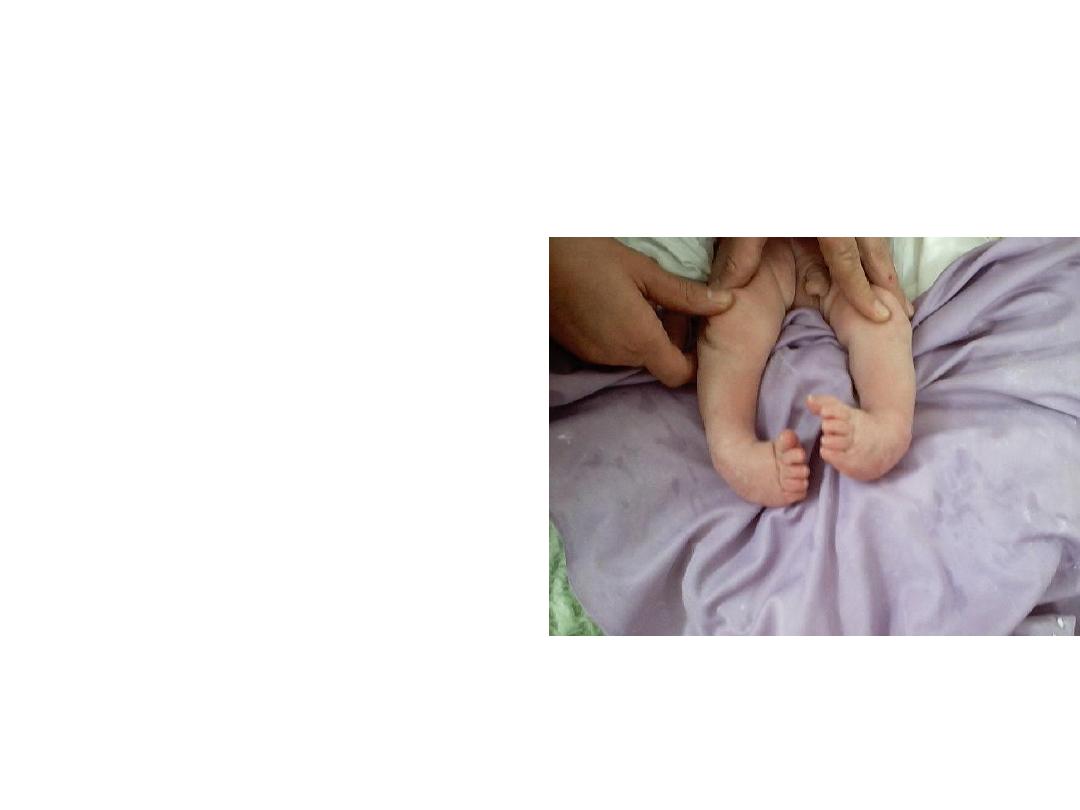
• Describe
the
clinical
finding of this patient?
• If you discover him early
how you can treat him?
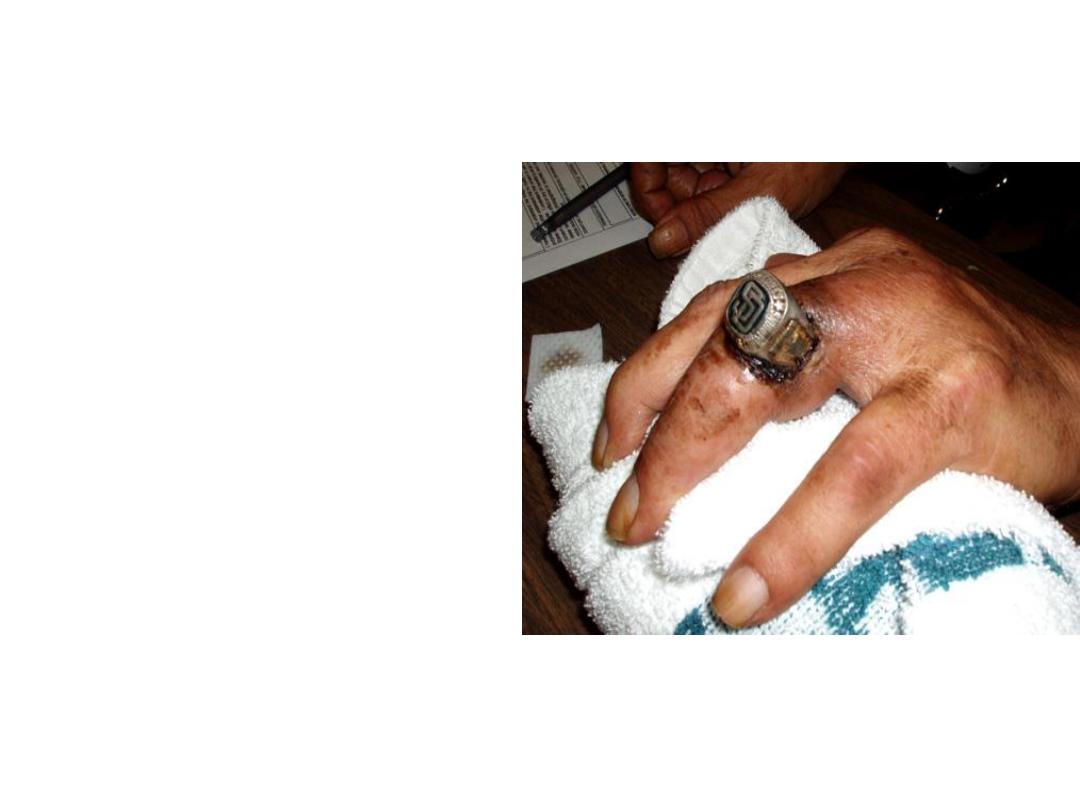
• What is this?
• How you remove such a
ring?
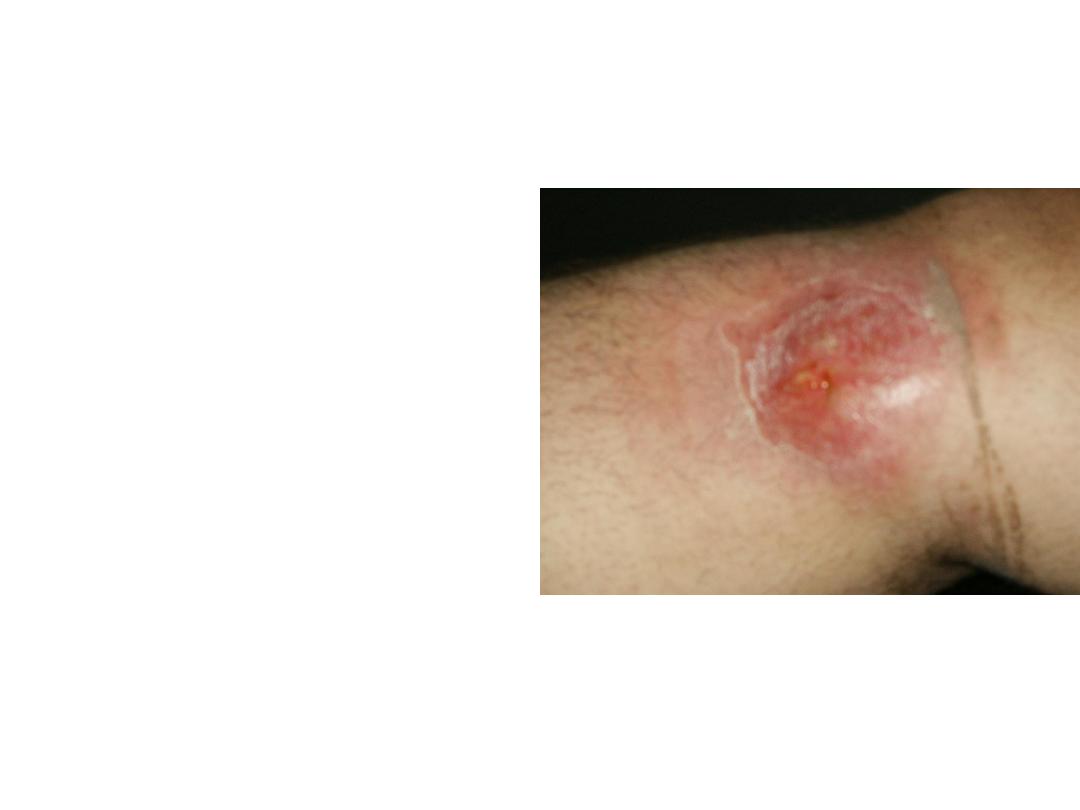
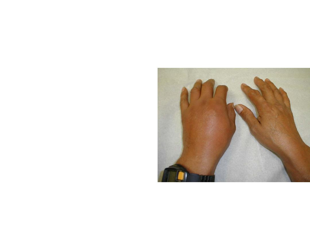
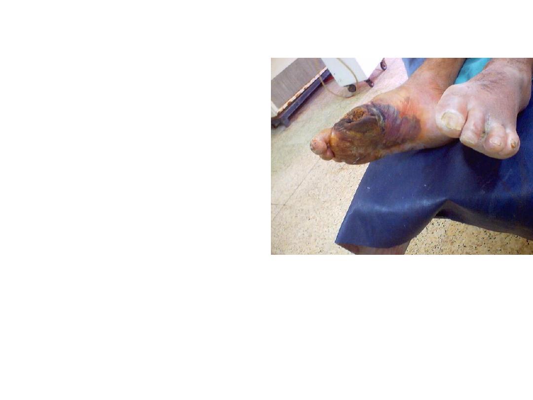
• This patient presented
with this changes.
• Describe it ?
• What are the possible
differential diagnosis?
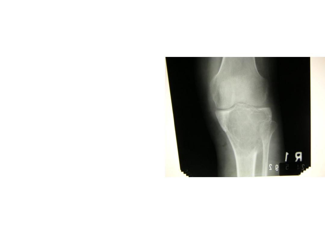
• Describe this knee x-
ray?
• What should you do
before treating such
patient?
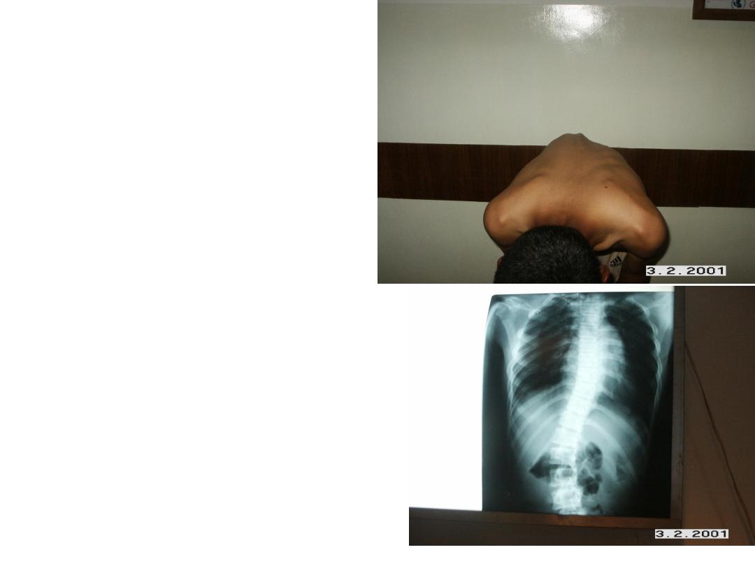
• This patient in forward
bending test for spine.
• Describe what you see
clinically and in x-ray?
• What is your diagnosis?
• You are going to examine
such an infant.
• Mention tests that had
been used?
• Describe one of them?
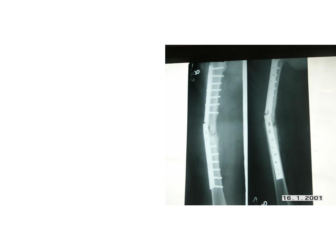
• This is femur with internal
fixation.
• What you can see?
• Mention
other
complication
might
occurred
in
internal
fixation?
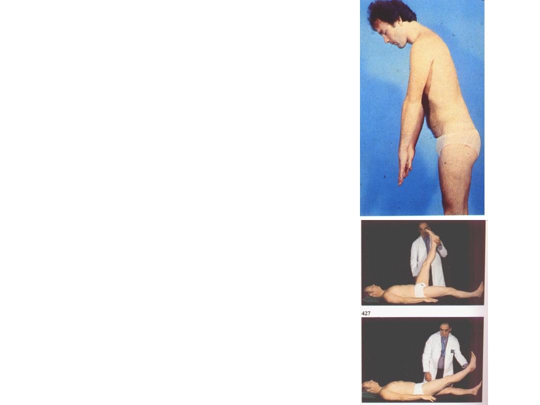
• This patient presented with
sever back pain?
• What is he doing?
• What you call this test?
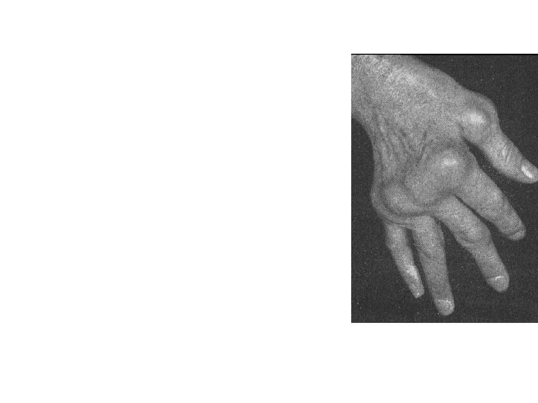
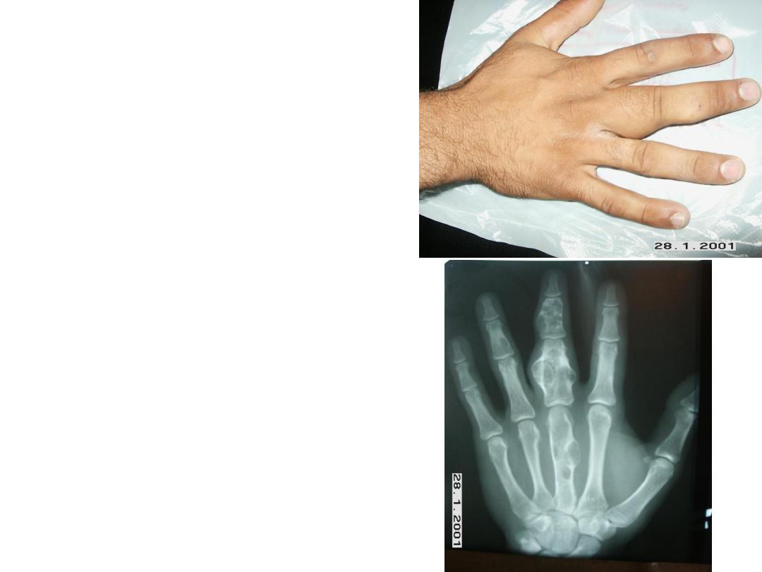
• What you can see in
this hand?
• This patient had chronic
history.
• What is your diagnosis?
• What is the radiological
finding you see?
• Mentioned
way
of
radiological
confirmation?
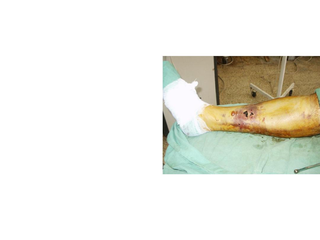
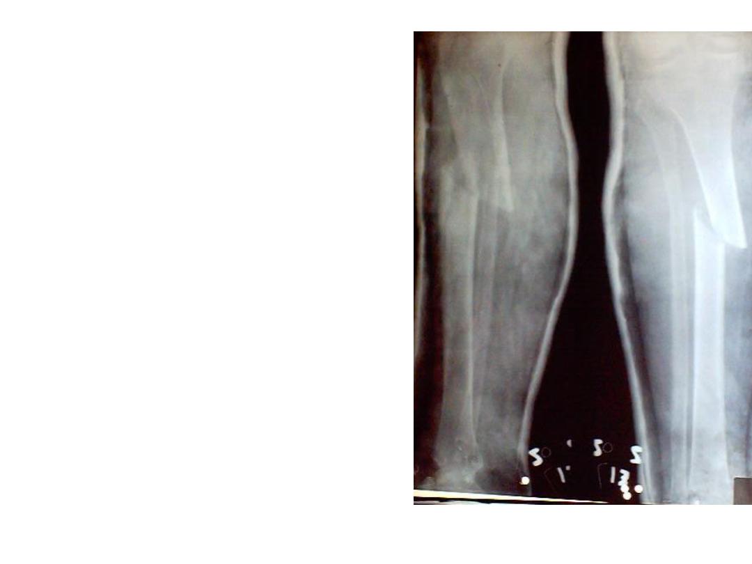
• This patient sustained
trauma to the leg.
• Describe this finding?
• How you confirm the
diagnosis?
• This patient presented with 10 days ,fever, swelling of lower thigh.
• Describe the clinical finding?
• How you should treat this patient?
• Describe
radiological
finding for such a patient
how had fever and swelling
of lower thigh?
• Mention
differential
diagnosis?
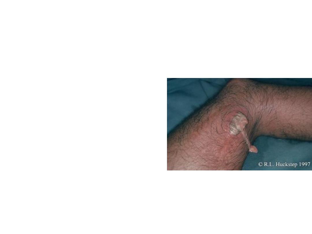
• This is an x –ray for a
patient how had history of
chronic sinus discharge.
• Describe this finding?
• What is your diagnosis?
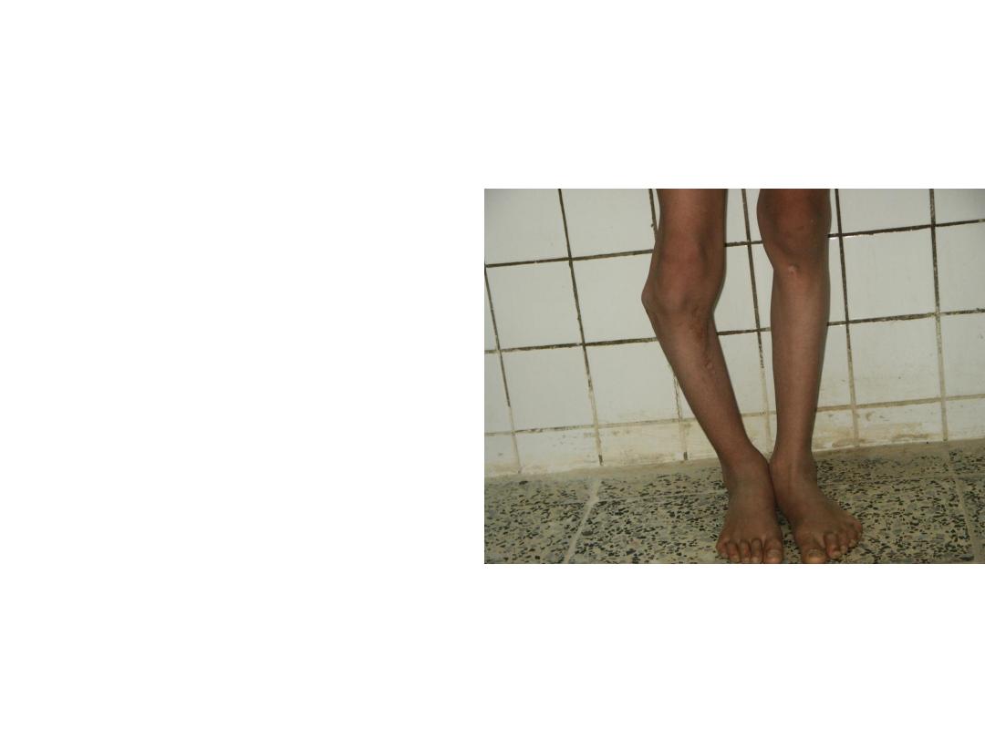
• This patient had along
history
of
acute
osteiomylites in the past.
• Describe this deformity?
• What other complications
might developed?
• This patient sustained car
accident .
• Describe the radiological
finding?
• What is your diagnosis?
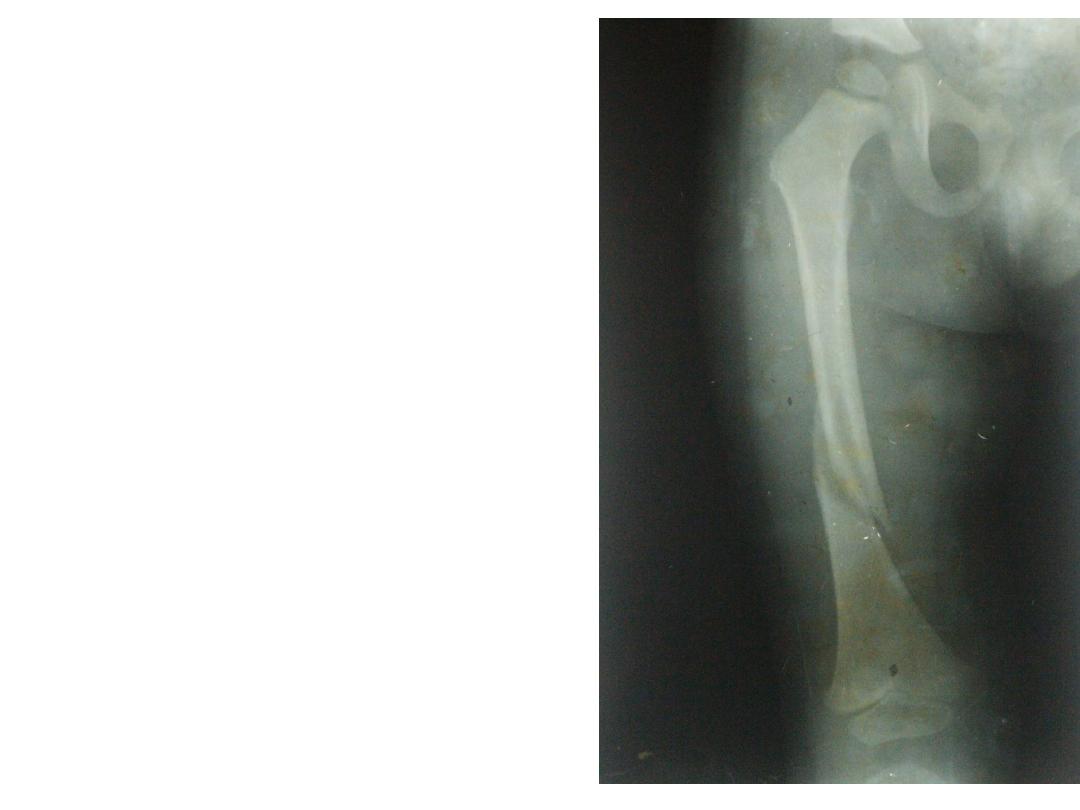
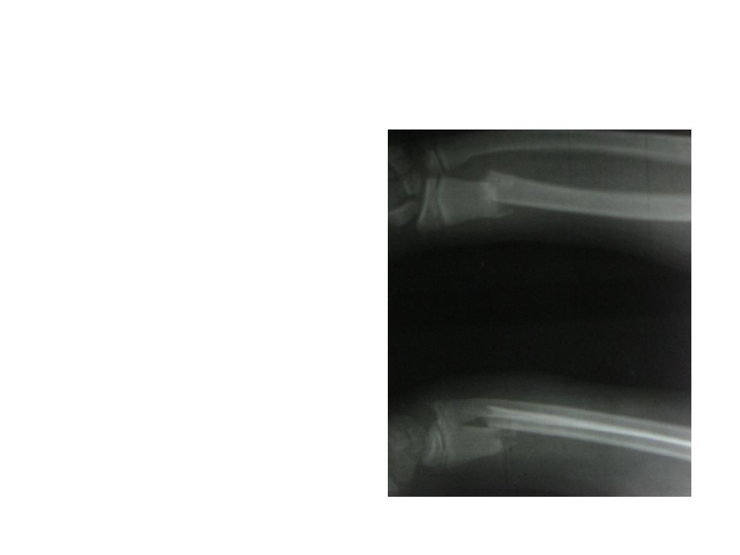
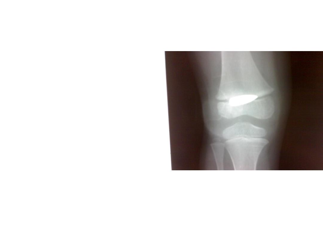
• This is an x – ray for a child
sustained trauma to the
lower limb
• Describe the radiological
finding?
• How long the duration of
healing you suspect?

• This is an x ray for an adult
man.
• Describe the radiological
finding?
• What is the important
early complication might
developed?
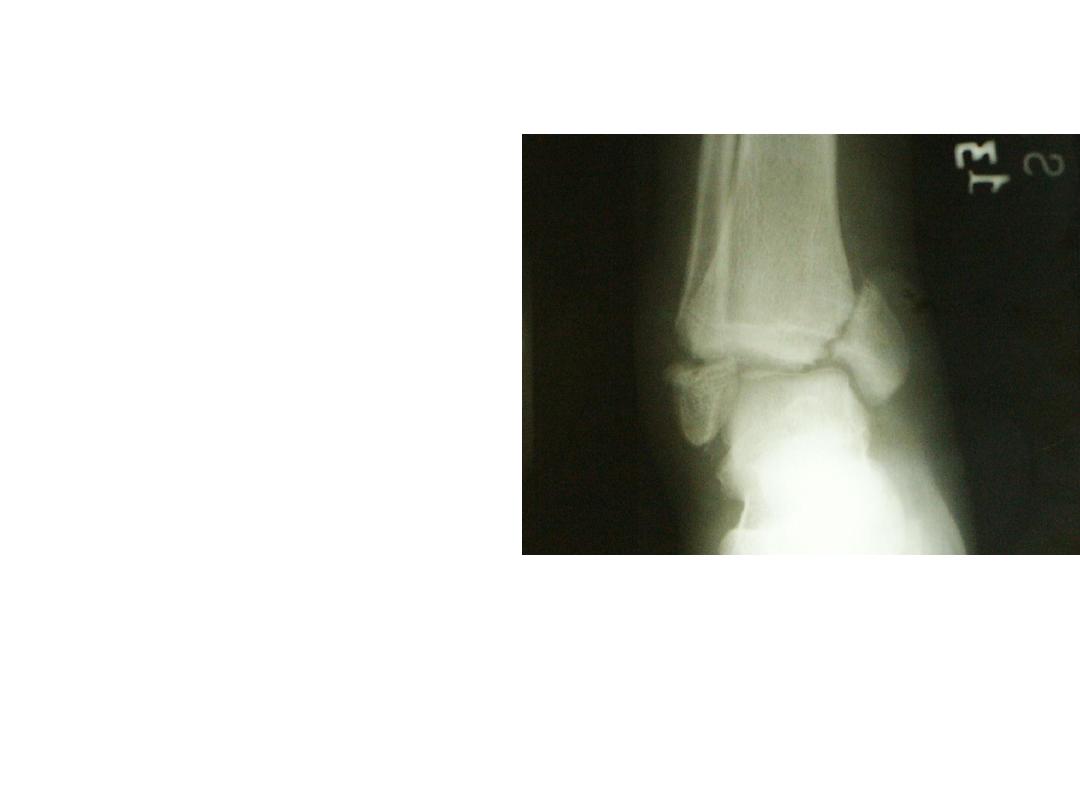
• Describe this radiological
finding?
• What are the method of
managing this patient?
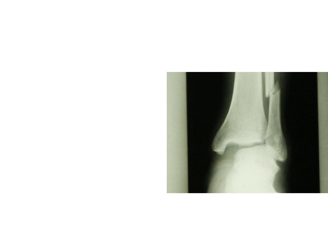
• Describe this radiological
finding?
• What is your diagnosis?
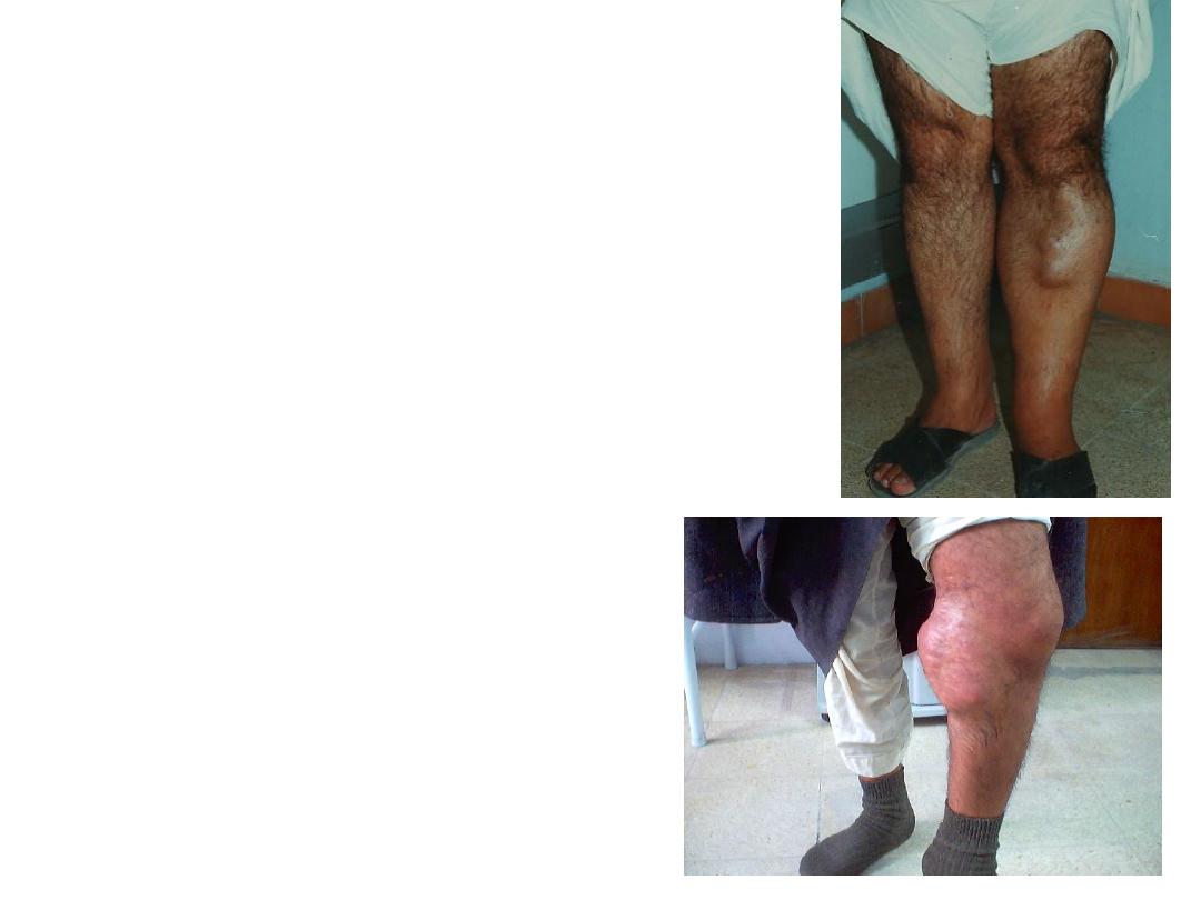
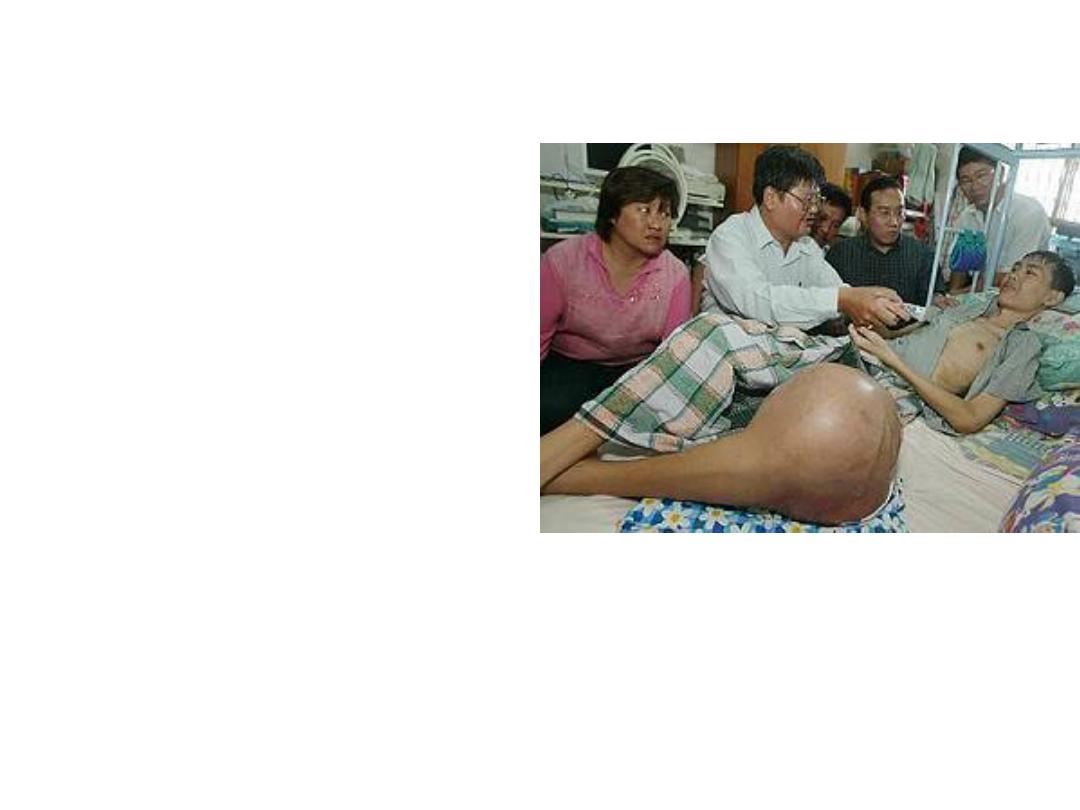
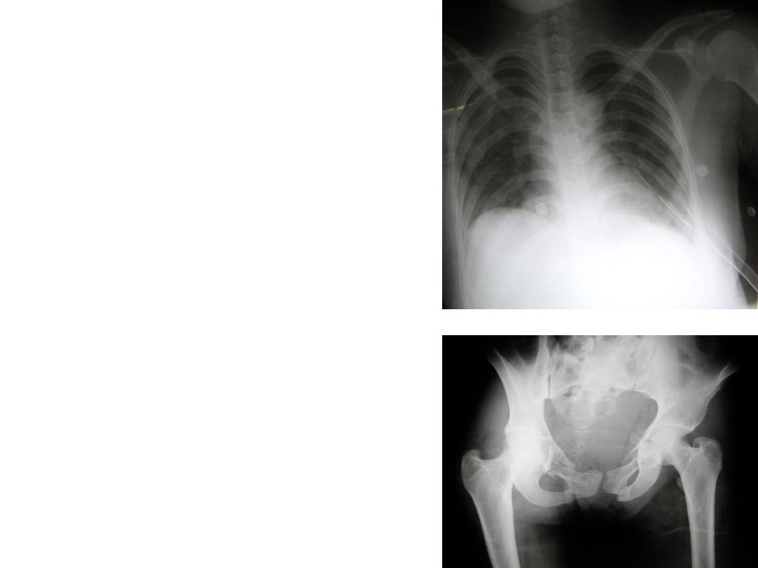
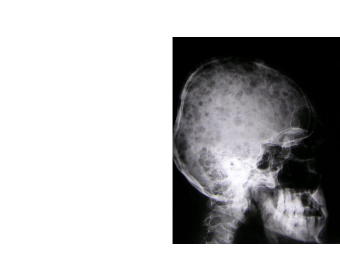
• Those two patients presented
with such a chronic swelling
with history of weight loss.
• Describe it?
• What
is
your
possible
diagnosis?
• Describe
this
radiological findings?
• What is your diagnosis?
• Describe
this
radiological findings?
• What is your diagnosis?
• Describe this
clinical
finding ?
• What are the causes of
this problem?
• Describe
this
radiological findings?
• What is your diagnosis?
• Describe
this
radiological findings?
• What is your diagnosis?
• Describe
this
radiological findings?
• What is your diagnosis?
• Describe the clinical
finding in this hand?
• How you can describe
the x –ray findings?

others
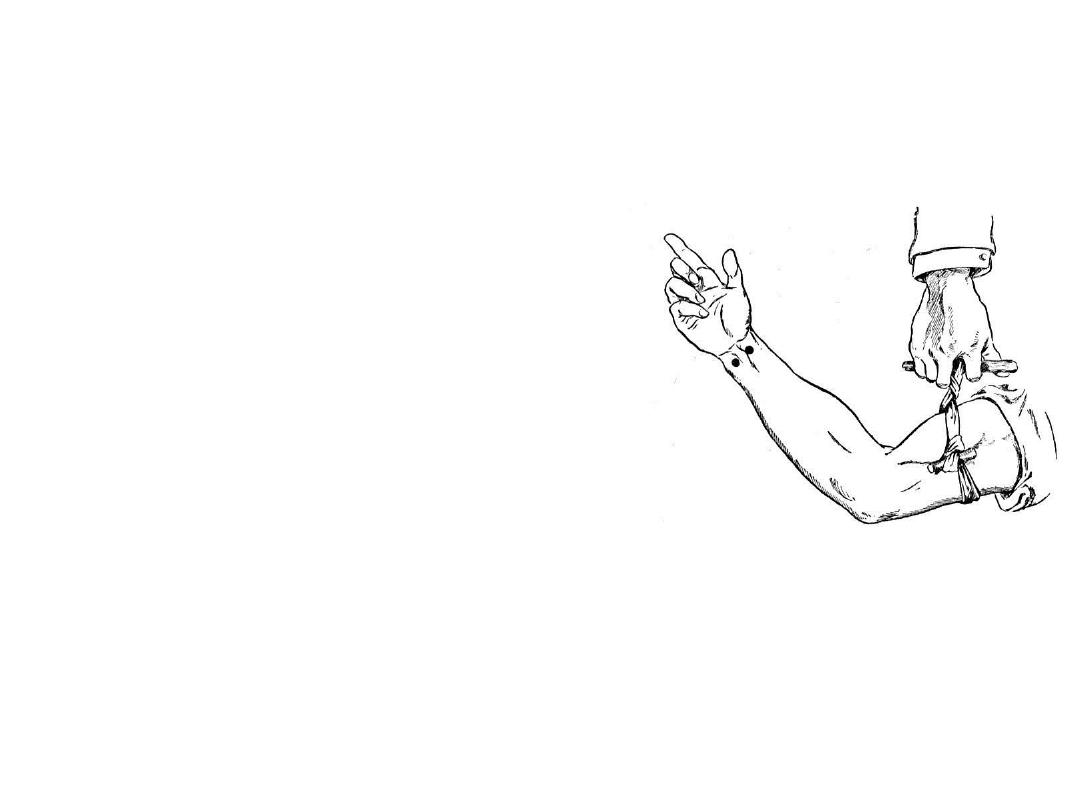
• What you call this
method of bleeding
stoppage?
• What precautions you
should take in?
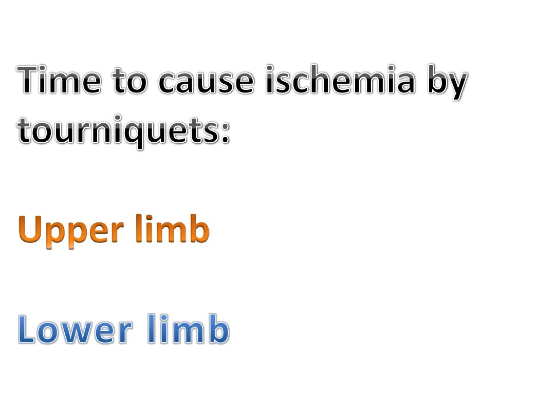
: 1-1:30 hour
: 2hr