
b
• What is this lesion?
• Corneal arcus(lipidis)
• What is the clinical
significance?
• It indicate hyperlipidemia in
young people which may causes
systemic diseases.
• What is the visual
prognosis?
• It never affect vision(never reach
cornea)
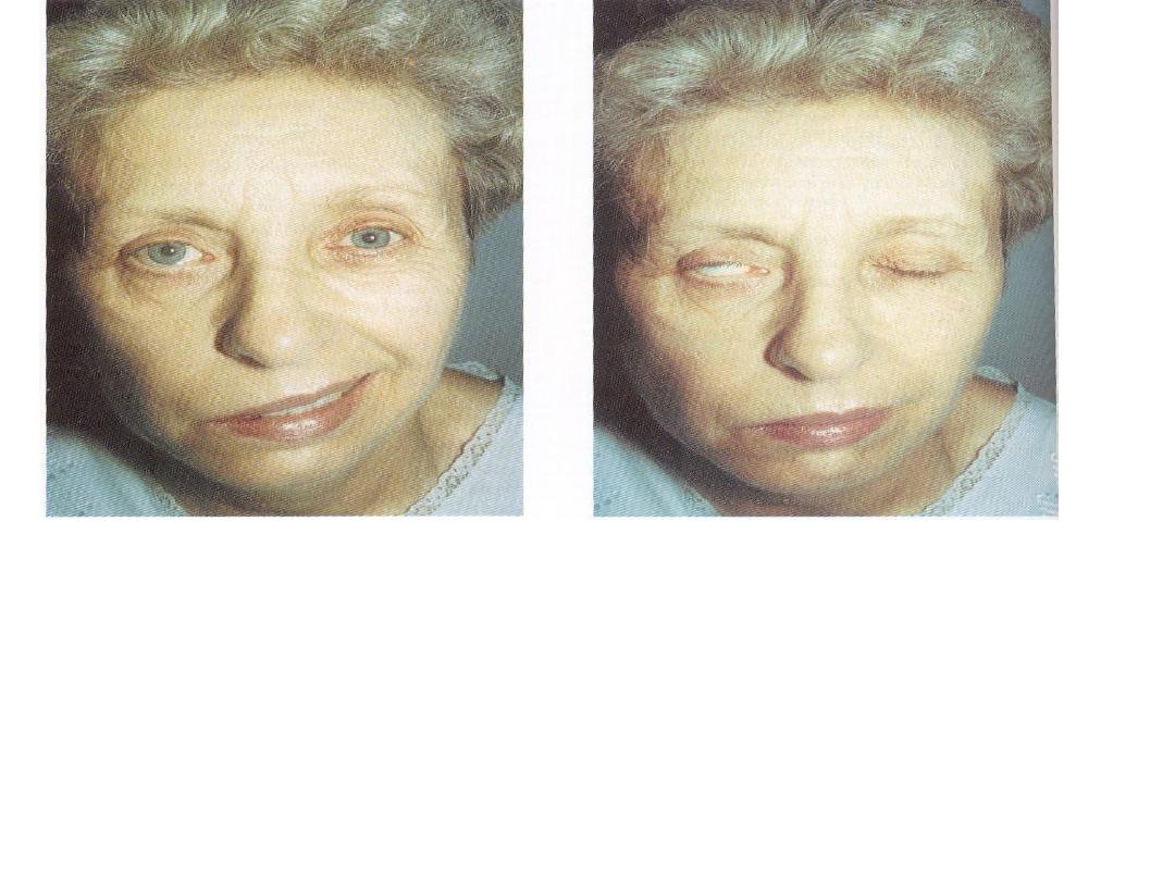
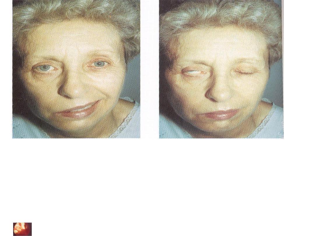
• What is your diagnosis?
• Fascial palsy on R. side with bells phenomenon
• Significance?
Exposure keratopathy
• How do you treat the eye of this patient?
• 1-artificial tears in days
• 2-closure of eye at sleep(taping)
• 3-If chronic do surgery(lateral tarsorraphy)
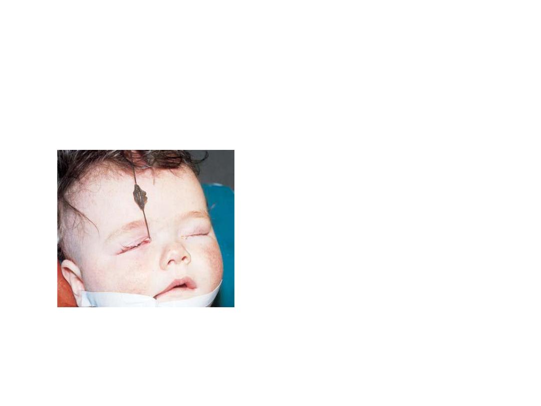
• Dx :
congenital failure of canalization
of right nasolacrimal duct
• What is the presentation of
this child?
• Lacrimation on right side(epiphora)
• What is the proper
management ?
• 1-reassurance of parents
• 2-topical AB with massage till the end of
the first year
• 3-if not opend(after the first year,most
open spontaneously during the first
year)probing and irrigation with
antiseptic solution
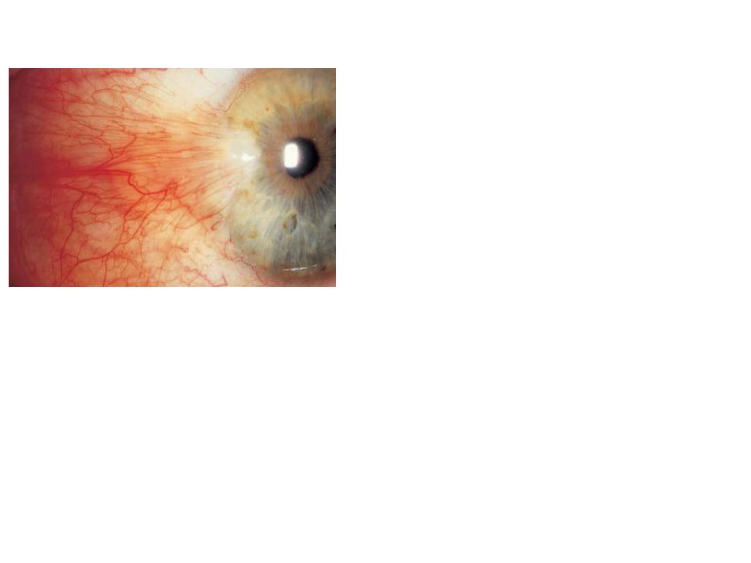
• Describe this lesion
• Localized redness located at nasal corner
of the eye,triangular in shape with the
apex extend to involve cornea due to
fibrovascular ingrowths
• Your diagnosis ?
• ptergyium
• causes ?
• 1-reflected or direct UV light exposure.2-
irritation
• Tx
?surgical removal with conjunctival
graft
• What are the indications of
surgery in this patient ?
1-affect
visual axis.2-severe discomfort.3-
cosmetic.
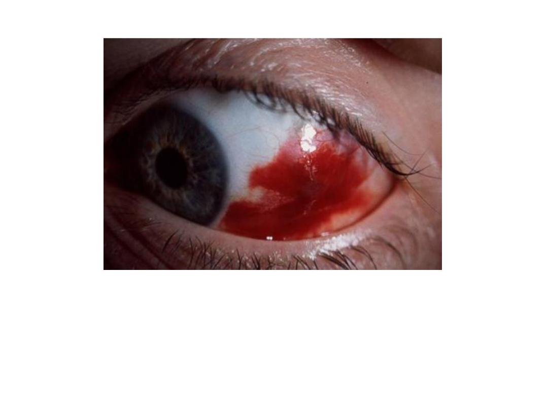
Dx
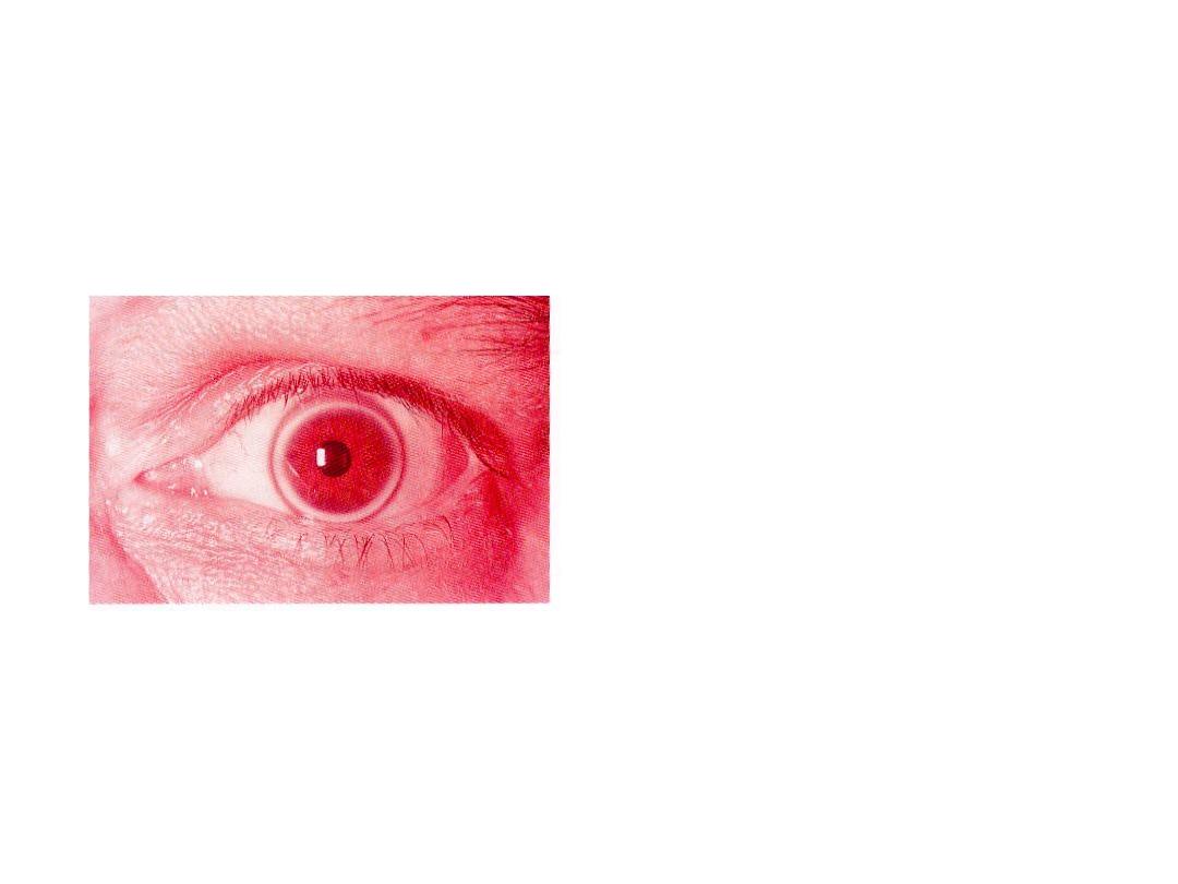
: subconjuctival haemorraghe
What will you ask in the Hx?
1-history of trauma , straining conditions.
2-history of HT.
3-bleeding tendency.
4-history of drugs:anticoaguants and NSAIDs
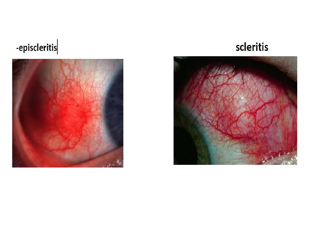
• What is the type of this
conjunctivitis?
• Membranous conjunctivits.
• What are the possible
causes ?
• 1-true membrane:diphtheria
• 2-pseudomembrane: most
bacteria like staph +adenoviral
infection
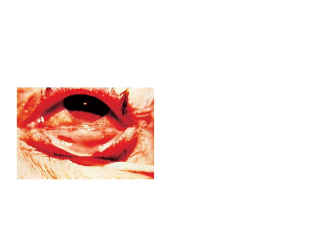
• What is your diagnosis?
• How do you treat the eye of this patient?
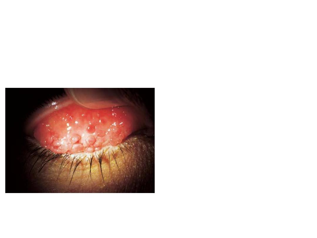
• What is this sign ?
• Papillae on the inner surface of
upper lid(giant cobblestones
papillae)
• What are its causes?
• Allergic conjunctivitis or chronic
conditions.
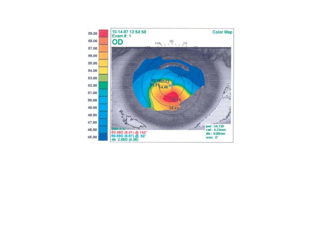
Corneal topography(color coded map) to study
corneal curvature
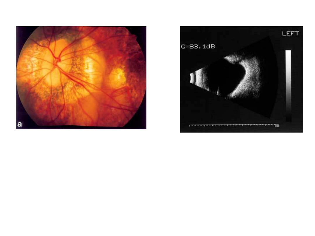
Staphyloma
Occur in high myobia
على االغلب
US in high myobia
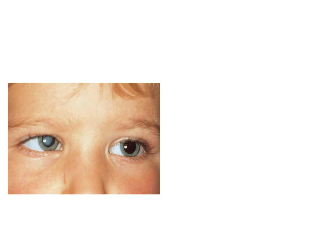
• Describe the lesion?.
• R.white pupil.
• Dx? :
leukocorea
• Enumerate the most
common causes
according to their
frequency?
• 1-congenital cataract
• 2-retinoplastoma
• 3-retinal fibrosis
• 4-persistent hyperplastic primary
vitreous
• 5-retinopathy of prematurity.