1
RESPIRATORY SYSTEM
Lecture 1
Dr. Mareb. H. Ahmad
/
Histology Department/ Faculty of Medicine/
2
The principle function
Exchange of gases (oxygen and carbon
dioxide) between air in alveoli of the lung &
blood in the capillaries.
3

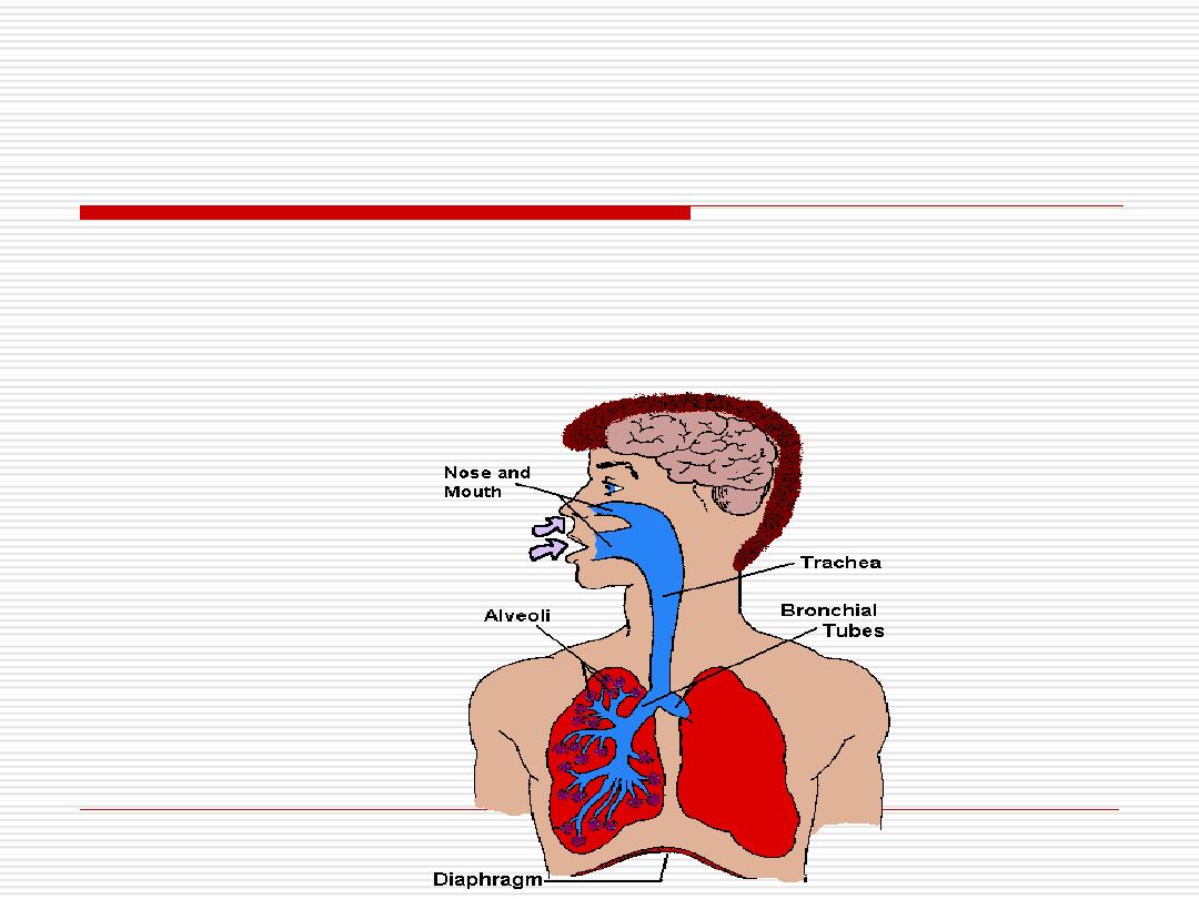
It includes:
a-
The lungs: the
site for gas exchange.
b
- A system of
tubes
:
links the lungs
with the external environment.
c
- A
ventilation mechanism
which is
important for the movement of air through
the conducting and respiratory parts of the
lungs.
It consists of the thoracic cage, intercostals
muscles, diaphragm, and elastic as well as
collagen components of the lungs.
4
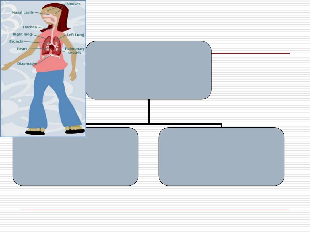
Respiratory system
Conducting part
Respiratory part
5
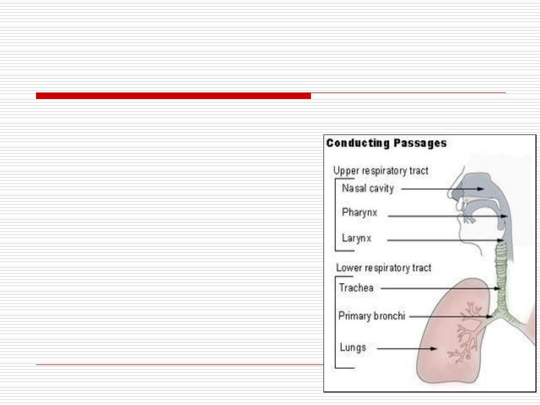
Conducting part
(upper respiratory tract):
by which air pass from the atmosphere to the lung
It consists of
The nasal cavity
The pharynx
The larynx
The trachea
Bronchi
Terminal bronchioles
6
Respiratory part
(lower respiratory system):
where gaseous
exchange took place
.
It consists of
Respiratory bronchioles
Alveolar ducts
Alveolar sacs
Alveoli.
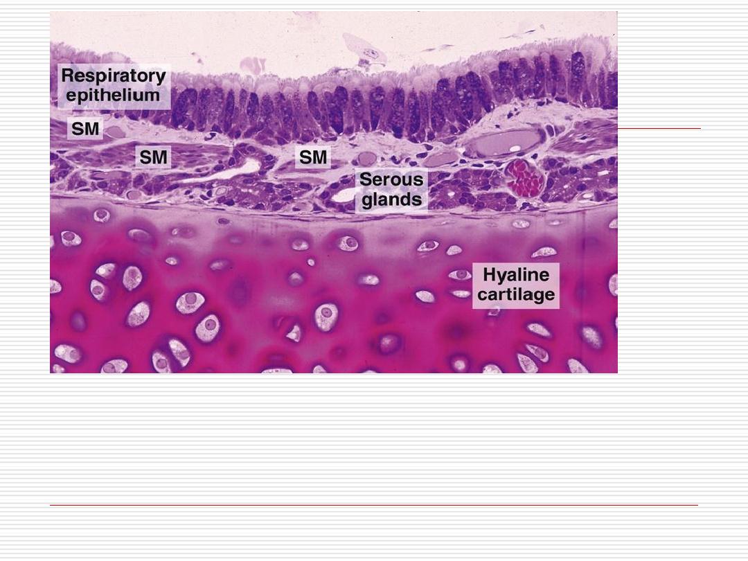
7

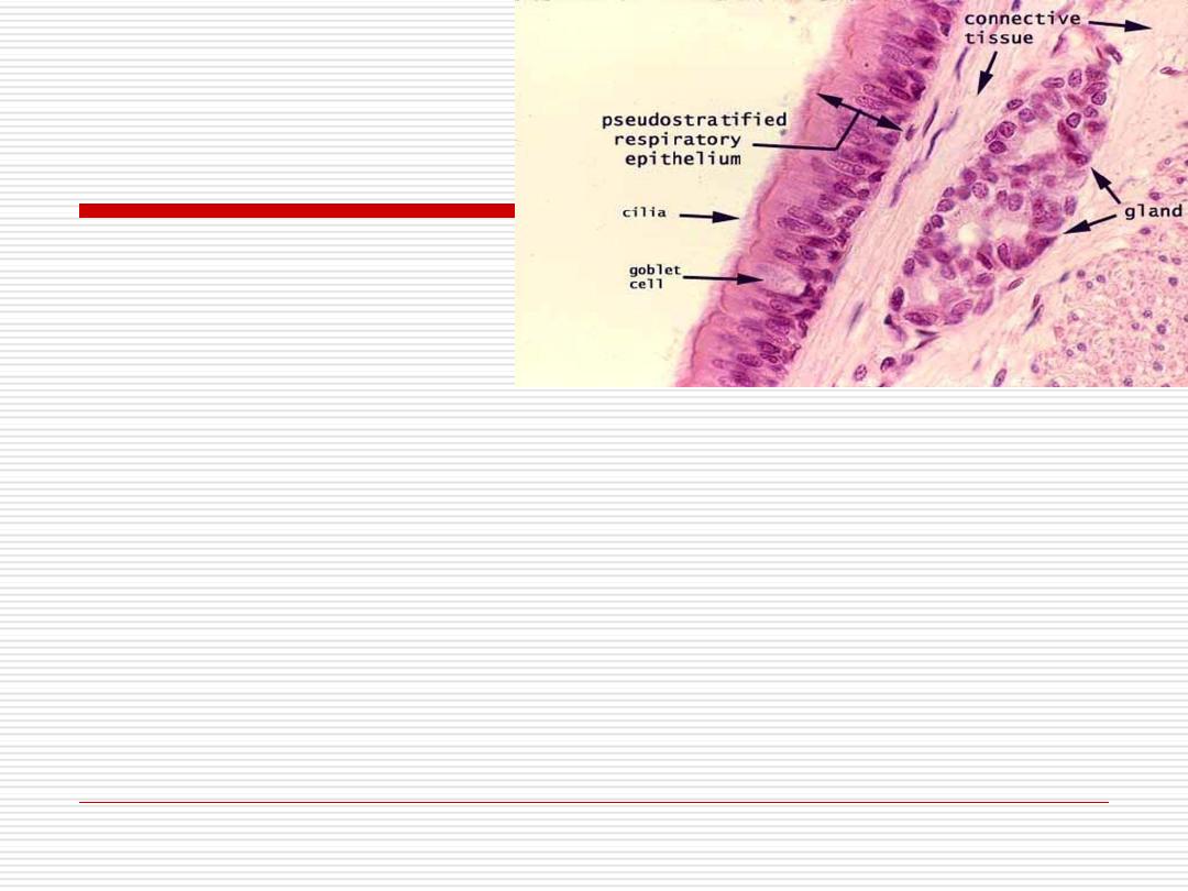
Conducting part
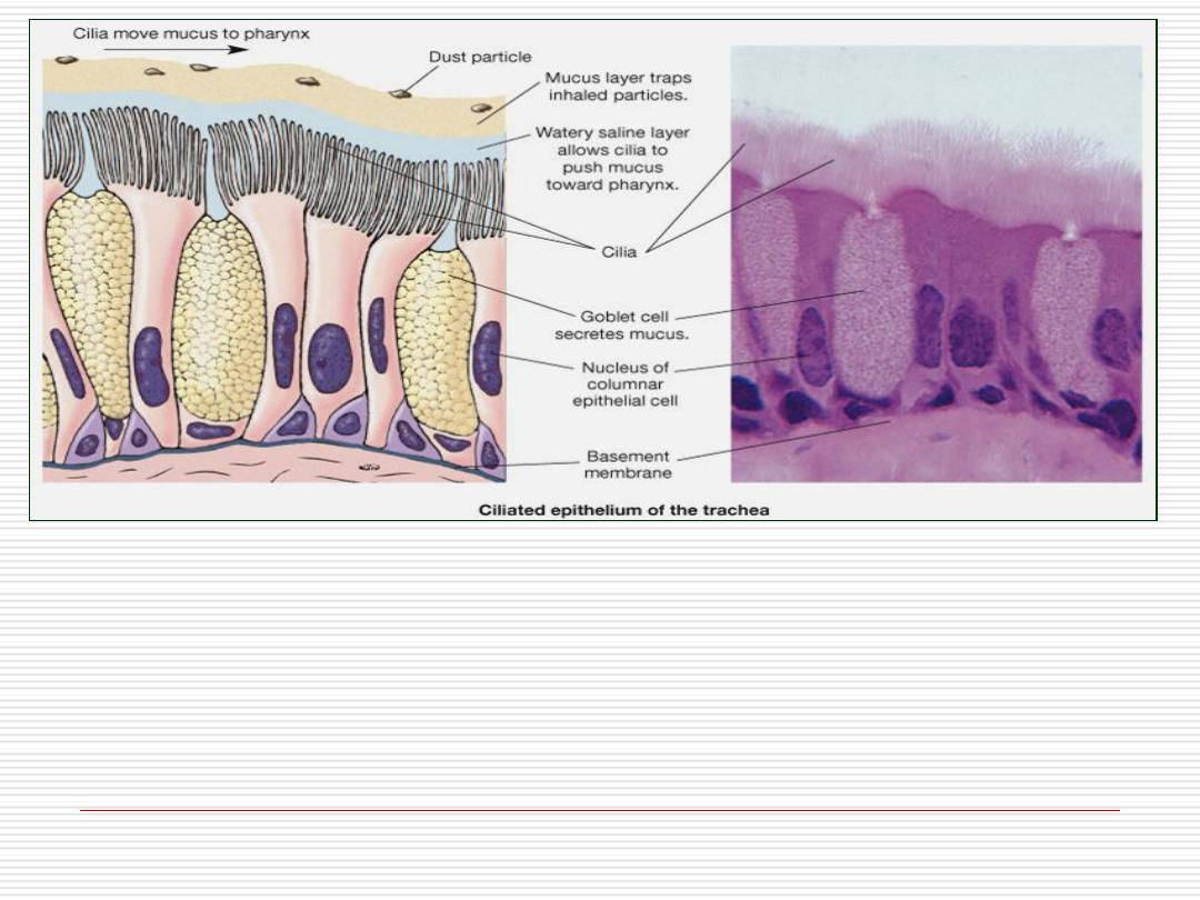
Has general structural characteristics :
Epithelium
: mostly pseudo stratified columnar ciliated
epithelium with abundant goblet cells (respiratory
epithelium).
Lamina propria
: This is the loose C.T, rich vascular supply,
abundant elastic fibers and seromucous glands.
Supportive layer
that keep the airway patent and protect it
from collapse.
It may be
bone & cartilage
(nasal cavity),
cartilage
(trachea & bronchi) or
smooth muscle
(bronchioles)
8

1- Conducts air to and from the lungs
2- Conditions the inspired air by:
- Cleaning: the mucous secreted by
goblet
cells entraps
the dust & then moves toward the mouth by the ciliary
activity which is facilitated by serous secretion from the
seromucous glands in lamina propria.
- Moisturing: by the
seromucous
secretion covering the surface.
- Warming: by the rich
blood vessels
in the lamina propria.
3- specialized structures that are involved in the perception of
smell & flavor (Olfactory mucosa) & the production of sound
(Larynx).
The conducting portion serves two functions:

Respiratory epithelium:
Most of the conducting portion is lined
by
pseudostratified columnar ciliated
epithelium with many
goblet
cells.
Typical respiratory epithelium consists
of
5
cell types as seen in the EM:
10

1- Ciliated columnar cells
The most
abundant
type
Each cell has
200-300 cilia
on its apical
surface.
There are numerous
mitochondria
beneath
the cilia to supply ATP necessary for ciliary
movement.
11
2-Goblet cells
The next abundant cells.
The
apical portion
is wide and contains
mucinogen granules.
They have
narrow basal
part that contains the
nucleus and organelles.
They secrete
mucous
that covers the epithelial
surface, traps particulate matter and absorbs
water soluble gases.
12
3- Brush cells:
Columnar cells that have
numerous
microvilli
on their apical surface.
They have afferent
nerve
endings on their
basal surfaces.
They are considered to be
sensory
receptors
.
13

4- Basal cells:
Small rounded cells that lie on the basal
lamina.
They are
short
and do
not
reach the
luminal surface.
They are considered as
stem
cells for other
cell types.
14
5- Endocrine cells:
Similar to basal cells.
They have small dense
granules
in the
basal
cytoplasm.
They belong to
DNES
(Diffuse Neuro-Endocrine
System).
They secrete amine and peptide hormones (e.g.
serotonin)
15
A smoker’s respiratory epithelium has :
?
?
?
16

NASAL CAVITIES
The nose is covered
externally
by
skin
with some large
sebaceous glands.
The cavity of the nose is divided by a midline septum
into right and left nasal cavities.
Each communicates anteriorly with the exterior by
anterior naris (nostril)
and
posteriorly
with the
nasopharynx
.
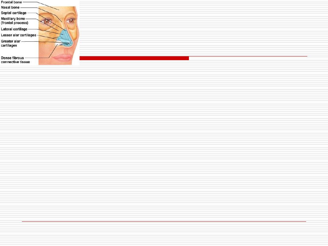
The wall of each nasal cavity has a rigid wall of hyaline
cartilage and bone.
17
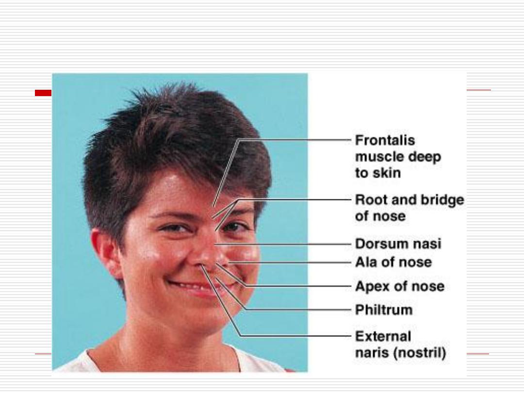
Structure of the Nose
18
Each nasal cavity is divided into:
1-
Vestibule
The most anterior and dilated portion of the nasal
cavity.
The nostrils are lined with
keratinized stratified
squamous
epithelium that covers a part of the
vestibule.
Deeper it is changed to
nonkeratinized
epithelium
then
respiratory
epithelium before entering the nasal
fossae.
It contains numerous sebaceous and sweat glands,
and thick short hair
(vibrissae)
that filter out large
particles from the inspired air.
19
Three
bony projections known as
conchae
extend from each
lateral wall.
The middle & inferior conchae, the floor of the nasal fossae
and most of the nasal septum are covered by respiratory
epithelium and are
called respiratory area
.
The superior conchae, roof of the nasal fossae and upper
part of the nasal septum are covered by olfactory
epithelium. This part is
called olfactory area
.
2- Nasal fossae
20

Olfactory epithelium
The olfactory chemoreceptors are present
in the olfactory epithelium covering the
olfactory area.
This is a
pseudostratified
columnar
epithelium composed of
3
types of cells.
21

supporting cells:
-
1
They are tall cells with apically located nuclei.
The apex of the cell is broad and the base is
narrow.
The cell has
microvilli
on its apical surface.
It contains a
yellow
pigment similar to
lipofuschin
pigment.
22
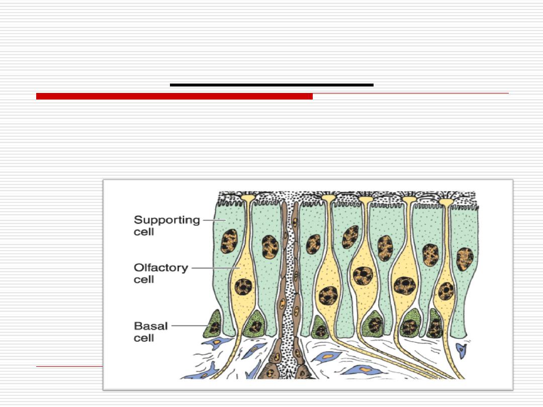
Basal cells
-
2
They are
short
and rounded, located at the
basal
region of the
epithelium.
They may act as
undifferentiated
stem cells or supporting cells
23

-They are
bipolar
nerve cells.
-Their nuclei lie below the level of nuclei of the supporting.
-The apical portion is a modified dendrite that ends at the surface of the
epithelium in a bulb
(the olfactory vesicle).
-This vesicle gives rise to several long
immotile cilia
(6-8) that act as
receptors.
-The afferent axons of these bipolar neurons unite & pass through the
cribriform plate of the ethmoid bone forming
olfactory nerve
-The cilia & the microvilli are submerged in a fluid layer consisting mainly
of
serous
secretion of glands in the lamina propria called
Bowman's glands
.
Olfactory cells
-
3
24
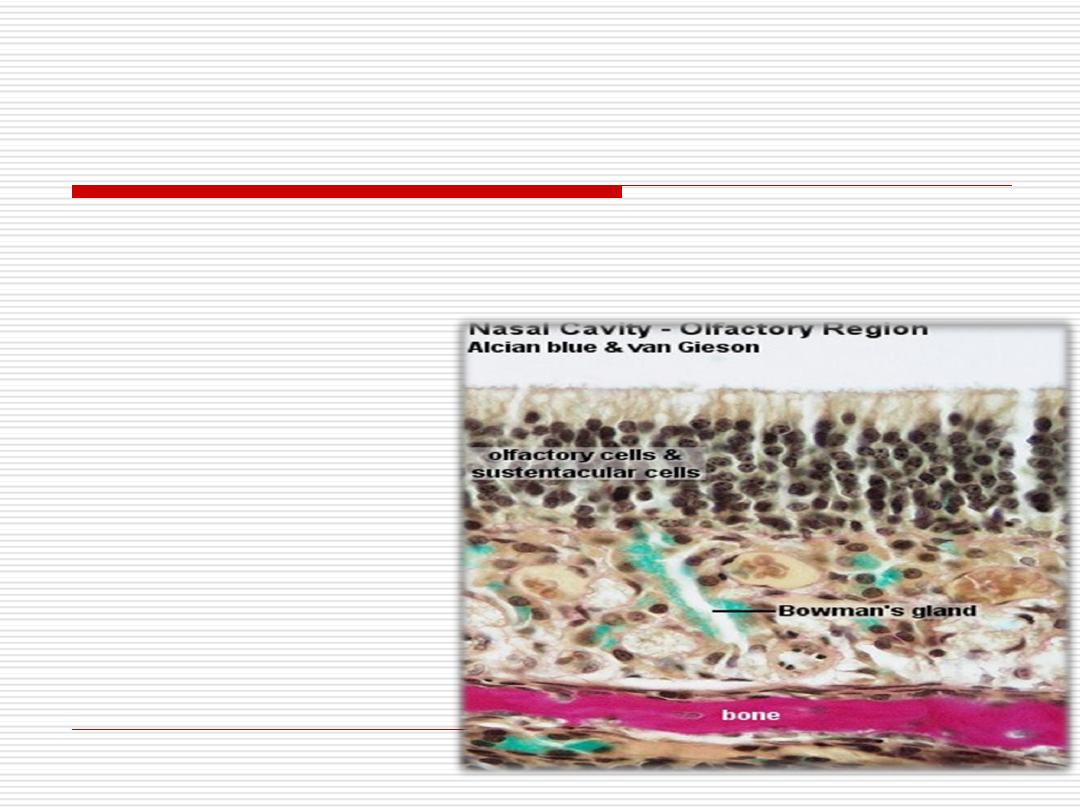
Functions of Bowman’s
glands
1-
Clear the surface of the olfactory epithelium
.
2- Serve as a solvent
for
odoriferous substances.
25
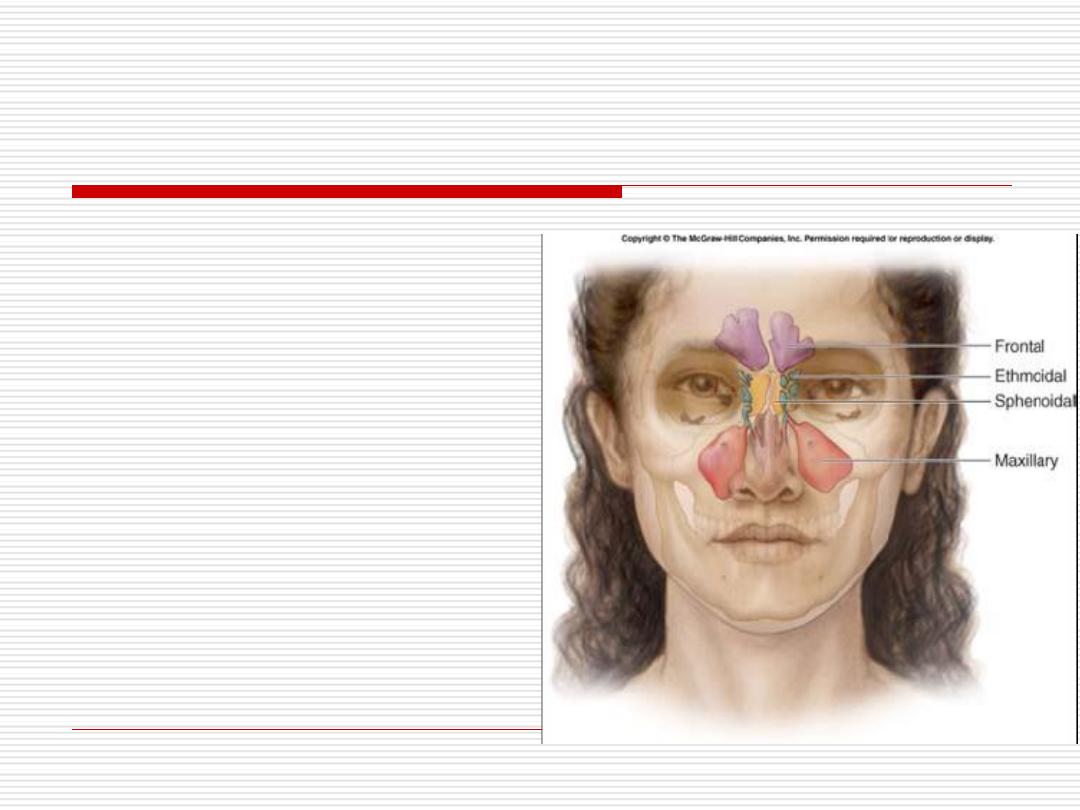
3- Para nasal sinuses
They are blind cavities
in the frontal,
maxillary, ethmoid
and
Sphenoid bones.
Reduce weight of skull
Resonating chamber –
modifies voice
26

They are lined by thin
respiratory epithelium, with
few goblet cells
.
The lamina propria contains few
small mucous
glands
.
They are connected with the nasal cavity by
small
openings
.
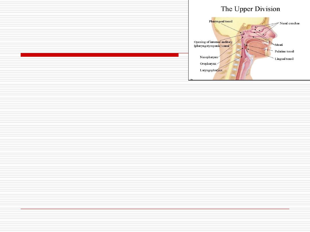
27
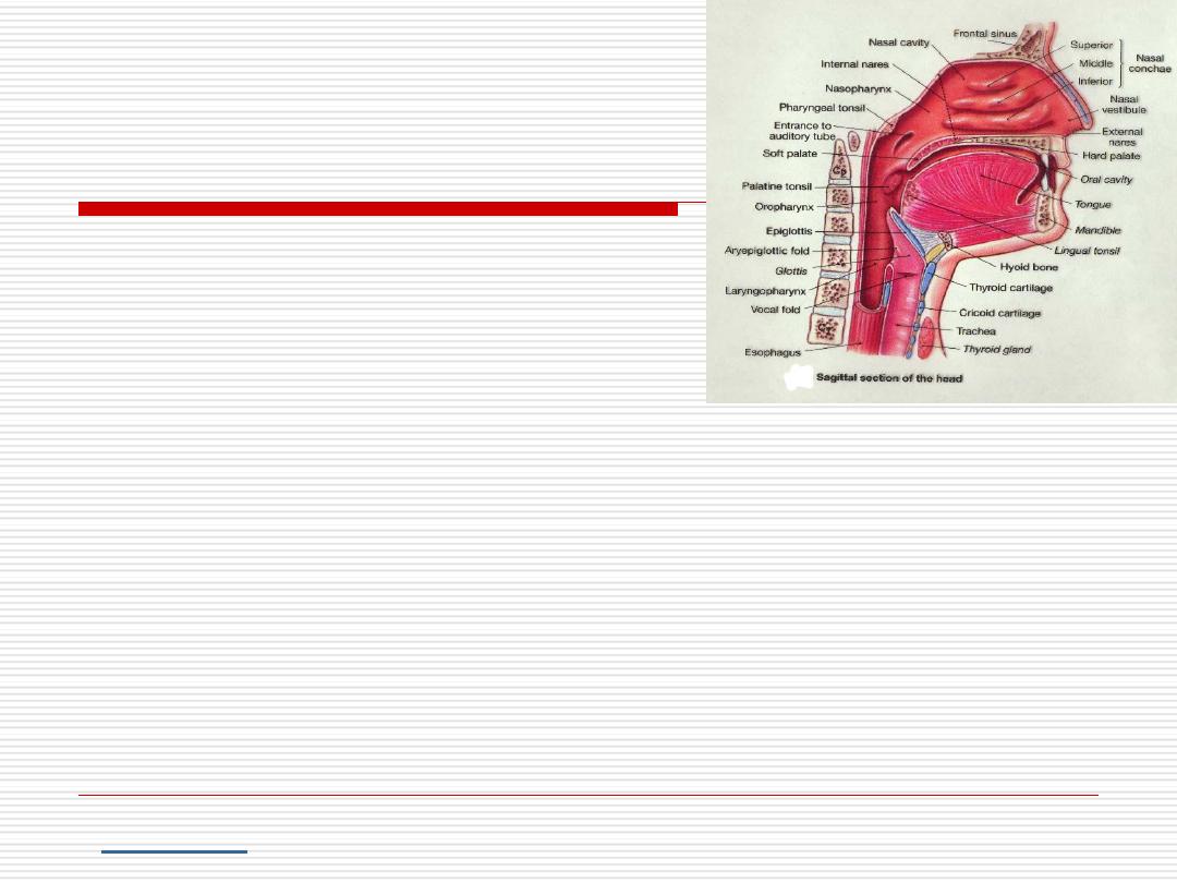
PHARYNX
It can be divided into
3 regions:
Nasopharynx
, lined by
respiratory epithelium
.
Oropharynx and laryngeal region
are lined by
stratified squamous epithelium
.
Lymphocytes frequently accumulate beneath the
epithelium of the pharynx.Accumulations of
lymphoid tissues surrounding the openings of the
digestive and respiratory passages form the
.
28
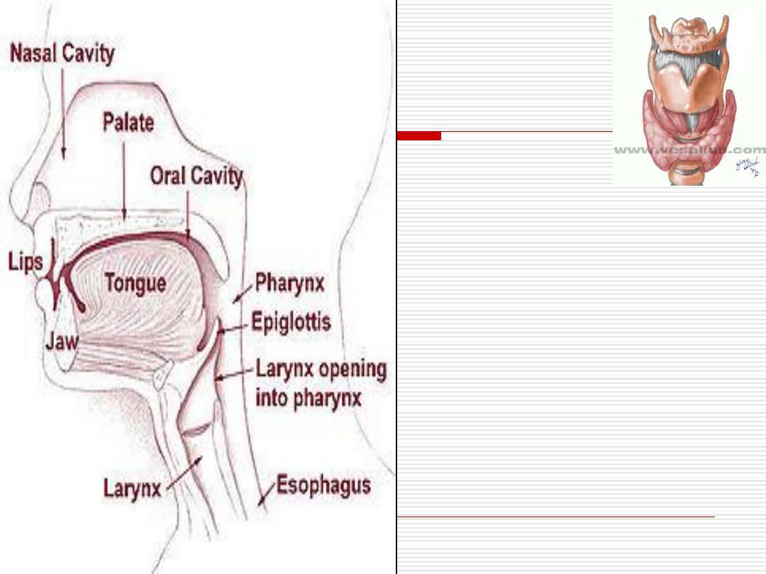
It is an
irregular
tube
Most of the larynx is
covered by
respiratory
epithelium
except
parts
of epiglottis and the
true vocal cords.
The lamina propria of
the
larynx
contains
seromucous
glands and
plates
of
cartilage
.
LARYNX
29

LARYNX
Large
hyaline
cartilages (thyroid, cricoid and most
of arytenoids).
Smaller
elastic
cartilages (epiglottis, cuneiform,
corniculate and the tips of arytenoids).
These cartilages are bound together by
ligaments
and the intrinsic
muscles
of the larynx
.
30

The larynx includes the epiglottis which is a valve like structure
preventing food from entering the respiratory passages.
has two surfaces,
lingual
(superior) and laryngeal
(inferior).
The lingual surface and the apical portion of the laryngeal
surface are covered with
non keratinized stratified
squamous epithelium.
The rest of laryngeal surface is covered by
respiratory
epithelium.
It is supported by elastic cartilage
.
31

Below the epiglottis the mucosa forms two pairs of folds
The upper pair
(False vocal cords)
covered by
respiratory
epithelium.
The lower pair
(True vocal cords)
covered by
stratified
squamous
epithelium.
Function
Maintaining the airway open
The larynx is the organ of phonation
LARYNX
32
Lecture 2
Dr. Mareb. H. Ahmad
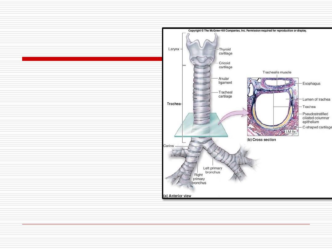
The trachea is
a rigid tube about
10cm long.
It extends from the
larynx to the upper
part of the thorax .
The wall is supported by 16-20
C shaped rings of
hyaline cartilage
joined at the free ends by bands
of dense connective
TRACHEA
33
The wall of the trachea consists of
4
layers.
1-Mucosa 2-Submucosa
3-supportive layer 4-Adventitia
34

1-Mucosa
A-Epithelium
: respiratory epithelium
resting on a thick basal lamina.
B- Lamina
propria
: of loose connective
tissue rich in elastic and reticular fibers
C-Elastic membrane
: separates the
lamina propria from submucosa
35
Submucosa
2-
Loose connective tissue laye containing
1- Numerous seromucous glands
2- Lymph follicles.
36

3-Supportive layer(cartilage and muscle)
It is the
outer
part which contains the C-shaped
hyaline
cartilage
.
The gap between the two ends of the C-shaped
cartilage is completed by bundles of
smooth
muscle
fibers (trachialis muscle).
Contraction of trachealis muscle in the cough
reflex, leads to ??????????
37
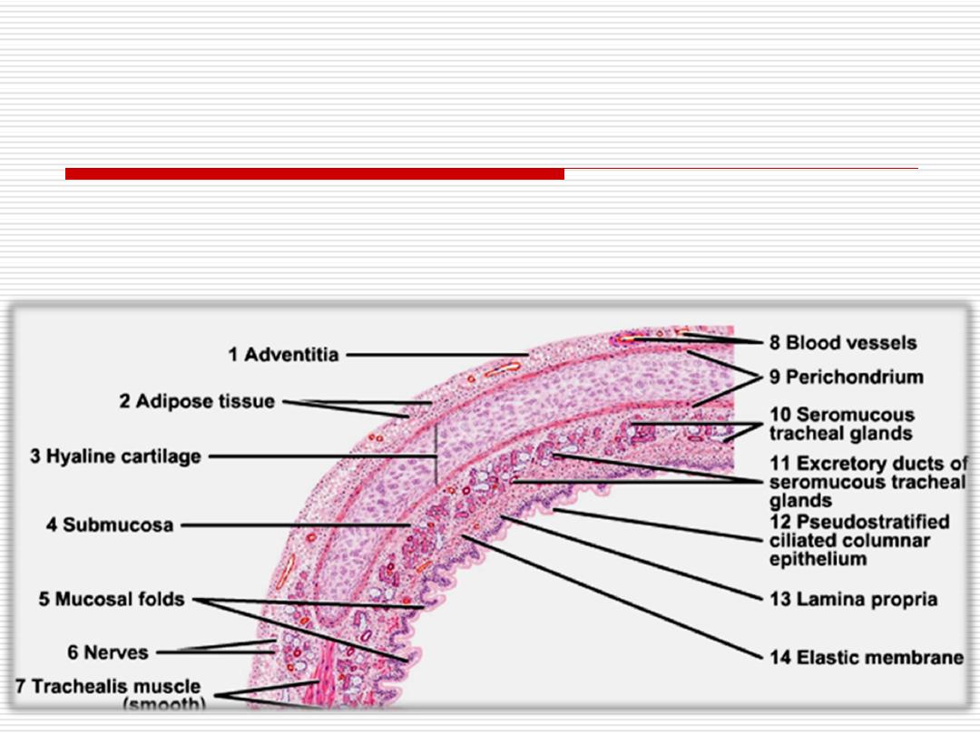
4. Adventitia
:
thin connective tissue layer that
surrounds the trachea.
38
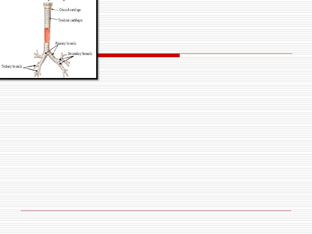
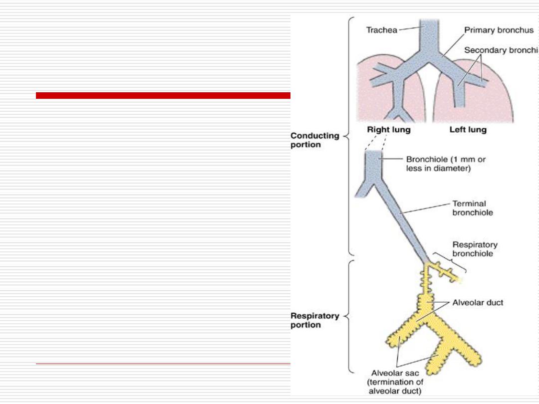
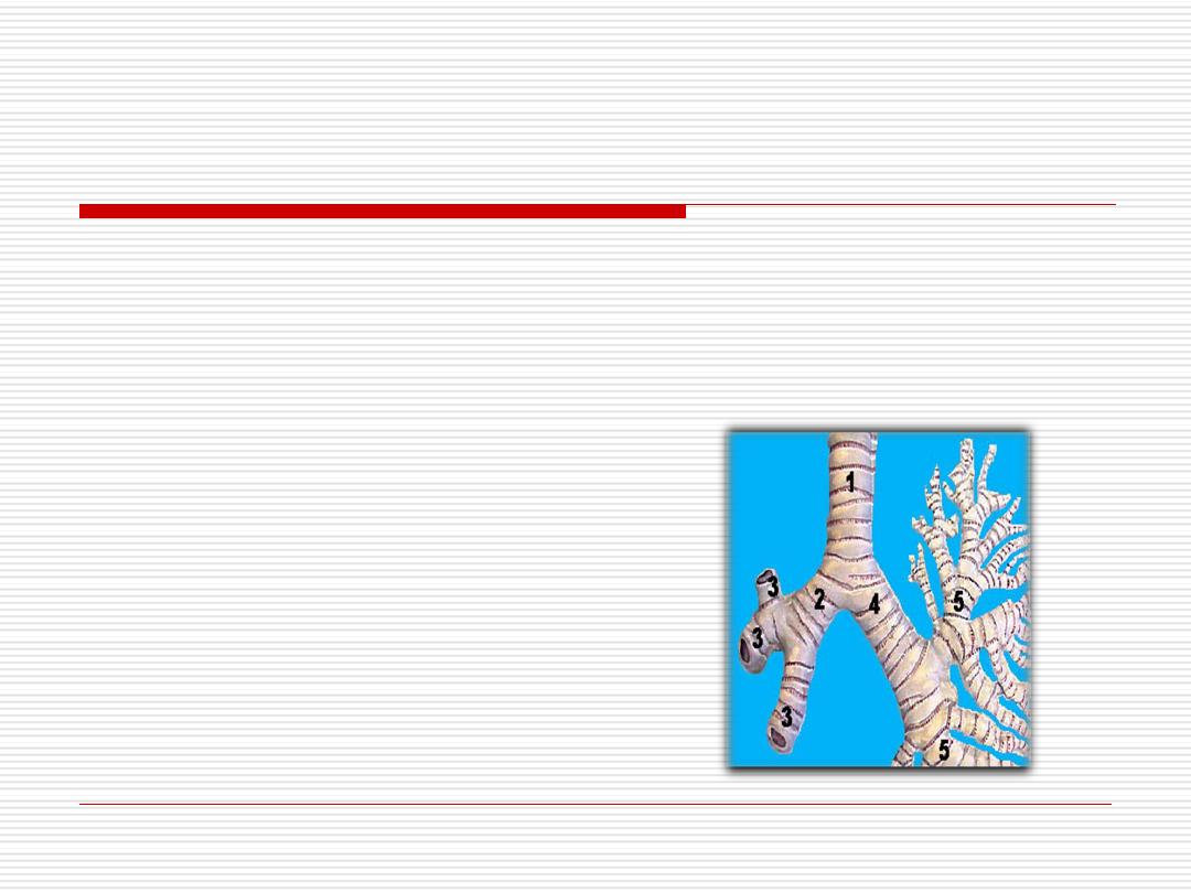
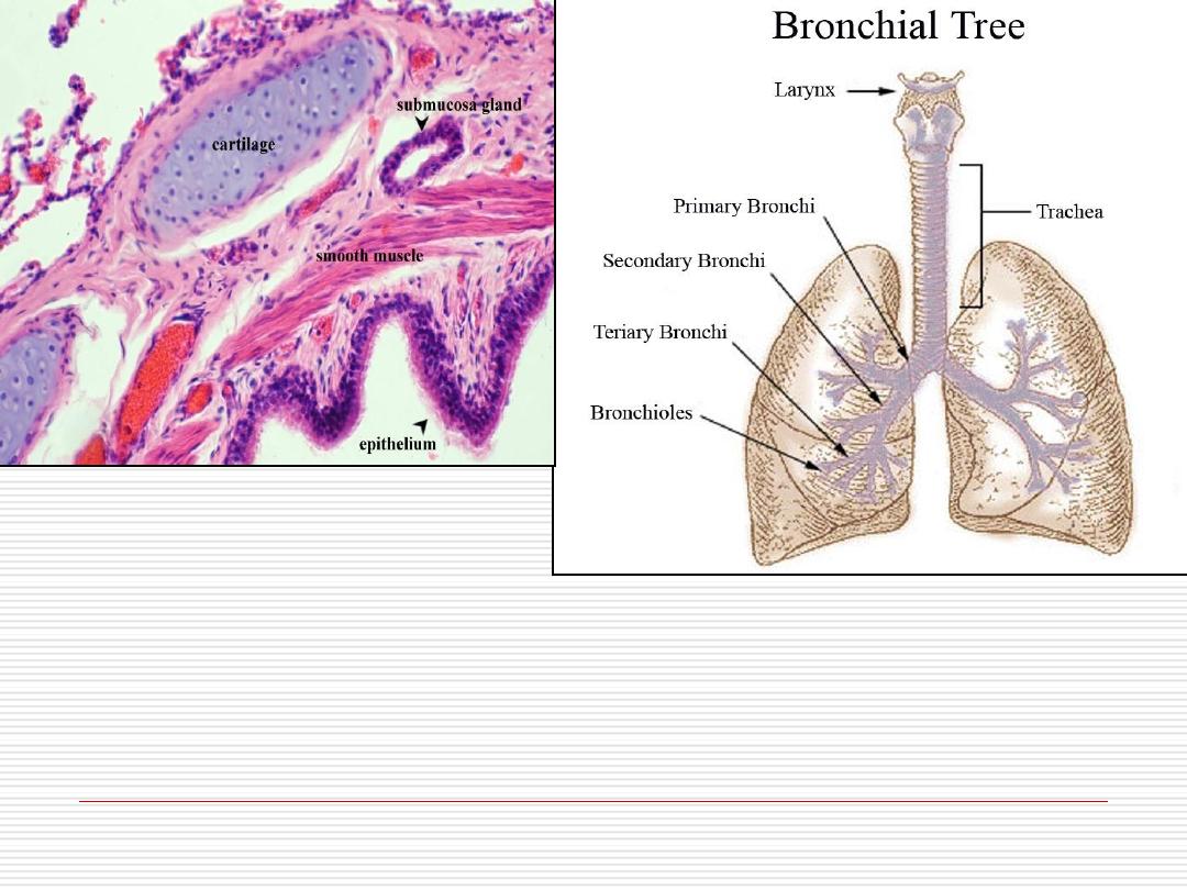
THE BRONCHIAL TREE
The trachea divides into 2 primary
bronchi
that enter the
lungs at the hilum with arteries, veins, lymphatics and
nerves (vagus and sympathetic).
In the lung, the primary bronchus divides into
secondary
(lobar) bronchi
, each enter a lobe, 3 in the right lung and
2 in the left one.
These lobar bronchi divide repeatedly giving rise to
smaller bronchi and then
bronchioles
.
Each bronchiole enters a pulmonary lobule where it
branches to 5-7
terminal bronchioles
.
39
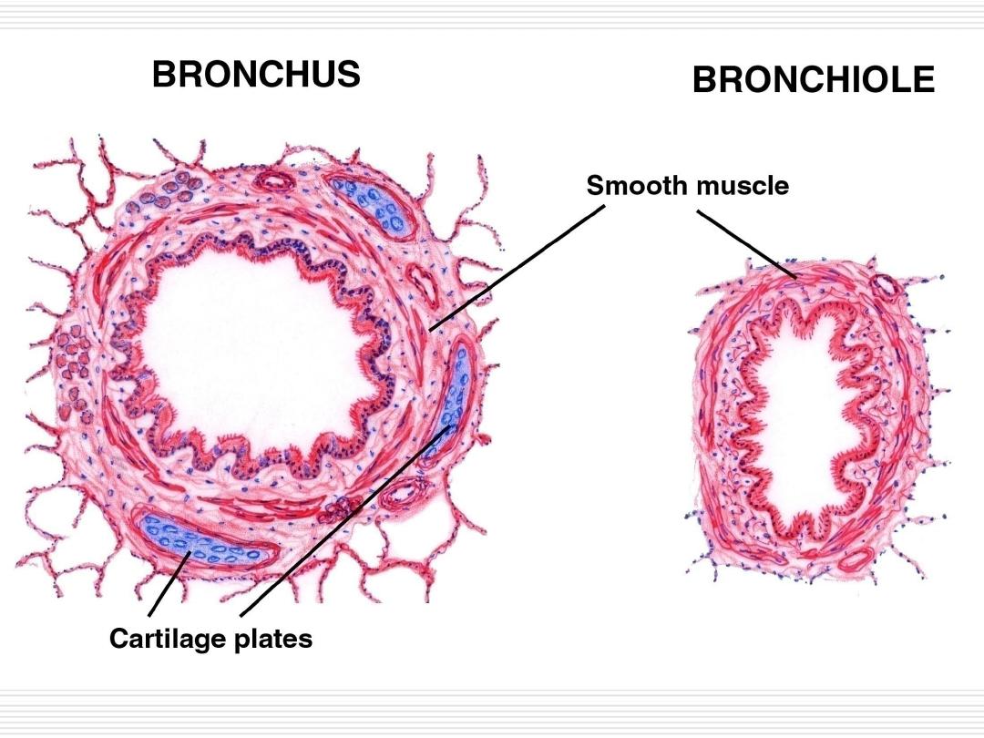
BRONCHI
The
extrapulmonary
primary bronchi have the same
structure of the trachea.
The
intrapulmonary
bronchi have some differences:
The epithelium is pseudostratified columnar ciliated but with
fewer
goblet cells.
A layer of smooth muscle fibers spirally arranged.
The C-shaped cartilage is replaced by
irregular
cartilage
plates that encircle the whole lumen.
These cartilage plates become
discontinuous
and smaller as
bronchi decrease in diameter.
The
seromucous
glands & lymph
follicles
are present in-
between the cartilage plates.
The submucosa layer becomes indistinct.
40
Foreign particles
are more likely to lodge in the
??????????primary bronchus
.
41
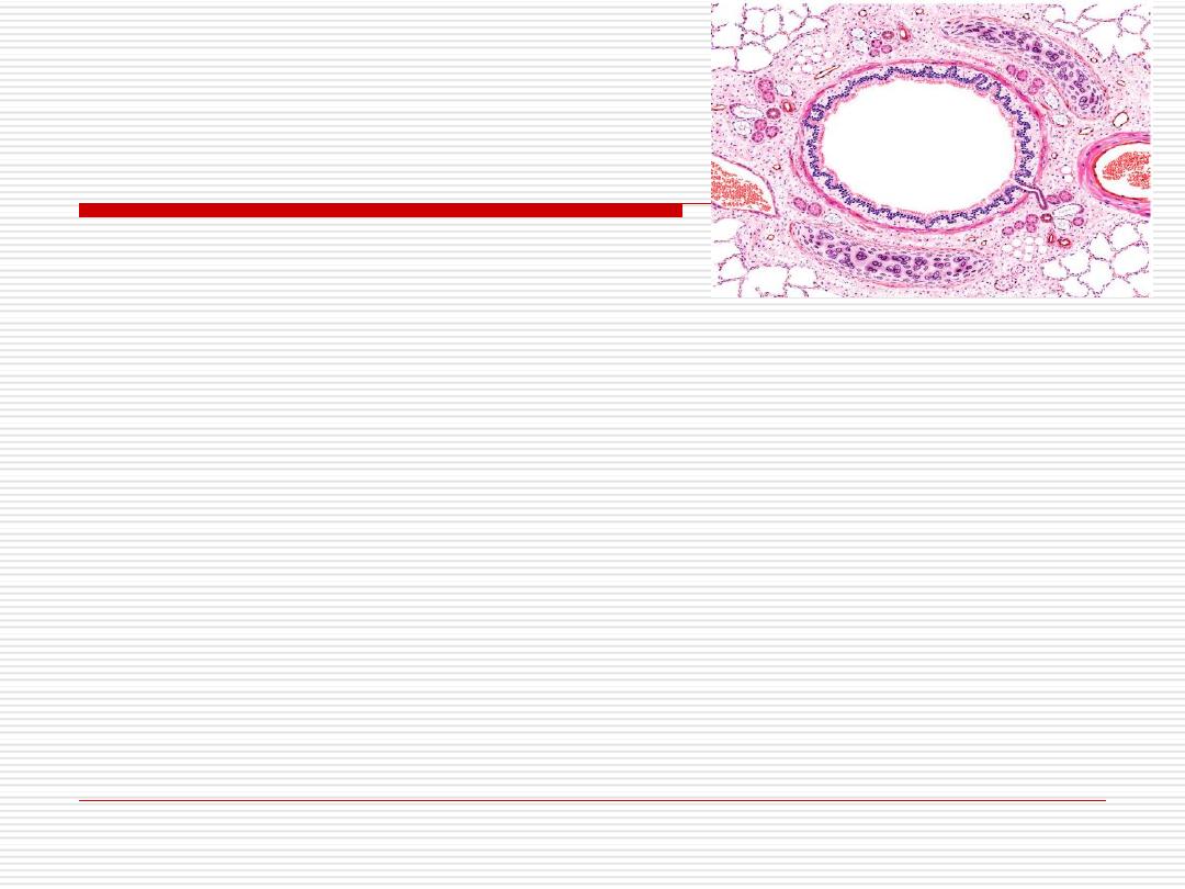
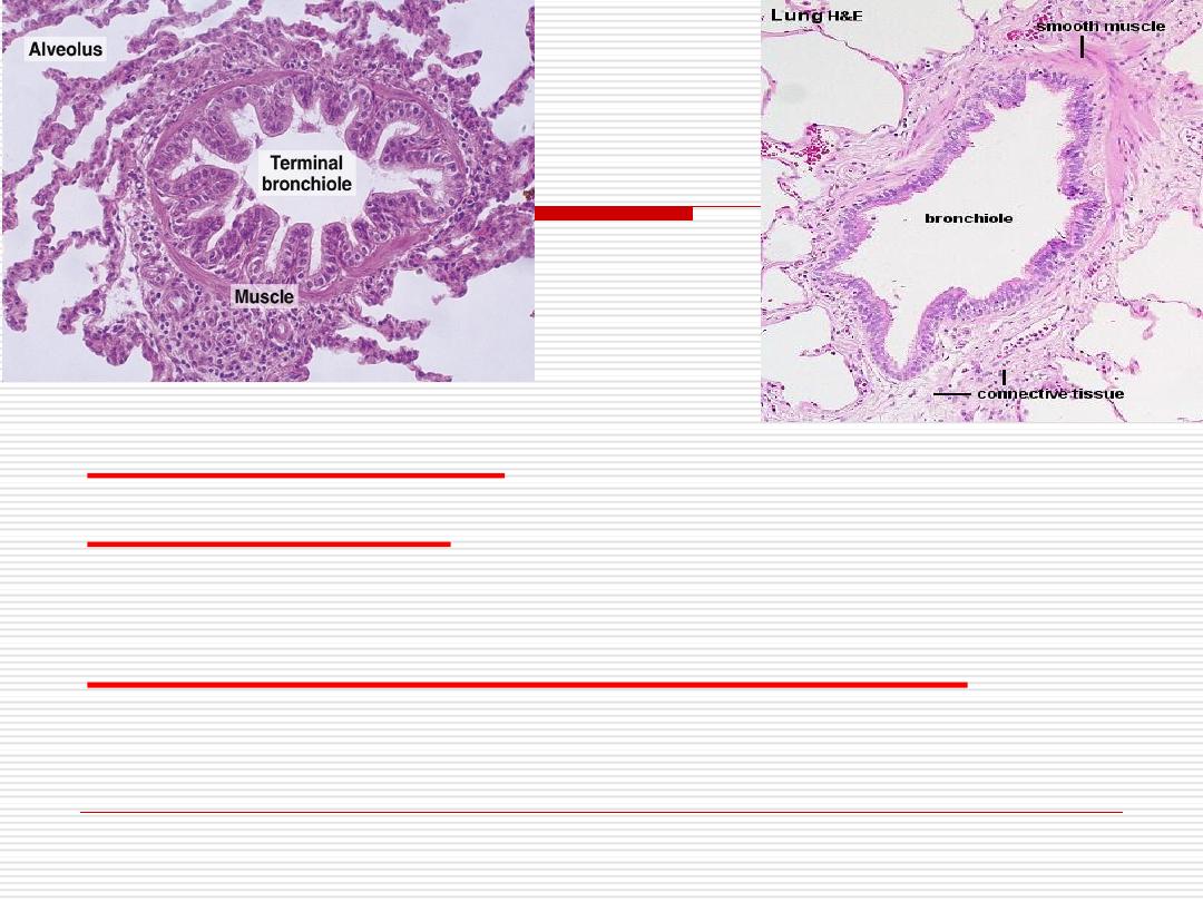
BRONCHIOLES
It is a conducting tube of 5mm diameter or less.
Their wall consists of:
Mucosa
-
Epithelium
is simple columnar ciliated,
decreases
to simple
cuboidal partially ciliated in terminal bronchioles.
-
Goblet
cells gradually
disappear
,
and are replaced in
terminal bronchioles
with
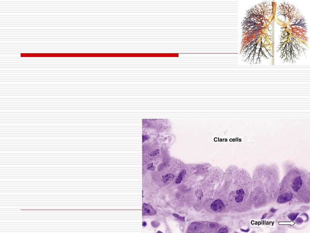
Clara cells.
42
43
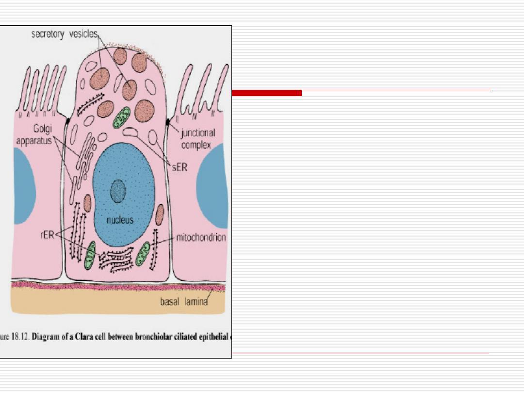
Clara
cells
have no cilia and contain
secretory granules in their apices.
They secrete proteins that
protect bronchiolar epithelium
against oxidative pollutants and
inflammation.
They may produce surfactant-
like material that reduces the
surface tension keeping the
bronchioles open
-
44
Lamina propria:
contains many elastic fibers
.
Muscle layer;
is well developed and spirally
arranged around the lumen
.
Outer connective tissue layer
;
has no
glands, no cartilage and no lymph follicles.
45
Lecture3
Dr. Mareb. H. Ahmad
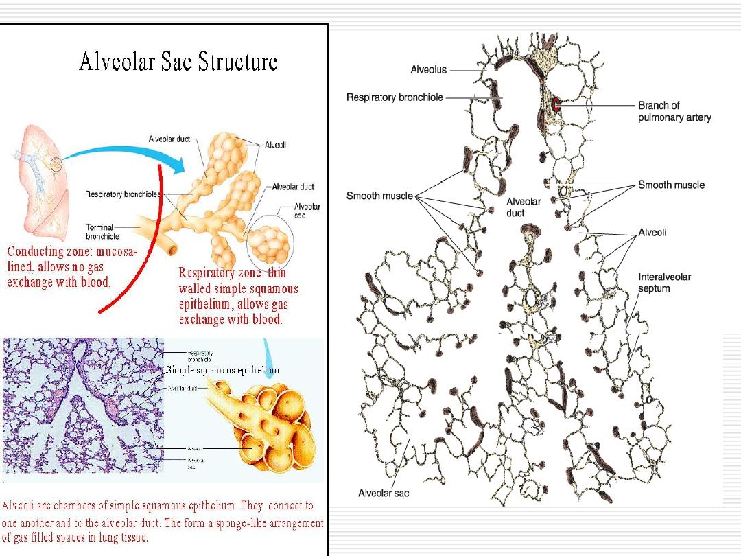
THE RESPIRATORY PORTION
46
THE RESPIRATORY PORTION
Each terminal bronchiole subdivides
into
two
or
more
respiratory
bronchioles, then into alveolar ducts,
alveolar sacs and alveoli.
Gaseous exchange occurs from the
respiratory bronchioles to alveoli.
47

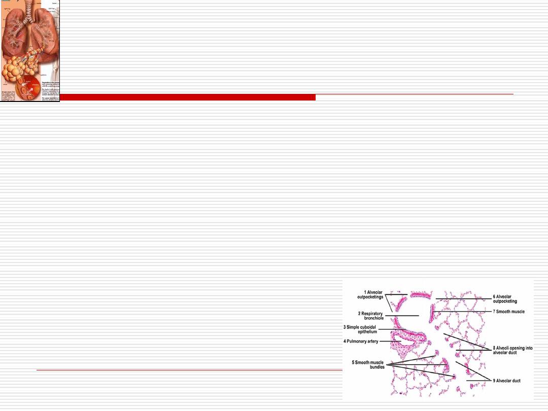
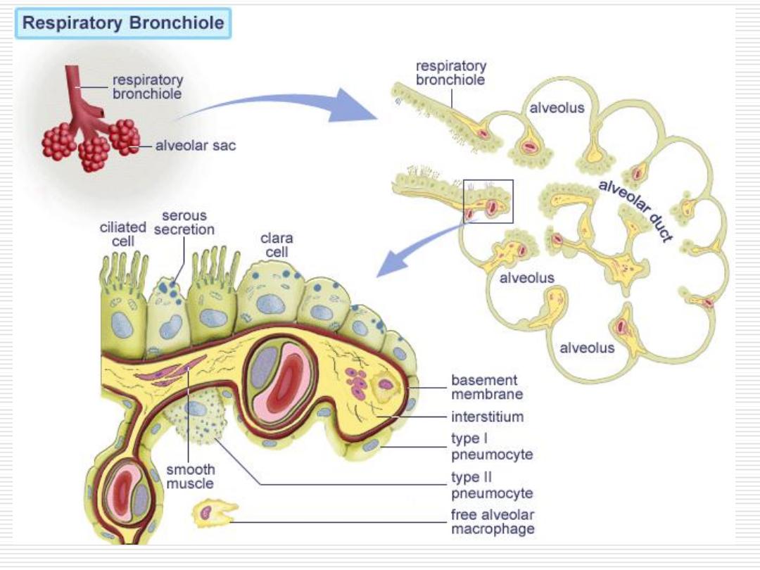
1-Respiratory bronchiole
-They are regions of transition between the conducting and
respiratory portions.
-The respiratory bronchiole is structurally
similar
to terminal
bronchiole
except
that its wall is interrupted by
numerous alveoli.
-The epithelium between the openings of alveoli is formed of
ciliated cuboidal cells and Clara cells
.
-In the distal portions, the epithelium is
simple cubical non-
ciliated
.
-Smooth muscle and elastic connective tissue support the
wall of respiratory bronchiole.
48

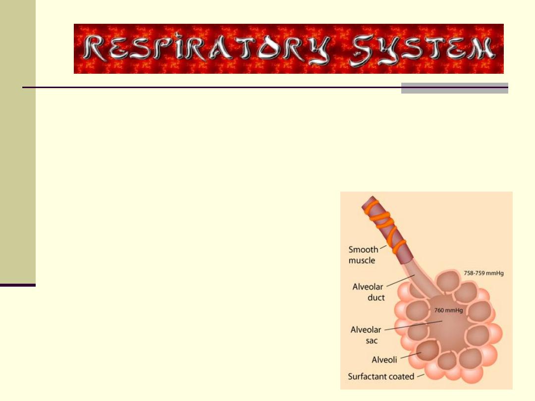
2- Alveolar ducts and
alveolar sacs:
When the wall of the respiratory bronchiole becomes
formed
only
of the opening of the alveoli, the thin walled
tube is termed
alveolar duct
.
Alveolar duct opens into a common chamber (atrium)
which communicates with two or more alveolar sacs.
Alveolar duct, alveolar sacs and alveoli are lined by very
thin squamous alveolar cells.
Their walls are supported by a matrix rich in
elastic
and
collagen
fibers.
49
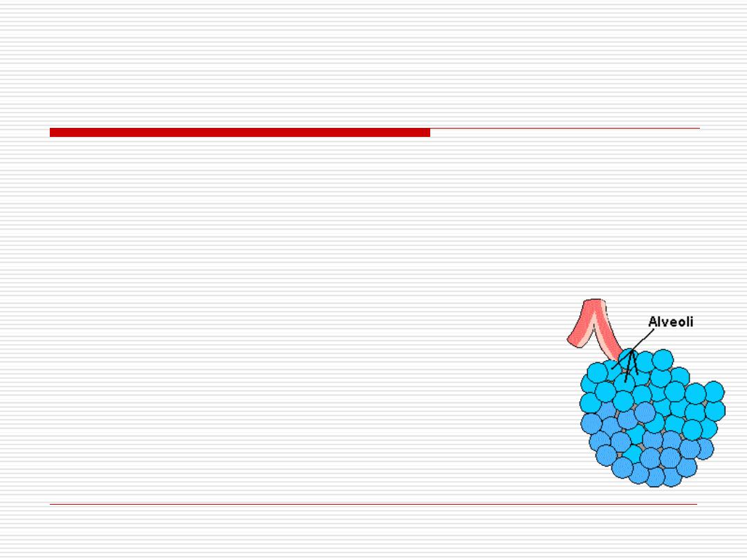
3- Alveoli:
-They are sac like evaginations of respiratory
bronchioles, alveolar ducts & sacs containing air
where gas exchange occurs.
-They are responsible for the spongy structure of
the lung.
-They resemble small pockets open on one side.
50
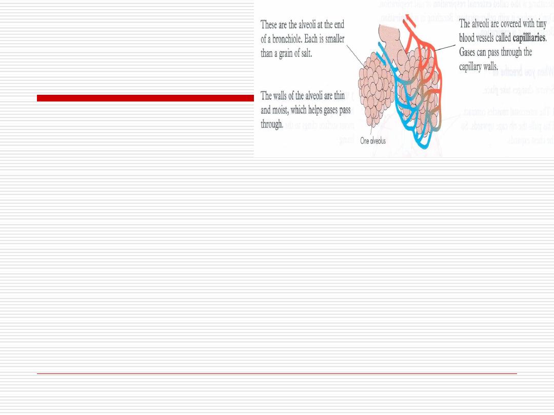
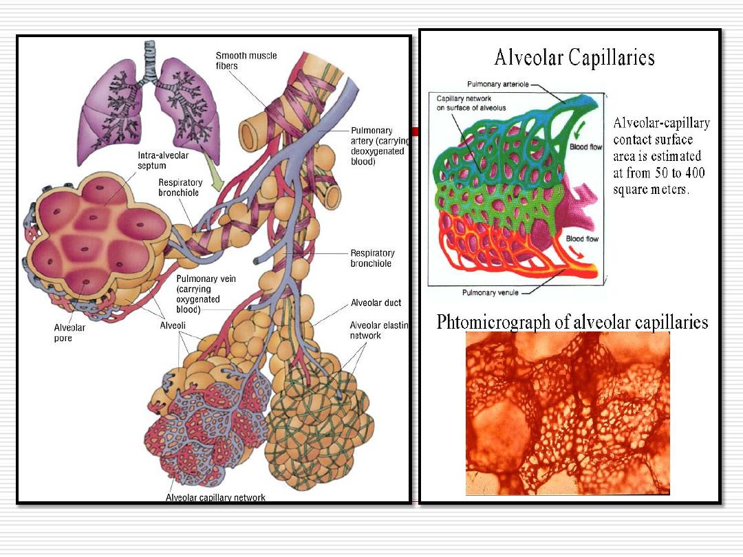
-The structure of the alveolar wall is specialized
for facilitating diffusion between air in the lumen of
the alveoli and blood in the very rich capillary
network in their walls.
~
300
million air sacs (alveoli).
Large surface area available for gas
exchange exceeds (140 m
2
).
51
52
-Generally, each wall lies between neighboring
alveoli, and so it is termed, interalveolar
septum.
-The alveoli are lined by two types of cells called
pneumocytes or alveolar cells.
-The surface of pneumocytes is covered by fluid
called surfactant, secreted by type II
pneumocyte.
53
54

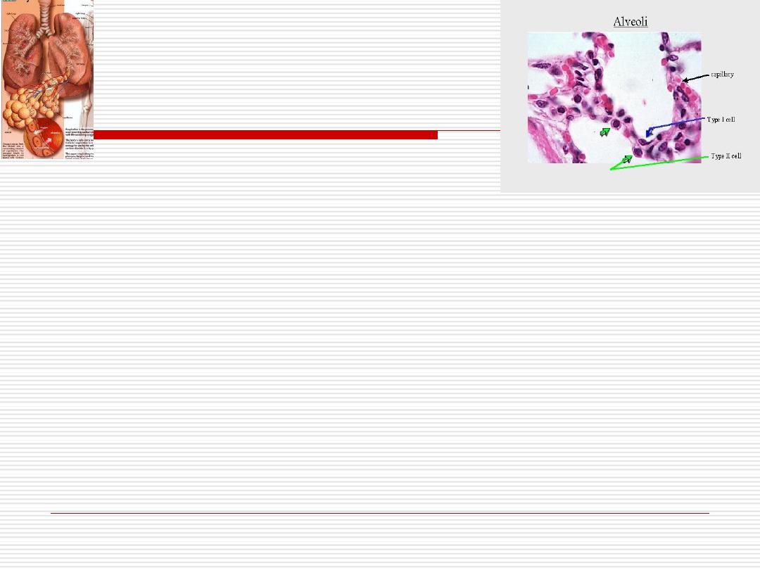
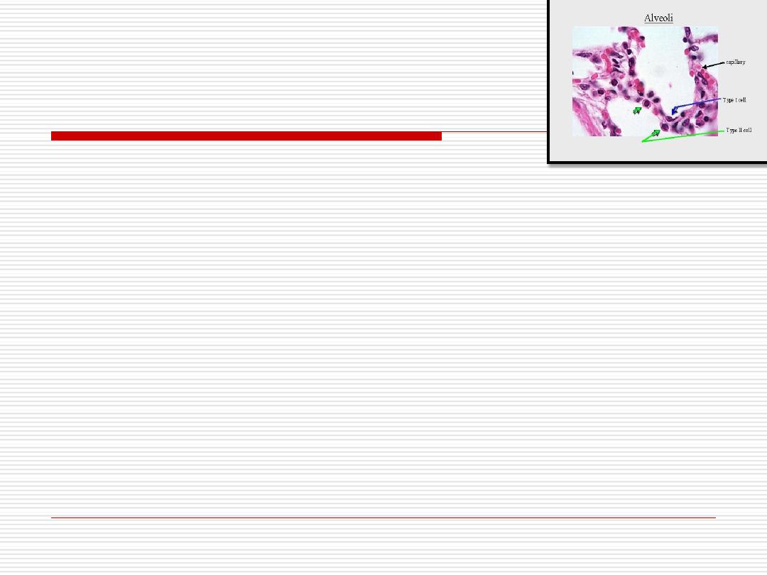
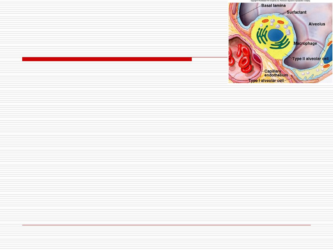
Lining epithelium of respiratory portion
(the alveolar epithelium)
Type I pneumocyte
(simple squamous cell)
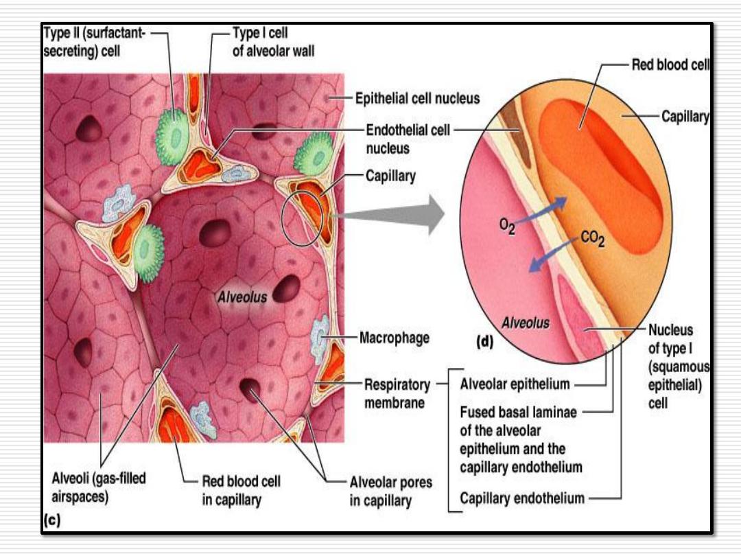
It lines 97% of the alveolar surface.
The cell organelles are grouped around the nucleus.
The cytoplasm in the thin portions is free of organelles, but
contains numerous pinocytotic vesicles.
It is responsible for gas exghange (O2 and CO2) between the
air in the alveoli and blood in the capillaries.
55
Type II pneumocyte
(secretory cell)
-
It is found between type I cells especially at the angles of
alveoli.
-It covers only 3% of alveolar surface.
-It is a cuboidal cell with foamy cytoplasm. It rests on the
basement membrane and has tight and desmosomal
junctions with type I cells.
-It resembles a typical secretory cell. By E/M it shows,
abundant RER, well developed Golgi, mitochondria and
irregular microvilli on their free surface.
56
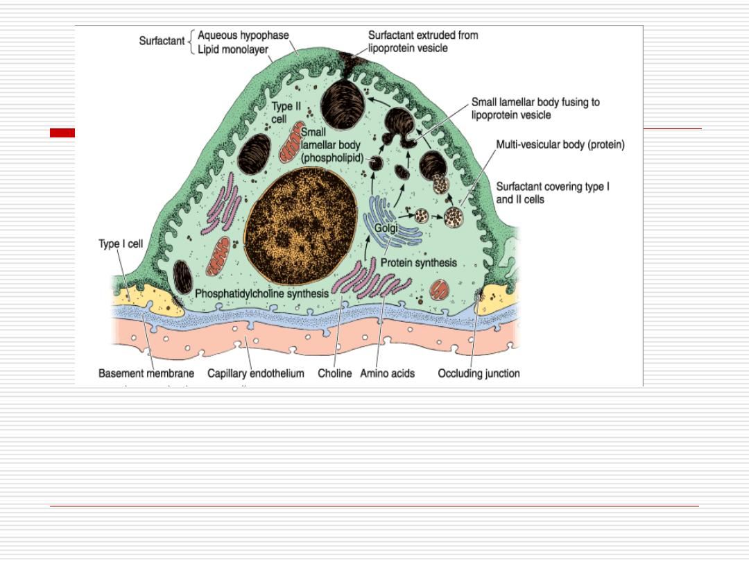
Type II pneumocyte
The cytoplasm contains characteristic vesicles called lamellar
bodies that are responsible for the foamy appearance of the
cytoplasm by L/M.
57
These
lamellar
bodies contain parallel lamellae formed of
phospholipids, glycosaminoglycans and proteins. They give rise
to a material called
surfactant
that spread over the surface of
alveoli.
The surfactant is continuously removed by ??
58

Function of surfactant
Lowers the alveolar surface tension, thus preventing
them from collapse during expiration.
Facilities inflation of alveoli in inspiration.
Surfactant has some bactericidal effect, so it prevents
bacterial invasion and clean the alveolar surface.
Type II alveolar cells act as stem cells, they divide and
form the two types of cells lining the alveoli.
59

Respiratory distress syndrome
RDS was previously called hyaline membrane disease.
In fetal development, surfactant appears in the last weeks of
pregnancy .
In infants born prematurely due to insuffient production of
surfactant there is respiratory distress due to difficulty in
expanding the alveoli.
Surfactant synthesis can be induced by administration of
glucocorticoids
60

Interalveolar septum
-
The septum between adjacent alveolar cavities consists of
two thin squamous epithelial layers
(pneumocyte type I),
between which lie the interstitium.
-The interstitium consists of blood capillaries and matrix of
connective tissue.
61
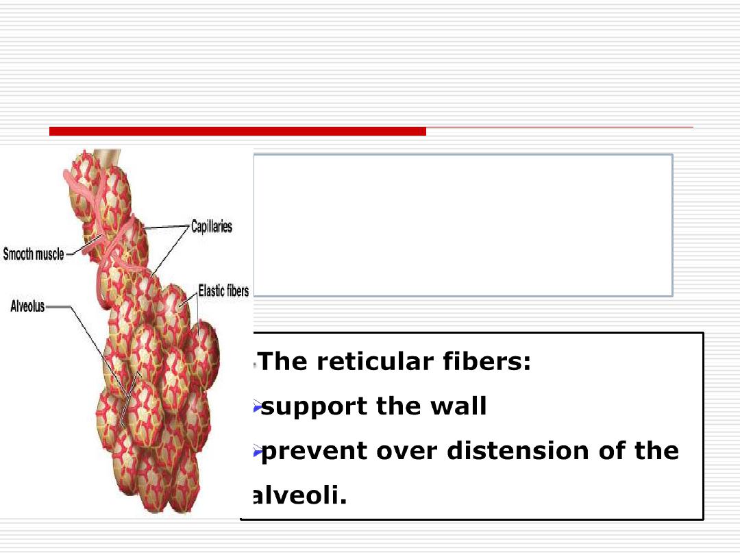
-The
elastic
fibers
permit expansion
recoil of alveolar wall.
-The connective tissue of the interstitium includes:-
=Fibers (elastic and reticular fibers)
62

1- The endothelial cells of the capillaries.
-
They are extremely
thin
.
-
They are continuous and
not
fenestrated.
-
The cell organelles are grouped around the nucleus.
-
The rest of the cell is very thin to increase the efficiency of
gas exchange. It contains numerous
pinocytotic
vesicles
.
-
The endothelium of pulmonary capillaries has some
metabolic functions as break down of serotonin and
conversion
of angiotensin I to angiotensin II.
The cells present in the interstitium
include:
63
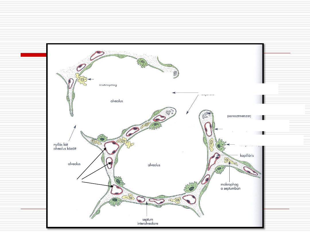
Schematic drawing of the alveolar wall:
Entrance to the alveolus
Elastic fiber in cross-section
Type I. pneumocyte
Type II. pneumocyte
Alveolar macrophage
capillaries
64
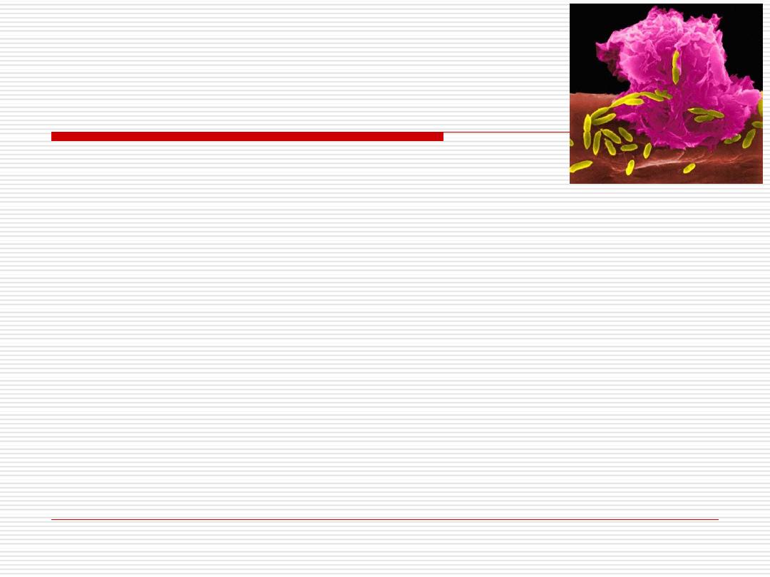
They are derived from
monocytes
.
They are found in the interstitium of the alveolar septum
and on the surface of the alveoli.
Their cytoplasm contains
phagocytosed
material like
carbon or dust.
In
congestive heart failure
, the cells contain iron pigment
from phagocytosed RBCS and are called
heart failure cells
.
2- Alveolar macrophages.
(Dust cells).
65
-They synthesize
- collagen type I
- collagen type III
- elastic
fibers &
glycosaminoglycans
.
3- Fibroblasts
66

-They are bound to the basement membrane of
the alveolar epithelium.
-They contain
actin
&
myosin
filaments.
In addition, the interalveolar septum contains
mast
cells and
leukocytes
.
4- Contractile interstitial cells
67
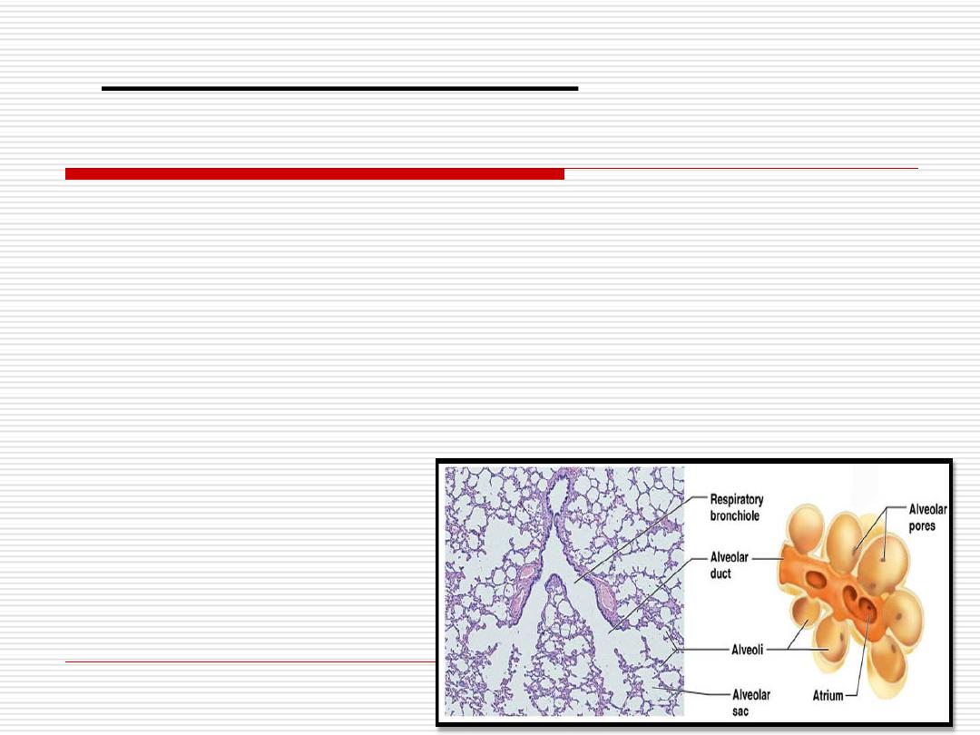
•The interalveolar septum may contain one or more
pores(8-12um in diameter) connecting neighboring
alveoli.
•They may equalize pressure in the alveoli and form
collateral ventilation.
.
Alveolar pores (of Khon):
68

Blood - air barrier
-
Air in the alveoli is separated from blood in capillaries
by:
1- The
surfactant
layer on the alveolar surface
2- The
cytoplasm
of the alveolar cells (type I).
3- The
fused basal laminae
of type I cell and endothelial
cells of the capillary.
4- The
cytoplasm
of the
endothelial
cells.
69
70
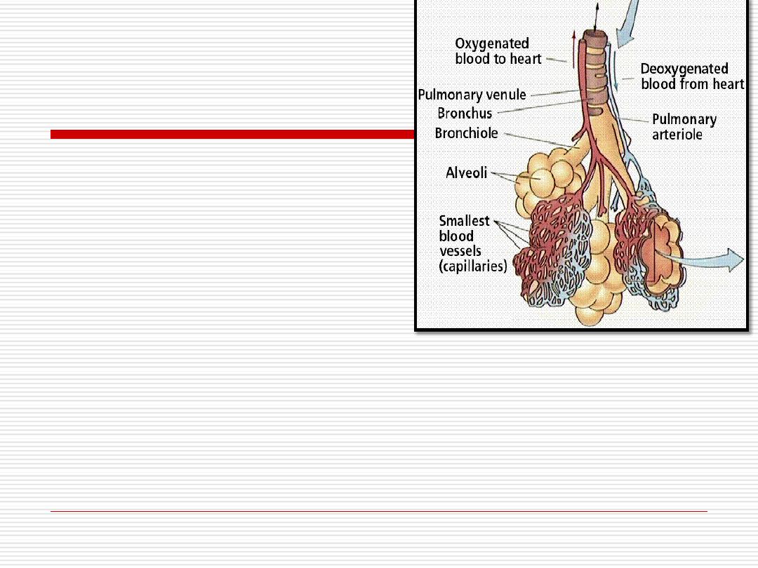
The lung has
two
types
of blood supply:
1-Nutrient (bronchial) vessels
,
Bronchial arteries from aorta
Bronchial veins carry the blood from lung to azygous vein.
2-Functional (pulmonary) vessels
Pulmonary arteries carry unoxygenated blood from right ventricle to the
lung capillaries for oxygenation
Pulmonary circulation
71
72

Pleura
It is the
serous
membrane covering the lung.
It consists of
two
layers;
parietal
and
visceral
which are
continuous in the hilum.
A
cavity
is present between the two layers and contains
fluid that acts as a lubricating agent .
Both membranes are composed of
mesothelial cells
resting on a fine connective tissue layer.
73

Defense mechanism in the
respiratory system:
The respiratory system has a large surface area
that is exposed to both blood and the external
environment so it is very susceptible to invasion
by bacteria and other non-infective agents
particles larger then (10 µm.) in the air are
trapped by hair in the nose
particles of (2-10 µm.) are trapped by the
mucous covering the respiratory epithelium
74

The
sneezing reflex
clears the nasal passages.
Cough reflex
can eliminates particles entering the
respiratory tract.
Smaller particles in the air or even bacteria are
removed by
alveolar macrophages
.
BALT
(Bronchus associated lymphatic tissue):
bronchi contain abundant lymphatic tissue mainly
in the form of lymphatic nodules.
75

If there is the will,
there’s the way.
Have a nice day!