1
Fifth stage
ENT
Lec-16
د.سعد
24/3/2016
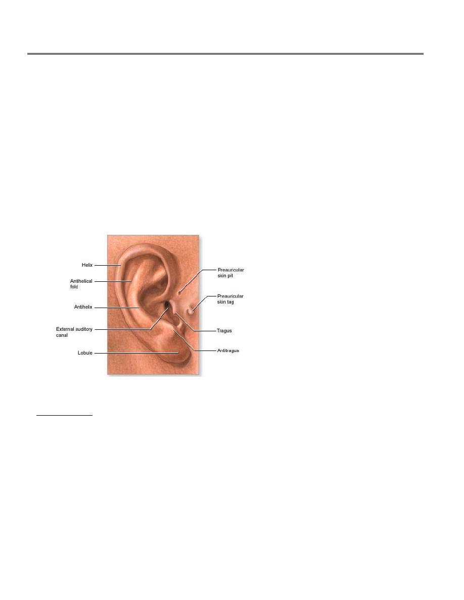
Diseases of the External Ear
Congenital Abnormality
1.
Preauricular sinuses and cysts.
2.
Anotia: Complete absence of the pinna.
3.
Microtia: Small pinna.
4.
Accessory auricles.
5.
Atresia of the external auditory meatus.
6.
Protruding ear (Bat ear).
Acquired Disorders
The Pinna
1.
Injuries to the pinna
A.
Simple laceration or partial or complete avulsion.
B.
Haematoma of the auricle (Haematoma auris):
Trauma to the pinna may cause swelling results from haematoma (extravasation of blood
between the cartilage and overlying perichondrium). The pinna appears swollen and blue. The
ear may be slightly tender with a feeling of heat and discomfort. If untreated, the pinna may
become distorted and thickened as the haematoma resolves. A `cauliflower ear` – so often seen
in boxers- may result.

2
Treatment; consist of drainage of the swelling followed by a firm pressure dressing in attempt
to discourage more blood from collecting.
2.
Infections of the pinna
A. Perichondritis
Is inflammation of the perichondrium which covers the auricular cartilage.
Causes;
Infection of haematoma or other injury.
Complication of severe otitis externa.
Complication of mastoid surgery.
Clinical Picture:
The pinna is uniformly enlarged and thickened and its surface is red and shiny.
Severe pain and tenderness.
Treatment:
Broad spectrum antipseudomonal antibiotic.
Application of soothing dressing such as glycerin and ichthamol.
A subperichondrial abscess may present and should be incised and drained if fluctuation
is definitely present (premature incision may result in further spread of infection and
more extensive cartilage necrosis).
B. Skin infections
Impetigo: (infection of superficial layers of skin by staphylococci) most commonly occurs
in young children secondary to an episode of otorrhea.
Treatment; topical antibiotic and control of otorrhea.
Erysipelas: streptococcal infection that produce raised red oedematus eruptions with a
characteristically sharply defined edge usually associated with severe malaise a high
temp.
Treatment; Systemic antibiotic (penicillin).
Dermatitis may complicate severe otitis externa.
Furunculosis (a staphylococcal infection of the hair follicle).

3
3.
Tumours of the pinna
Squamous cell and basal cell carcinomas occur on the pinna often on the edge. Sun
exposure with hyperkeratosis is a predisposing factor.
The external auditory meatus
1. Impacted Wax (Cerumen)
Wax is a mixture of secretions of ceruminous and sebaceous glands with desquamated skin
cells. The glands are situated in the cartilaginous portion of the EAM. Normally, it is expelled
outside the canal by movement of chewing and by underlying epithelial migration.
Function: Protects the skin by:
-
Acidic reaction
-
Lyzozyme activity
-
It is nature provision for removal of dust and foreign bodies from EAM.
Plug formation is encouraged by excessive wax formation and its retention by stiff hairs. Also
attempts of the patient to clean his ear will push the wax medially
Clinical Picture:
1. Deafness and discomfort in the ear; the onset deafness is often sudden following washing
or bathing which cause the wax to swell.
2. Pain and irritation especially if the wax presses on the drum.
3. Tinnitus and disturbance of balance; also caused by pressure of the wax on the drum.
4. Reflex cough due to stimulation of the auricular branch of the vagus.
Examination:
Otoscopic examination shows brown, yellowish or black plug obscuring the tympanic
membrane.
Treatment:
1. Syringing with water at body temperature. If too hot or too cold, a caloric response may
be induced in the labyrinth causing vertigo.
4
It is contraindicated in cases of perforated tympanic membrane or laceration of external ear
canal.
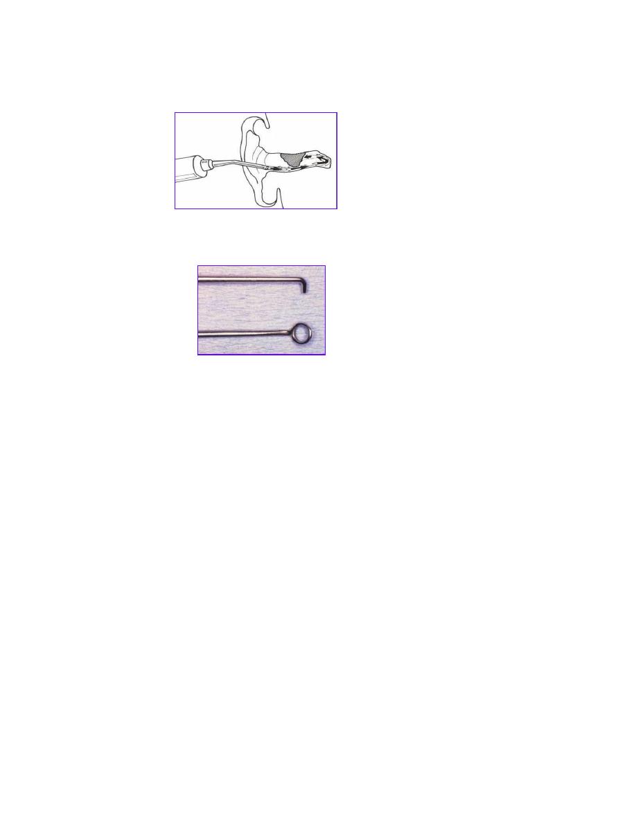
2. Removal with a hook ring probe or forceps is often better with old, dry and hard plugs.
3. Suction through the operating microscope when a perforation in the tympanic membrane
is present or in previous ear surgery.
If the wax is hard then preliminary softening by the use of repeated instillation of drops of
5% sodium bicarbonate in 30% glycerin, olive oil or diluted hydrogen peroxide.
2. Keratosis Obturans
In this condition the meatus on one or both sides become blocked in its deep portion by a
mass consisting of wax, desquamated epithelium and cholesterol. This mass causes excessive
erosion and expansion of the bony meatus and in this, it resembles a cholesteatoma of the
middle ear. Keratosis obturans frequently associated with bronchiectasis and sinusitis in young
patients.
Aetiology; is unknown but hyperaemia of the meatal skin and instability of the epidermis are
possible factors.

5
Clinical Picture:
1. Pain and deafness.
2. Tinnitus and discharge.
3. In severe cases the facial nerve may be exposed leading to facial palsy.
Examination:
Otoscopy reveals pearly-white glistening mass occluding the bony meatus.
Treatment:
Removal of the keratotic mass under general anaesthesia. Regular observation is advised as the
keratosis may reform.
3. Foreign Body
Type of patient:
Commonly children
Mentally retarded adult
Type of FB:
1) Animate FB: Flies, larvae, fleas, mosquito...
2) Inanimate FB:
a) Non- vegetable: bead, button, disc battery
b) Vegetable: bean and pea
Symptoms
- History
- Hearing loss if the FB obstructs the canal
- Severe irritation and noise in the ear with animate FB
Signs:
The FB can be seen by otoscopy

6
Treatment:
1. Animate FB: Kill by alcohol or oil remove by ear wash or instruments.
2. Inanimate FB:
-Non vegetable: remove by ear wash or instruments BUT PLEASE DO NOT WASH IF THE
FB IS DISC BATTERY
- Vegetable: Remove by instruments and avoid ear wash because it may swell by water
and become more impacted
General anesthesia may be needed in impacted FB and uncooperative children.
Complications: Injury of the external canal or drum by the FB or during removal
3. Inflammatory conditions of the external auditory canal (Otitis Externa)
Bacterial:
1- Diffuse OE {* OE = otitis externa}
2- Localized OE: frunculosis
3- Malignant OE (Necrotizing OE)
Viral ( bullous- Herpes)
Fungal: (Otomycosis)
Non infective (Allergic OE, Seborrhoic OE)
Bacterial otitis externa:
1- Diffuse OE
Definition: Diffuse inflammation of the skin lining of the external auditory canal
So don’t wash if:
The FB is vegetable
The FB is disc battery

7
Predisposing factors:
1. Skin laceration:
Self inflicted.
Iatrogenic: Ear wash or instruments.
2. Skin maceration:
Hot humid atmosphere.
Swimmer ear.
Discharge of chronic suppurative otitis media.
Symptoms:
1) Earache:
- Severe because the skin is tightly adherent to the underlying perichondrium &
periostium.
- Increase on moving the jaw because the external canal lies immediately behind
temporomandibular joint.
2) Deafness when edema is severe obstructs the canal.
Signs:
External exam:
o Tenderness on moving the auricle or pressure on the tragus.
o Tender pre and post auricular LN
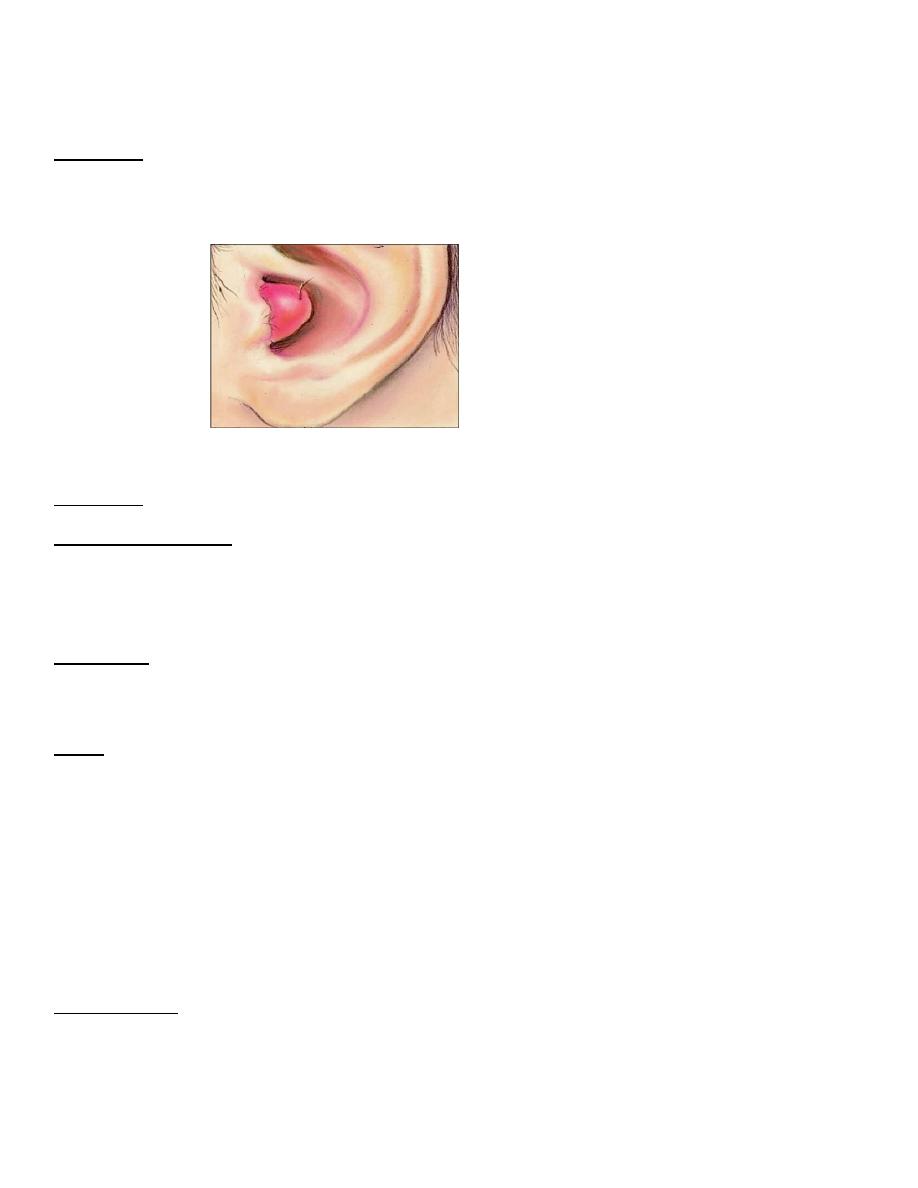
Otoscopic exam :
o Redness, edema, tenderness of the skin of the external canal
o Scanty discharge, serous or purulent
Treatment:
• Meticulous cleaning followed by antibiotic steroid drops.
• If the canal is very swollen, a strip of ribbon gauze (wick) impregnated with 8% aluminium
acetate or ichthamol and glycerin, and these may need to be changed daily.
8
2- Frunculosis
Definition: Localized suppurative inflammation of a hair follicle in the skin of the outer
cartilaginous part of external auditory canal.
Organism: Staph aureus
Precipitating factors:
- Scratching of ear canal
- DM
Symptoms:
1. Earache: Severe and. increase on moving the jaw.
2. Deafness when edema is severe obstruct the canal
Signs:
External:
o Tenderness on moving the auricle or pressure on the tragus.
o Tender pre and post auricular LN.
Otoscopic.
o It is difficult to examine the external canal by otoscope because there is localized area
of tenderness in the skin of the outer part of the external canal.
o No or scanty purulent otorrhea (never mucoid as there is no mucous glands)
Investigations:
Blood glucose level especially in recurrent and bilateral cases.

9
Differential diagnosis: acute mastoiditis
Treatment:
Antibiotics ( flucloxacillin for 5 days ).
Analgesics and heat application to the ear by a covered hot water bottle.
Aural toilet: removal of ear discharge when the furuncle has burst.
Incision of a boil should be delayed until it is clearly pointing to the skin.
Aural pack: by gauze strip soaked in ichthamol and glycerin and changed daily.
3- Malignant otitis externa (Necrotizing otitis externa)
Definition: invasive potentially fatal infection of the external canal which extends to the base of
the skull.
Necrotizing otitis externa should be suspected when patients with diabetes
mellitus (or another condition that compromises the immune system) complain
of persistent otitis externa that causes severe pain, especially at night
Incidence: elderly uncontrolled diabetic patient.
Organism: pseudomonas aeuruginosa.
Symptoms: Ear discharge and severe earache which does not respond to analgesics.
Signs:
External examination:
o Tenderness on pulling the auricle or pressure on the tragus.
o Tender pre and post auricular lymph node.
Otoscopic examination:
o Granulations at the floor of the external canal at the attachment of bony and
cartilaginous part.(This is very important sign)
o Scanty, sanguineous and purulent otorrhea.
Investigations:
-
Blood glucose level

10
-
CT scan of the temporal bone& skull base
-
Radio-isotope scan ( Gallium &Technetium) to assess severity & prognosis
-
Biopsy
-
Culture &sensitivity
Complications:
a) Osteomyelitis of the temporal bone &skull base
b) Facial nerve paralysis at the stylomastoid foramen
c) Last 4 cranial nerves paralysis at the jugular foramen
Treatment:
1) Medical:
a) Control of diabetes
b) Systemic antibiotics; Gentamycin , Quinolones , 3
rd
generation cephalosporins
c) Local antibiotic ear drops
d) Analgesics
e) Aural toilet
2) Surgical: Removal of granulations and debridement of necrotic tissue.
Viral otitis externa:
Herpes Zoster Oticus
Etiology: Herpes zoster virus
Clinically:
-
Pain in and around the ear
-
Vesicles on the auricle and external canal
-
Ramsay-Hunt syndrome:
Vesicles+ facial nerve palsy+SNHL& Vertigo
Treatment:
1. Antiviral drugs
2. Corticosteroid if there is affection of VII & VIII nerve.

11
Fungal otitis externa: or Otomycosis:
Fungal infection of the skin of the exernal canal.
Organism:
Aspirigillus Niger.
Candida albicans.
Symptoms:
Itching is usually the only symptom.
Pain if there is secondary infection.
Deafness if the external canal is obstructed.
Signs:
-
The external canal contains whitish mass with black spots like wet newspaper.
Treatment:
-
Aural toilet: removal of the fungal mass by suction.
-
Antifungal: nystatin, or salicylic acid (2%) as a keratolytic in alcohol as fungicidal. Regular
attendance for treatment lasting 3 or 4 weeks is necessary for elimination of the infection.
5- Neoplasm of External Auditory Canal
Benign;
Exostosis
Definition: is a bony outgrowth from the wall of the EAM. It may be composed of cancellous or
compact bone. They are usually multiple and bilateral affecting chiefly the anterior and posterior
wall.
Incidence: The commonest benign tumor of the external canal and is more common in
swimmers.
Symptoms:
Usually asymptomatic.
Hearing loss if the external canal is obstructed by large exostosis or wax.
Signs: Bilateral smooth bony swelling
Treatment: If obstructing the canal excision

12
Malignant;
Squamous cell carcinoma
Incidence; Rare & more common in elderly males.
Clinical Picture:
1. Otological:
-
Deep seated earache
-
Bloody stained otorrhea
-
Fleshy friable mass in the external canal
-
Progressive hearing loss, initially CHL then SNHL
2. Neurological: paralysis of VII& last 4 cranial nerves.
3. Cervical: Enlarged preauricular, postauricular and upper deep cervical LN
Investigations:
-
CT scan and MRI to assess tumor extension and lymph nodes involvement
-
Biopsy
-
Metastatic work up; Chest X ray, abdominal ultrasound, bone scan, CT scan of brain.
Treatment:
Surgical resection of the temporal bone + postoperative radiotherapy.