Phenylketonuria (PKU)
Classic inborn error of metabolism
PKU is an autosomal recessive metabolic genetic disorder characterized by a mutation in the gene for the hepatic enzyme phenylalanine hydroxylase (PAH), rendering it nonfunctional.
This enzyme is necessary to metabolize the amino acid phenylalanine (Phe) to the amino acid tyrosine.
Patients who are diagnosed early and maintain a strict diet can have a normal life span with normal mental development.
• When PAH activity is reduced, phenylalanine accumulates and is converted into phnylpyruvate and phenylacete acid (also known as phynyletone), which can be detected in the urine which give a strong musty or mousy odor of the urine of affected infants
• Untreated PKU can lead to intellectual disability (Mental Retardation) decrease skin pigmentation with eczema and can’t talk or walk.
• The mainstream treatment for classic PKU Patients is a strict PHE-restricted diet supplemented by a medical formal containing amino acids and other nutrients even for maternal PKU.


Cystic Fibrosis
also known as mucoviscidosis the incidence is 1/4000 live birthsThis is a recessive genetic disorder that affects most critically the lungs, and also the pancreas, liver, intestine and reproductive system.
It is characterized by abnormal transport of chloride and sodium across an epithelium leading to thick, viscous secretions.
• CF is caused by a mutation in the gene for protein cystic fibrosis trans membrane conductance regulator (CFTR), this protein is required to regulate the component of sweat, digestive fluid and mucus.
• This disorder result in abnormally viscid mucus secretion which obstruct organ passages causing pulmonary infection, pancreatic insufficiency, hepatic cirrhosis intestinal obstruction and male infertility, recently CF increase the risk of GIT cancer.
• In most cases, the Diagnosis of CF in based on persistently elevated of sweat electrolyte concentration.
Sudden infant death syndrome (SIDS)
Tumor & tumor-like lesions of infancy & childhoodBenign
Malignant
Sudden infant death syndrome (SIDS)
The usual history is a healthy infant suddenly turn blue, stops breathing, & become limp without emitting a cry or struggling.It is generally accepted that it is a multifactorial condition including mother, infant or environment causes
This is a sudden death of an infant under 1 year of age which remains unexplained after a thorough case investigation, including performance of a complete autopsy, examination of the death scene & review of the clinical history.
Infant usually dies while asleep, hence the pseudonyms of crib death or cot death
It is the most common cause of mortality in postnatal infants in the United States, while only 1 to 5 death per 1000 live births around the world
Approximately 90% of all SIDS occur during the first 6 months of life, most between the age of 2 & 4 months.
Most of these infants die at home, usually during night after a period of sleep
Maternal causes
Young less than 20 years of ageShort inter-gestational intervals
Low socioeconomic group
Smoking
Drug abuse
Infant
PrematurityLow birth weight
Male sex
Product of a multiple birth
SIDS in a prior sibling
Inborn error of metabolism
Apnea
Environmental
Prone sleep
Thermal stress
Tumor & tumor-like lesions of infancy & childhood
Benign tumor are even more common than malignant tumors.Most common benign tumor is hemangioma
Only 2% of all malignant tumors occur in infancy & childhoodCancer (including leukemia) is the leading cause of death from the disease in United State in children between 4 & up to 14 years
Heterotopia
Is applied to microscopically normal cells or tissues that are present in abnormal locationsExample a rest of pancreatic tissue found in the wall of stomach or small intestine
It is of little significant, but they can be confused clinically with neoplasmsHamartoma
refers to an excessive focal overgrowth of cells and tissues native to the organ in which it occurs.
Hamartomas can be thought of as the linkage between malformations and neoplasms
The frequency of these lesions in infancy and childhood and their clinical behavior give credence to the belief that many are developmental aberrationsBenign Tumors and Tumor-Like Lesions
Hemangiomas:are the most common tumors of infancy.
Architecturally, they do not differ from those encountered in adults.
In children, most are located in the skin, particularly on the face and scalp, where they produce flat-to-elevated, irregular, red-blue masses;
some of the flat, larger lesions are referred to as port-wine stains.
Types: Capillary hemangioma, cavernous hemangioma, arteriovenous hemangioma, port-wine hemangioma

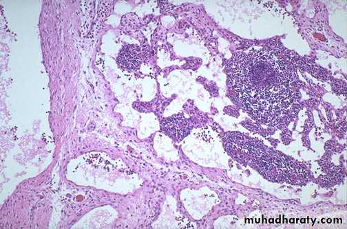
Lymphangioma:
Abnormal dilated lymphatic channels
In skin particularly neck, axilla, mediastinum and retroperitoneumAppears as spongy, doughy, skinned color mass
Types: Cavernous lymphangioma, cystic hygromaMajority are congenital, very rarely acquired


Example :
• Kidney – Wilms tumor• Neural crest – neuroblastoma
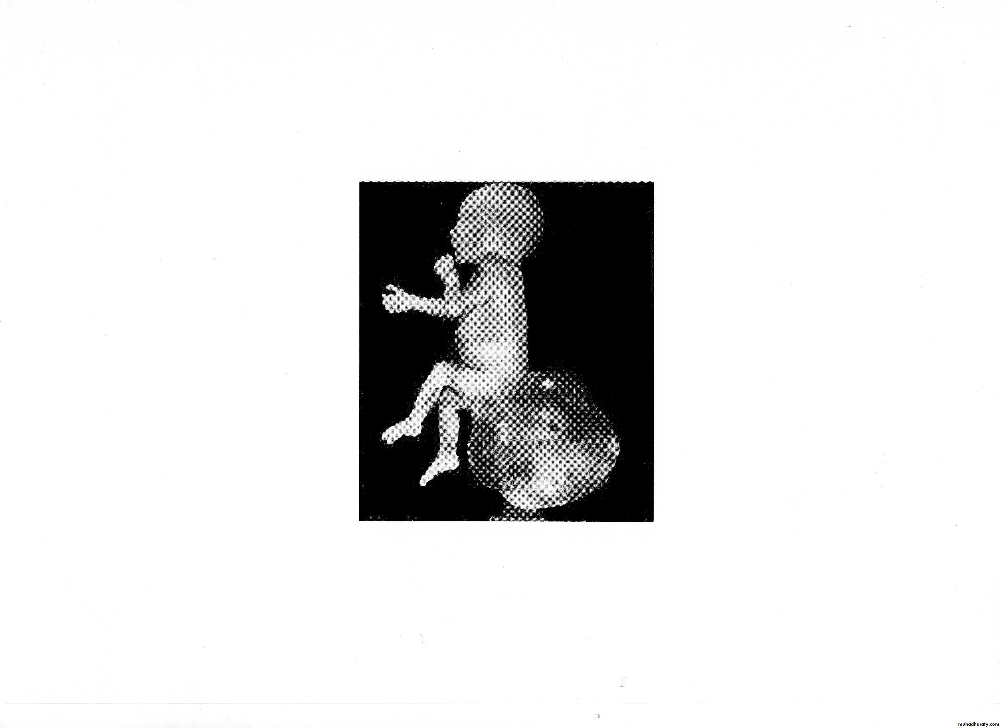
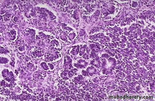
• Eye – retionoblastoma
• Bones – osteosarcoma + Ewing / PNETs
Small, round, blue cell tumours


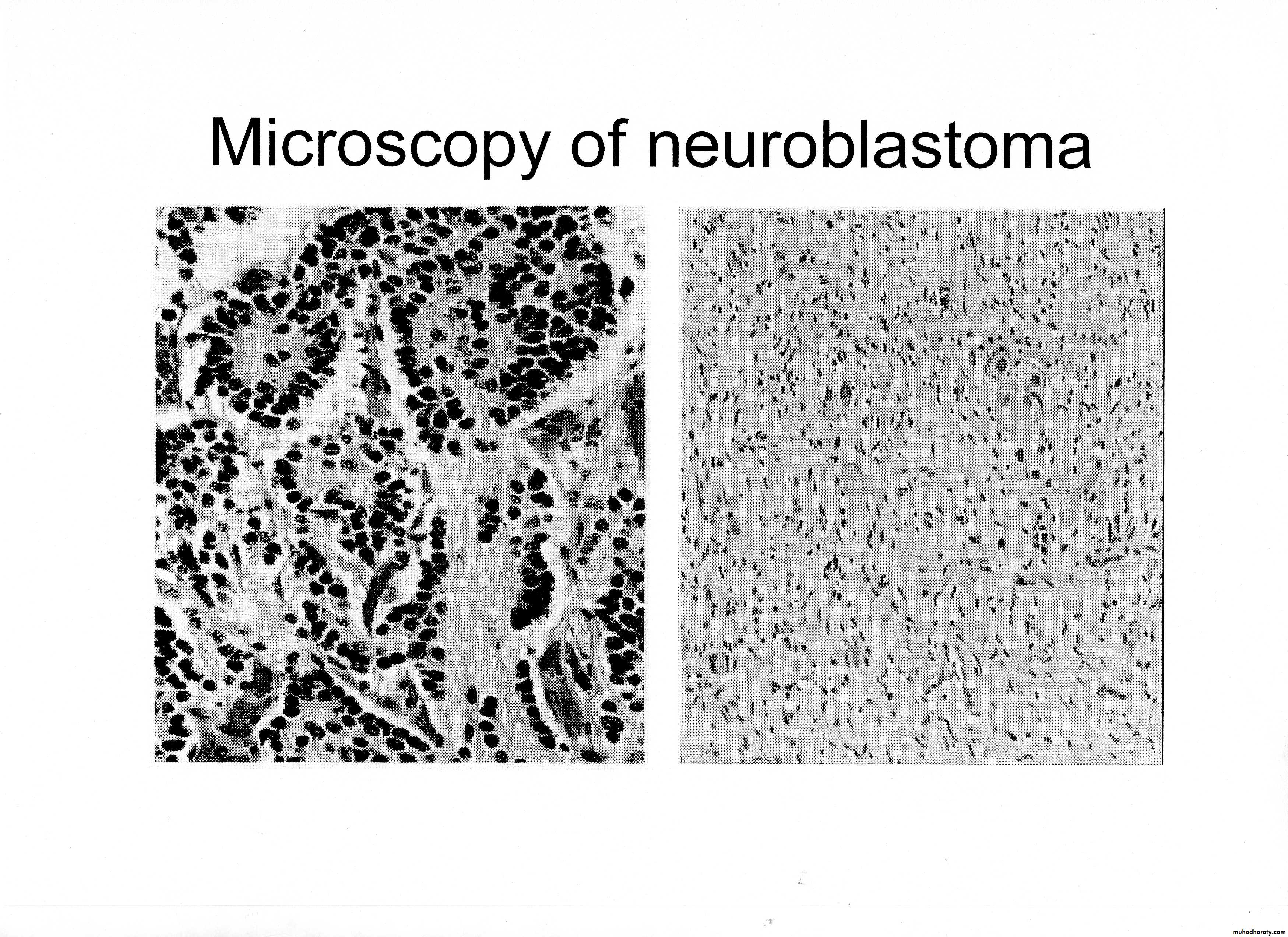
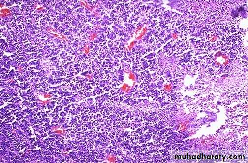
Neuroblastoma