
1
Fifth stage
Pediatric
Lec-4
.د
رياض
4/4/2016
Infectious Diseases-2
Mumps,Measles,Rubella
Differential diagnosis of Fever and rash syndrome:
1-Maculopapuler;measles,roseola,fifth disease, EB virus, scarlet fever, ricketsiae.
2-Diffuse erythema; scarlet fever, toxic shock syndrome(staph. aureus).
3-Urticarial; mycoplasma, EB virus
4- vesiculobullous; Herpes simplex, varicella, staphyllococcal bullous impetigo.
5.petechial; meningococcemia
Mumps
Mumps is an acute self-limited infection, now unusual in developed countries because of
widespread use of vaccination. It is characterized by fever, bilateral or unilateral parotid
swelling and tenderness, and the frequent occurrence of meningoencephalitis and orchitis.
Although no longer common in countries with extensive vaccination programs, mumps
remains endemic
Transmission
Mumps is spread from person to person by respiratory droplets. Virus appears in the saliva
from up to 7 days before to as long as 7 days after onset of parotid swelling. The period of
maximum infectivity is 1-2 days before to 5 days after onset of parotid swelling
CLINICAL FEATURES
The incubation period is 16-18 days resulting in clinical presentation ranging from
asymptomatic to the typical illness associated with parotitis. The typical patient presents
with a prodrom 1-2 days and consisting of fever, headache, and vomiting. Parotitis then
appears and may be unilateral ,then becomes bilateral in about 70% of cases .The parotid
gland is tender, and may be accompanied by ear pain on the ipsilateral side. Sour foods or
liquids may enhance pain in the parotid. As swelling progresses, the angle of the jaw filled
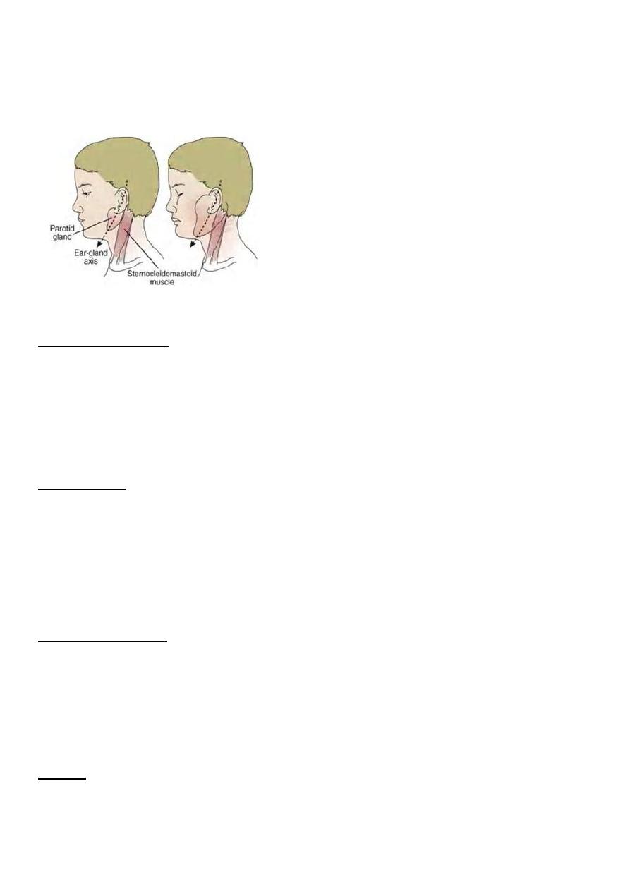
2
and earlobule is pushed outward. The opening of Stensen duct may be red and edematous.
The parotid swelling peaks in approximately 3 days, then gradually subsides over 7 days.
Submandibular salivary glands may also be involved or may be enlarged without parotid
swelling. Edema over the sternum due to lymphatic obstruction may also occur
Differential diagnosis
Purulent parotitis, is usually caused by Staphylococcus aureus, unilateral, extremely tender,
and is associated with an elevated white blood cell count, and may involve purulent
drainage from Stensen duct. Submandibular or anterior cervical adenitis due to a variety of
pathogens may also be confused with parotitis.
Complications
The most common complications of mumps are meningitis, with or without encephalitis,
and orchitis. Uncommon complications include deafness, facial palsy, pancreatitis, and
thrombocytopenia.
Maternal infection with mumps during the 1st trimester of pregnancy results in increased
fetal loss. No fetal malformations have been associated with intrauterine mumps infection.
Meningoencephalitis
Symptomatic m.e. occurs in only10-30% of mumps cases, but CSF pleocytosis has been
found in 40-60% of patients as subclinical more than apparent meningitis. The
meningoencephalitis is usually benign, may occur before, along with, or following the
parotitis
Orchitis
Involvement in young boys is rare, but in adolescent and after puberty, orchitis occurs in
30-40% of male cases. Atrophy of the testes may occur, but sterility is rare even with
bilateral involvement.

3
Prevention
Antibody develops in 95% of children after 1 vaccine dose.. As a live- vaccine, MMR should
not be administered to pregnant women or to immunodeficient child.
Measles
Measles is highly contagious disease. Owing to widespread vaccination, transmission is
limited .
RNA virus in the family Paramyxoviridae and genus Morbillivirus
Transmission
The portal of entry of measles virus is through the respiratory tract or conjunctivae
following contact with aerosol droplets in which the virus is suspended. Patients are
infectious from 3 days before -to 4-6 days after the onset of rash
Clinical manifestations
Measles is a serious infection characterized by high fever, and maculopapular rash.`
The incubation period is 8-12 days.
The prodromal phase (2-4) days begins as conjunctivitis with photophobia, coryza, cough,
and increasing fever
Koplik spots represent the enanthem and are the pathognomonic sign of measles,
appearing 12-24 hours before the onset of the rash and it last for 1-2 days after rash
appearance. They first appear as sandy white spots on minute red lesions in the inner
aspects of the cheeks at the level of the lower premolars. Koplik spots present in 50-70%.
The rash begins on the forehead (along the hairline), and behind the ears as a red
maculopapular eruption. It then spreads to the face and neck and downward to the trunk
and limbs, and reaching the palms and soles.The rash last 5-6 days, then fades over about 7
days in the same manner as it evolved, often leaving a fine desquamation of skin. Of the
major symptoms of measles, the cough lasts the longest, often up to 10 days. generalized
lymphadenopathy may be present, with cervical and occipital lymph nodes enlargement.
Diagnosis
Mainly clinical but confirming serological test can be done by high antibody IgM level .

4
Blood and urine samples for viral culture is the most commonly WHO protocol for
detection of virus of the illness.
Differential diagnosis ; include other fever and rash illnesses like rubella , roseola, erythema
infectiosum, scarlet fever
Complications
Pneumonia is the most common cause of death in measles. It may manifest as giant cell
pneumonia caused directly by the viral infection or as superimposed bacterial infection..
Vomiting and diarrhea even bloody.
Encephalitis 1:3000 of cases infection may be fatal. Rarely Fatal Hemorrhagic measeles
(black measles) leading to hemorrhagic skin lesions. Subacute sclerosing panenecephalitis
is rare . It is slow virus infection infect the CNS developed in 7-10 years after measles and it
is fatal.
Treatment
Mainly supportive as antipyritics and rehydration and respiratory suppport ..
Vitamin A
defeciency is common in developing countries and is associated with high mortality and
morbidity in measles, so it is recommended to the patients with measles.
Prevention
Exposure of susceptible individuals to patients with measles should be avoided during
period of infectivity .
A 2-doses schedule (with MMR) is recommended for full immunity.
The first dose is recommended at 12-15 mo of age; the 2nd is recommended at 4-6 yr of
age. For immune deficient if exposed to a case immune globulin I M should be given.
RUBELLA
Rubella (German measles or 3-day measles) is a mild, often exanthematous disease of
infants and children.
Adult also can get the infection.
Its major clinical significance is transplacental infection when pregnant get it , and fetal
damage as part of the congenital rubella syndrome .

5
Clinical Manifestations
Postnatal infection rubella is a mild disease . Following an incubation period of 14-21 days,
a prodrome consisting of low-grade fever, sore throat, red eyes , lymphadenopathy
;Suboccipital, postauricular, and lymph nodes are most prominent. In children, the 1st
manifestation of rubella is usually the rash, which is variable and not distinctive. It begins
on the face and neck as maculopapular, and it spreads to involve the trunk and extrimities
.The duration of the rash is generally 3 days, and it usually resolves without desquamation
Congenital rubella syndrome
Deafness , Cataracts , Patent ductus arteriosus , pulmonary artery stenosis mental
retardation ,Neonatal purpura, Death( intrauterine) %35.
As part of TORCHS syndrome
Poliomyelitis
It is one of the causes of acute flaccid paralysis syndrome causing paralysis of the muscles
of the limbs caused by; either wild strain PV , or by oral vaccine induced virus. It is
transmitted from person to person via feco-oral route.
poliovirus is RNA enterovirus.
Patterns of polio infection
• Basically three forms of infection
• A. minor illness (abortive) not affecting muscle power, presents as flu like fever, sore
throat ,vomiting.95%
• B. aseptic meningitis as headache, neck stiffness,fever. 2-3% of cases, no paralysis
• C. paralytic form 1% cause acute flaccid paralysis of the limbs , even bulbar or
bulbospinal paralysis.
Diagnosis and differential diagnosis
• Acute flaccid paralysis syndrome defined as Any acute limping less than 15 days
onset with weakness and absent reflexes should consider acute flaccid paralysis AFP
syndrom (polio,GB,TM) so send stool sample to the preventive medical center for
polio virus detection by; culture ,and by PCR to detect type of polio virus.

6
• Guillain-Barre syndrome GB is another cause for AFP ,so it should be excluded . In GB
syndrome is acute ascending bilateral symmetrical paralysis,while in polio it is
unilateral paralysis.
• Transverse myelitis TM also should be excluded causing paraplegia.
EPSTEIN-BARR VIRUS INFECTION
• INFECTIOUS MONONUCLEOSIS is the best known clinical syndrome caused mostly by
Epstein-Barr virus (EBV).it also called glandular fever. The virus is related to herpes
group; DNA virus. It causes 90% of infectious mononucleosis syndrome.
• Other 10% caused by CMV and Toxoplasmosis, even adenovirus.
Clinical picture
• The virus transmitted by saliva. It is DNA herpes virus.
• Incubation period 1-2 months. Presentation as triad of fever for 1-2 weeks with
lymphadenopathy of the back of the neck, axilla, groin, and sore throat simulate
exactly follicular tonsillitis due to streptococcal infection. Hepatosplenomegaly may
be found They regress in 2-3 weeks. Fever resolve in 1week. Most of cases develops
body maculopapular rash when ampicillin or amoxicillin are given.
• Spontaneous improvement within 2-4 weeks without special treatment.
Complications
• Splenic rupture if exposed to trauma, in 0.5%of cases.
• Other complications are rare includes; hepatitis and jaundice, encephalitis ,Guaillain-
barre syndrome, hemolytic anemia,, thrombocytopenia, carditis, Burkett lymphoma,
and upper air way obstruction due to oropharyngeal swelling.
Diagnosis
• CBC shows high leukocytosis mainly due to lymphocytosis; with 20-40% of
lymphocyte count are atypical (reactive cells) seen in the blood film Throat swab
culture ; negative for strept.bact,
• Definite diagnosis for EBV infection is to detect EBV- IgM antibody in the serum by
the lab.
• Differential diagnosis of EBV infection are CMV, adenovirus,, and toxoplasmosis, all
these shows lymphocytosis, and even atypical lymphocytes, but negative EBV
serology tests.

7
Management
• Usually no need for specific treatment. It remits spontaneously within 1-2 weeks. Just
supportive like antipyretic, but in case of complications like upper respiratory
obstruction due to oropharyngeal or laryngeal edema corticosteroids may be used.
Hand foot mouth disease
• It is a frequent viral infection in children below 5 years due to infection by
enterovirus mostly coxackie A16. It is highly infectious. It transmitted by coughing
and sneezing or feco-oral . It cause fever and sores and vesicles involving the mouth
and pharynx, and the hand and arms and feet and buttocks. It resolves
spontaneously by 1 week. Cold ice fluid may help the sore mouth.
Herpes simplex
• HSV-1 and HSV-2. infections.
• Hsv-1 called oral virus commonly cause lip sore. HSV-2 is genital cause genital area
sore and can infect the newborn during delivery and may lead to encephalitis.
• Common infections by herpes includes encephalitis in older children by HSV1.Other
forms like;
• Whitlow infection of fingers,
• eczema herpeticum; with severe infection of eczema lesion site,
• and Gingivostomatitis ; is mouth and gingiva infection that may needs oral acyclovir
and local anesthetic gell,and even I.V FUID due to difficult feeding.
Kala azar visceral leishmaniasis
• Parasitic infection endemic in Baghdad.india,sudan,africa.caused by many types of
leishmania well known one; L. donovani, transmitted by Sand fly.cause prolonged
irregular fever hepatosplenomegaly,lymphadenopathy,
• Anemia loss of weight,alopecia infections due to neutropenia,bone marrow invasion
and suppression with thrombocytopenia. Diagnosis depend on serological for
antibodies followed by PCR.
• MORE DIAGNOSTIC definite TEST IS TO SEE THE PARASITE IN THE BONE MARROW
ASPIRATE 65%.splenic needle aspirate give95% positive.treatment is by IV sodium
stiboguluconate(pentostam),for 3 weeks.

8
Scarlet fever:
It is streptococcal infection of tonsil or surgical scarlet fever (skin infection).
Group A – b hemolytic strain (42,46).
Clinical features: tonsillitis follicular, skin erythema, fever, pastia lines (in skin), goose skin,
branny desquamation.
Complications:
Tonsillar infection may lead to rheumatic fever and post-streptococcal
glomerulonephritis.
Skin infection may lead to post-streptococcal glomerulonephritis.
Treatment:
Antibiotic.
This treatment not eliminate the possibility of complication.