
4/14/2011
Laser in Ophthalmology1
GOOD MORNING
LASER in OphthalmologyLaser is an acronym for Light Amplification by Stimulated Emission of Radiation .
Laser are used in the management of many Ophthalmic conditions , particularly because so many ocular structures can be easily visualized & because of precision of laser delivery .4/14/2011
3
Laser in Ophthalmology
• Stringent safety regulations must be applied because of the risk of laser damage to the eyes of patient , the operator and the bystander , so it is recommended to wear safety goggles when entering the Laser room.
4/14/2011
4
Laser in Ophthalmology
The Physics of Laser :
Energy is applied to a potential light source ,the applied energy excites atoms raising their electrons to a higher energy level.When an electron fall back to the lower energy level , it emits a photon of light.
4/14/2011
5Laser in Ophthalmology
In a Laser instrument ,the process of excitation & photon release is controlled & synchronized so that an extremely bright light is emitted in which photons are of identical wavelength , are in phase ( at the same stage of wave cycle at any given point), and travel in parallel.
4/14/2011
6
Laser in Ophthalmology
Laser- Tissue interaction
1- Photocoagulation: conversion of Laser energy to heat ,with subsequent thermally induced structural changes in the target, e.g. Laser for diabetic retinopathy.2- Photodisruption
High-peak-power pulsed laser to ionize the target & rupture the surrounding tissues e.g. Nd-YAG Laser for peripheral iridiotomy.
4/14/2011
7
Laser in Ophthalmology
3- Photoablation:
A high powered ultraviolet laser pulses can precisely etch the cornea , e.g. Excimer Laser (193 nm ) used in refractive surgery.
4/14/2011
8
Laser in Ophthalmology
In summery:
Laser light is coherent : all photons have the same wavelength & in phase.The Laser beam is also collimated i.e. the waves of light are parallel & monochromatic.
4/14/2011
9
Laser in Ophthalmology
Clinical applications:
1- Diabetic RetinopathyLaser treatment of proliferative diabetic retinopathy( PDR) , and macular edema has revolutionized the progress of these diseases.
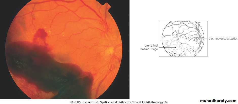
In PDR : ischemic retina stimulates the growth of abnormal “ New Vessels” which can bleed & cause retinal detachment ( RD).
4/14/2011
10
Laser in Ophthalmology
Proliferative diabetic retinopathy
4/14/2011
11Laser in Ophthalmology
Diabetic macular edema

4/14/2011
12Laser in Ophthalmology
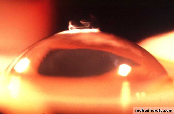
Sever rubeosis iridis

4/14/2011
13Laser in Ophthalmology
Laser for diabetic retinopathy

4/14/2011
14Laser in Ophthalmology
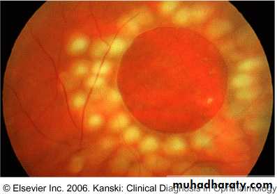
Pan retinal photocoagulation ( PRP )

4/14/2011
15Laser in Ophthalmology
Pan retinal photocoagulation PRP
4/14/2011Laser in Ophthalmology
16


Argon or Diode Laser ablation of ischemic areas ( pan retinal photocoagulation) PRP , causes regression of neovascularization.
Certain types of Diabetic macular edema respond to gentle Argon Laser treatment.
Same principle applied in cases of Retinopathy of prematurity ( ROP ).4/14/2011
18
Laser in Ophthalmology
Retinopathy of prematurity (ROP)
4/14/2011Laser in Ophthalmology
19

4/14/2011
Laser in Ophthalmology
20

2- Glaucoma :
Nd- YAG Laser peripheral iridotomy in angle closure glaucoma.Argon Laser trabeculoplasty in open angle glaucoma.
Diode Laser as a cyclodestructive procedure in Rebuotic glaucoma which occur in response to ischemic retina of PDR , and retinal vein occlusion.
4/14/2011
21
Laser in Ophthalmology
Argon Laser trabeculoplasty ( ALT)
4/14/2011
22Laser in Ophthalmology
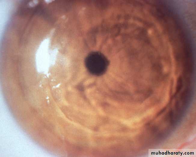
3- Posterior capsule opacification:
Posterior capsule thickening , also known as “ After cataract ” , is a common late complication of cataract extraction.
It occur as a result of proliferation & metaplesia of residual lens fibers attached to the capsule , symptoms include poor vision & glare.
The Nd-YAG Laser is used to create a central defect in the posterior capsule.
This does not affect the position or integrity of the Intraocular Lens Implant (IOL) .
4/14/2011
23
Laser in Ophthalmology
Posterior capsule opacity ( PCO )

4/14/2011
24Laser in Ophthalmology
• 4- Age–related Macular Degeneration ( AMD):
• The growth of abnormal vascular tissue from the choroid into the subretinal spaces cause rapidly progressive sight loss.• This tissue can be destroyed by Argon Laser.
4/14/2011
25
Laser in Ophthalmology
Choroidal neovascular membrane(CNV) in AMD
4/14/2011
Laser in Ophthalmology
26

5- Retinal Detachment ( RD ) :
A retinal tear or hole without RD can be surround with laser to induce Adhesion & prevent RD.During RD Surgery , Laser is some times used as an alternative to cryotherapy to promote retinal adhesion.
4/14/2011
27
Laser in Ophthalmology
Retinal hole surrounded by laser
4/14/2011
28Laser in Ophthalmology
6- Refractive Errors :
The Excimer Laser , applied with the computer assistance , very precisely remove corneal tissue in the management of low-moderate degree of myopia , hypermetropia & astigmatism combined with creation of hinged flap of cornea ( LASIK).
Larger Refractive Errors can now also successfully treated .
4/14/201129
Laser in Ophthalmology

Photorefractive keratectomy ( PRK )
IndicationsStable myopia up to 6D with astigmatism no more than 3D
Hypermetropia up to 2.5D
Main complication
Subepithelial haze which
usually resolves after 1-6
months
Reshaping of cornea by excimer laser ablation of
Bowman layer and anterior stroma

Technique
Laser in-situ keratomileusis (LASIK)Indications - similar to PRK but corrects higher degrees of myopia
Thin flap of cornea fashioned
Bed treated with excimer laser
Flap repositioned
Complications
Wrinkles in flap
Cellular interface proliferation

Technique
7- Miscellaneous uses :A- ablation of intraocular & adnexal tumors.
B- Division of intraocular post- inflammatory adhesions.
C- Destruction of aberrant lashes.
D-Removal of superficial corneal scars & calcific band keratopathy ( Excimer laser ) .
4/14/2011
32
Laser in Ophthalmology
Vitreous Body
4/14/2011
33
Laser in Ophthalmology
The transparent vitreous body, or hyaloid is one of the most delicate connective tissues in the body.
A. It occupies the posterior or larger compartment of the eye, filling the globe
between the internal limiting membrane of the neural retina and the posterior lens capsule.
B. The structure is composed of a framework of extremely delicate collagen filaments closely associated with a large quantity of water binding hyaluronic acid.
Anatomy of vitreous
Features:Virtually acellular viscous content of the globe.
Framework of collagen fibrils reinforced with hyaluronic acid molecules
98 % water.Volume = 4-5 ml in emmetropic eye.
4/14/2011
35Laser in Ophthalmology
Anatomy o the vitreous body

4/14/2011
36Laser in Ophthalmology
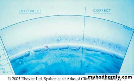
Attachments:
Vitreous base3-4 mm annular attachment
Very strong
Extends across ora serrata.
4/14/2011
37
Laser in Ophthalmology
The ora serrata

4/14/2011
38Laser in Ophthalmology
Ageing changes:
Dissociation of hyaluronic acid from fibrils
Fibril degeneration & reduced elasticity.Drainage of hyaluronic acid into retrovitreal space ( producing posterior vitreous detachment PVD).
4/14/2011
39
Laser in Ophthalmology
Vitreous opacities:
1- Muscae volitantes: remnants of hyaloid system.2- Syneresis: the Weiss ring ( posterior vitreous detachment PVD )
3- Hemorrhage
4- Asteroid hyalosis
Appears in 1 in 200 eyes
Composed of calcium soaps adherent to fibrils
Does not settle at rest
More common in diabetic people.
4/14/2011
40
Laser in Ophthalmology
Asteroid hyalosis

4/14/2011
41Laser in Ophthalmology
5- Synchisis scintillans
6- Inflammatory cells: Pars planitis, Chorioretinitis7- Neoplastic
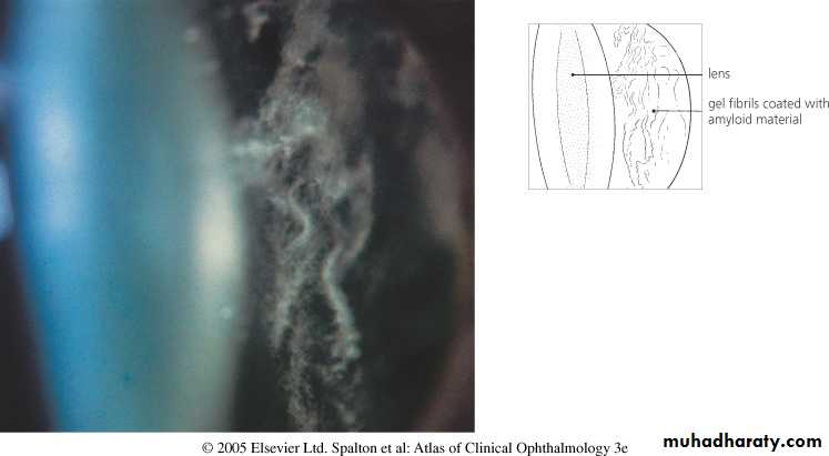
8- Amyloid
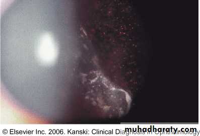
9- Tobacco dust in retinal detachment :pigment cells.
4/14/2011
42
Laser in Ophthalmology
Amyloid deposit in the vitreous
4/14/2011
43Laser in Ophthalmology
Tobacco dust ( pigment cells in the vitreous)
4/14/2011
44Laser in Ophthalmology
Vitreous degeneration
1- Syneresis:Vitreous liquefaction
Aggregation & condensation of collagen fibrils
Associated with floaters
Causes:
Myopia , senescence , trauma , inflammation , hereditary e.g. Stickler’s disease
2- Detachment
Collapse of vitreous gel
Associated with floaters and photopsia
Causes:
Senile , myopia , post inflammatory, postvitrous hemorrhage , and diabetic retinopathy.
4/14/2011
45Laser in Ophthalmology
Posterior vitreous detachment (PVD )

4/14/2011
46Laser in Ophthalmology
Vitreous hemorrhage ( VH )
1- causes:Proliferative retinopathy: DM , retinal vein occlusion, sickle cell retinopathy and retinopathy of prematurity.
Posterior vitreous detachment PVD
Trauma
Disciform macular degeneration
Blood dyscrasias
Subarachnoid hemorrhage ( Terson’ s syndrome)
4/14/2011
47
Laser in Ophthalmology
Vitreous Hemorrhage

4/14/2011
48
Laser in Ophthalmology
2- Complications
SyneresisInflammation & fibrosis : leads to traction detachment
Haemosiderosis
Synchisis scintillans:cholestrol crystals ; settles inferiorly at rest.
4/14/2011
49
Laser in Ophthalmology
Indications for Vitrectomy
1- Anterior segment conditions:Incarceration of vitreous in cataract section
Vitreous touch causing bullous keratopathy
Accidental vitreous loss during surgery
Lensectomy e.g. for secondary cataract in childhood arthritis
Malignant glaucoma
Glaucoma drainage surgery in aphakic eye.
4/14/2011
50
Laser in Ophthalmology
2- posterior segment conditions:
Diabetic disease:
Persistent hemorrhage, rubeosis with poor view, tractional RD involving macula.
Trauma: foreign body retrieval , dislocated lens, vitreous hemorrhage, giant tears
Complicated RD:gaint tear, proliferative vitreoretinopathy, large retinal breaks.
Endophthalmitis
Vitreous biopsy e.g. Amyloid.
Idiopathic macular hole.
4/14/2011
51Laser in Ophthalmology

4/14/2011
Laser in Ophthalmology52
Thank you