Intracranial Tumours
Intracranial neoplasm
Intracraneal neoplasm
All neoplasms arising from the skull, meninges, blood, blood vessels, pituitary and pineal glands, cranial nerves, brain tissue, or congenital rests, as well as metastatic tumors.
General Considerations
Although either benign or malignant, almost all brain tumours are malignant in the sense that they may lead eventually to death if not treated.Epidemiology
Brain tumors are responsible for 2% of all cancer deaths.
The incidence varies with age.
In adults, metastatic brain tumor is the most common intracranial neoplasm comprising about 15-20% of them.
Among the primary, the most frequent are gliomas followed by meningiomas.
Glioma more frequent in males and meningiomas are more common in females.
In children, brain tumors are the second most common after leukemia. 70% are infratentoreal. The most frequent are cerebellar astrocytoma, brainstem glioma, medulloblastoma and ependymoma.
In neonates and in infants, brain tumors are rare mostly congenital.
Classification of brain tumours
The classification of brain tumours is determined by their cell of origin.Over 50% are neuroepithelial in origin, 15% metastatic, 15% meningioma and 8% pituitary tumours.
The World Health Organization (WHO) classification of brain tumours is outlined in the next slide.
(WHO) Classification of brain tumours
• Neuroepithelial Tumours:• Nerve sheath tumour: acoustic neuroma
• Meningeal Tumours: Meningioma
• Pituitary Tumours
• Germ cell tumours: e.g. Germinoma, Teratoma
• Lymphomas
• Tumour-like malformations:
• Metastatic Tumours
Neuroepithelial Tumours
• Gliomas:
• Astrocytomas
• Oligodendrogliomas
• Ependymoma
• Choroid plexus tumour
• Pineal Tumours
• Neuronal Tumours
• 4) Medulloblastoma
Tumour-like malformations
• Craniopharyngioma• Epidermoid tumour
• Dermoid tumour
• Colloid cyst
Aetiology of Brain Tumors
The etiology of brain tumors is still not clearly understood.Radiation associated with meningioma , glioma and fibrosarcoma
Although there is no genetic predisposition, chromosomal abnormalities have been noted in many CNS tumors (e.g. von Recklinghausen's).
Viruses, EBV associated with Burkitt s lymphoma and nasopharyngeal carcinoma
Immunosuppression markedly increase the incidence of primary CNS lymphoma.
Mutation in the p53 tumor suppressor gene is the most common gene alteration and is found in astrocytoma and meningioma
Clinical Features of Brain Tumours
This will result from one or a combination of:
Focal neurological deficits
Raised intracranial pressure
Seizures, and
Endocrine dysfunction, or
Incidental findings (i.e. symptomless).
a) Focal Neurological Deficits
• Frontal lobe lesions: presents with personality changes, gait ataxia, and urinary incontinence, contralateral hemiparesis if posterior frontal and expressive dysphasia if involving the dominant inferior frontal gyrus.• Parietal lobe lesions: contralateral sensory impairement, dressing apraxia, asteriognosis and, if on the dominant side, acalculia, agraphia, left-right disorientation and finger agnosia.
• Temporal lobe lesions: olfactory and auditory hallucination, memory disturbances, contralateral superior quadrantanopia or hemiparesis and, if on the dominant side, receptive dysphasia.
• Occipital lesions: associated with visual field deficits, most commonly an incomplete contralateral homonymous hemianopia.
a) Focal Neurological Deficits
• 5. Subfrontal lesions: may involve the olfactory nerves (anosmia).• 6. Sellar and parasellar: tumours present with visual field problems due to compression of the optic chiasm, hypopitutarism and oversecretion syndromes such as Cushing's disease and acromegaly.
• 7. Cerebellar lesions: Tumours involving the cerebellar vermis cause truncal ataxia, whereas tumours in the hemispheres produce appendicular signs such as incoordination and nystagmus.
• 8. Brainstem and Cerebellopontine angle lesions: may result in cranial nerve palsies, long tract signs and secondary hydrocephalus.
• 9. Ventricular system tumours: may obstruct CSF drainage and result in hydrocephalus, compounding raised ICP.
b) Raised Intracranial Pressure
Direct mass effect.
Vasogenic oedema.
Obstructive hydrocephalus.
c) Seizures
New onset of seizure in an adult is suggestive of an intracranial mass until proven otherwise.The type of seizure may give a clue to the location of the tumour:
Parietal lesions cause simple partial seizures, which may become secondarily generalized.
Medial temporal lesions will cause complex partial seizures.
c) Seizures
Late onset epilepsy, particularly over the age of 30 years, should prompt investigations to exclude an intracranial neoplasm.New onset of seizure in an adult is suggestive of an intracranial mass until proven otherwise.
The type of seizure may give a clue to the location of the tumour:
Parietal lesions cause simple partial seizures, which may become secondarily generalized.
Medial temporal lesions will cause complex partial seizures.
Personality Changes
Headache
VomitingPapilloedema
AtaxiaInvestigations of Brain Tumours
• Plain Skull x-ray• Computed Tomography (CT-scan)
• Magnetic Resonance Imaging (MRI)
• Angiography
• Positron Emission Tomography (PET).
Investigations of Brain Tumours
I. Plain Skull x-ray:a. May be normal
b. May show changes due to raised intracranial pressure.
c. Local changes may occur with specific tumours: like
1. Suprasellar calcification in craniopharyngiomas
2. Hyperostosis and skull thickening in meningioma.
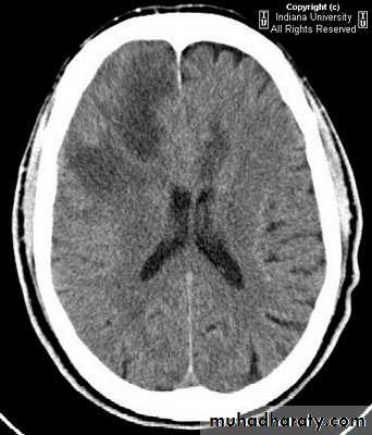
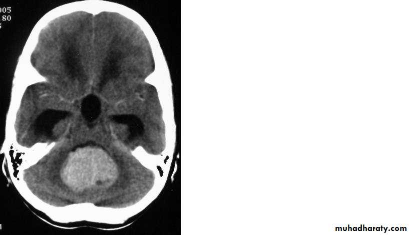
Investigations of Brain Tumours
• Computed Tomography (CT-scan):
• Site and size of the tumour.
• Boundaries of the tumour.
• If there is surrounding oedema or not.
• Shift of the midline structures by the mass.
• Associated hydrocephalus.
• Bony changes.
• Nature of the tumour (solid, cystic, necrotic or calcified), and so its pathology.
Investigations of Brain Tumours
• 3. Magnetic Resonance Imaging (MRI): same as CT• 4. Angiography: Used to evaluate the blood supply and vasculature of the tumor.
Investigations of Brain Tumours• 5. Positron Emission Tomography (PET): can study the brain metabolism.
Used to characterize the most malignant component of a tumour.
Assess prognosis of brain tumour.
Differentiate recurrent tumour from radiation necrosis.
Management of intracranial tumours
1. If the patient shows signs of compression, or raised intracranial pressure, treat with measures that lower ICP.
2. If an intracranial tumour is associated with hydrocephalus, the hydrocephalus is dealt with first by a ventriculoperitoneal shunt before surgery.
• CT or MRI guided stereotactic surgery : tumour is deeply seated in the brain.
• Surgical excision or debulking for accesssible tumours.
5. In cases of malignant brain tumours or metastases : adjunctive therapy : Radiotherapy and/or Chemotherapy are supplementary to surgical excision.
Management of intracranial tumours
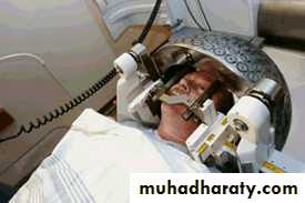
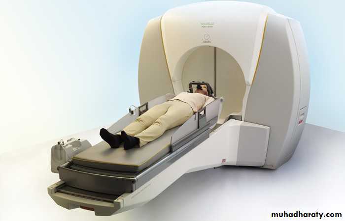
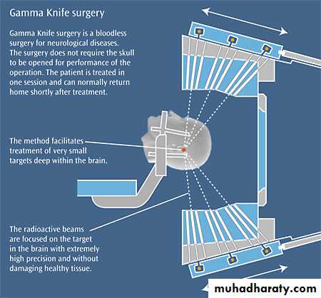
Radiotherapy either conventional radiotherapy or radiosurgeryFor small tumor less than 3 cm which is of many types
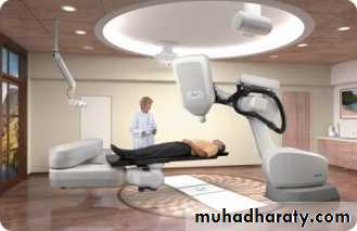
Linear accelerator
Gama knife
Cyber knife
Cyber knife
Stereotactic Neurosurgery
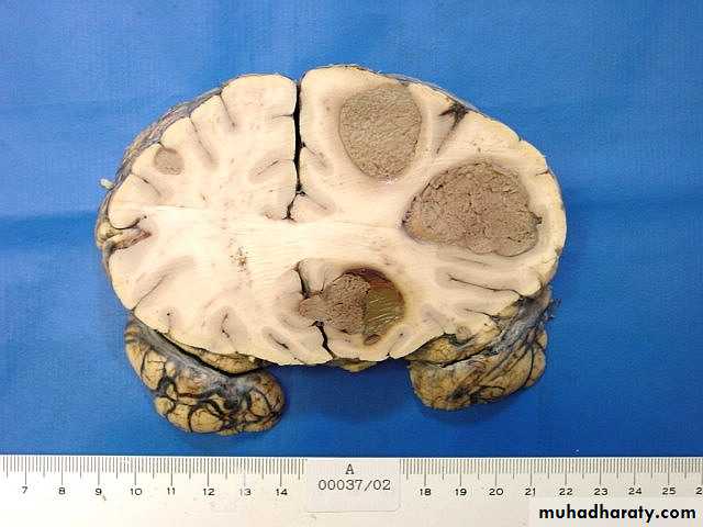
Metastatic Brain Tumours• Constitute 15% of intracranial tumours.
• The commonest sites of origin are the lung (40%) and breast (10-30%), in addition to melanoma (5-15%), kidney and colon.
• In 15% of cases, a primary source is never found.
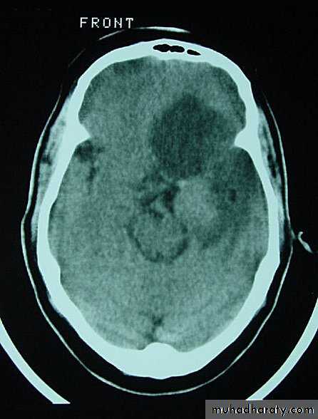
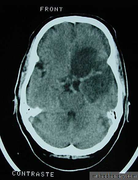
• On CT, they only show well with intravenous contrast.
• Steroids may help reduce peritumour oedema.
• Surgery may be appropriate for an isolated metastasis.
CSF shunt used for lesions causing Hydrocephalus.
• Radiotherapy can be used for multiple metastases.
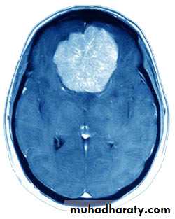
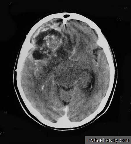
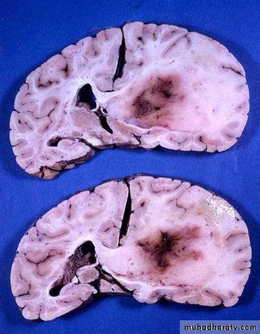
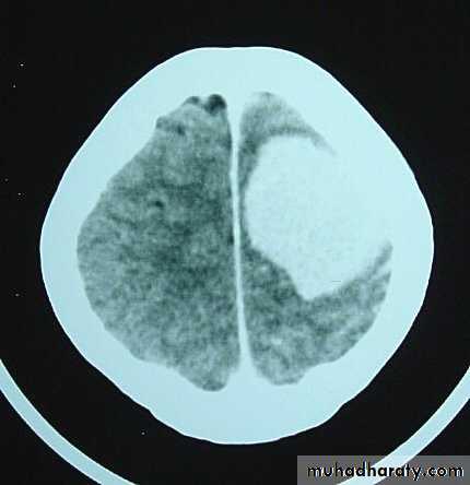
Metastatic Brain Tumours
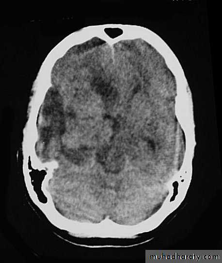
Metastatic Brain Tumours Pre contrast CT
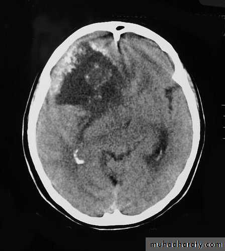
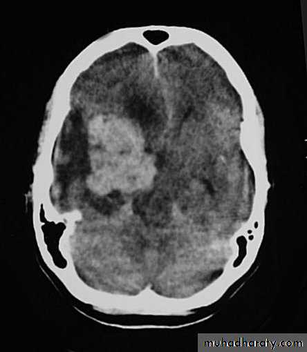
Metastatic Brain Tumours Post contrast CT
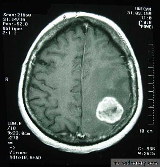
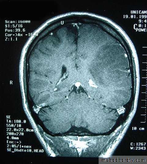
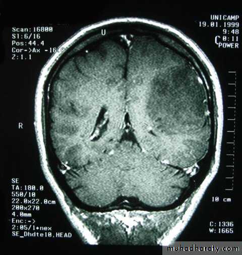
Metastatic Brain TumoursMRI
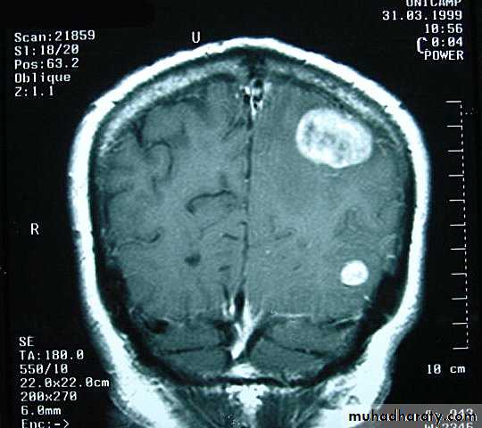
Metastatic Brain TumoursMRI
Gliomas
They are derived from cells of glial origin.• They are the commonest of primary CNS neoplasms.
• They form 50% of adult intracranial tumours.
• They are of four types: astrocytoma, oligodendroglioma, ependymoma and choroid plexus papilloma. The most common of which is astrocytoma.
• Survival is closely associated with grading.
• Treatment involves surgery and radiotherapy and/or chemotherapy.
Glioma
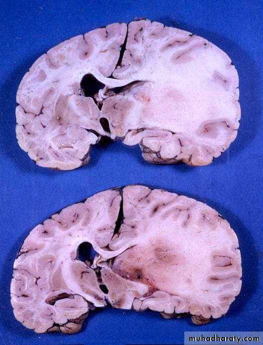
Astrocytoma
AstrocytomaPre contrast CT
AstrocytomaPost contrast CT
Astrocytoma Pre contrast MRI
Astrocytoma Post contrast MRI
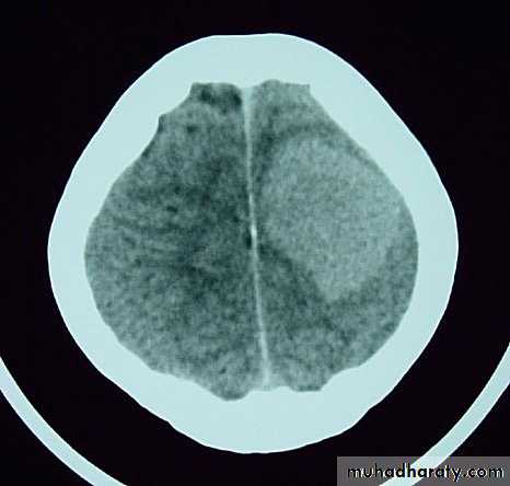
Malignant GliomaPre contrast CT
Malignant Glioma Post contrast CT
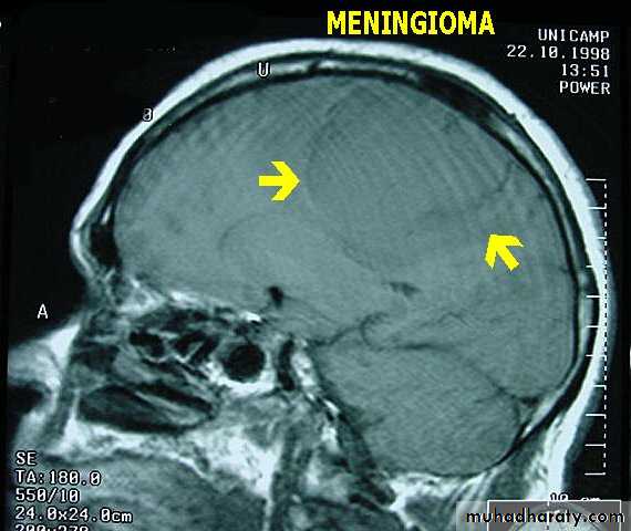
Meningiomas
• These account for 15% of intracranial neoplasms, and are the most common benign neoplasm.• They occur more frequently in women than in men, and their incidence peaks in middle age.
• The tumour arises from meningothelial cells of the arachnoid villi.
Meningiomas
• They classically arise from a broad base along the dura.• They may invade bone.
• They derive their blood supply from the external carotid circulation.
• Malignant meningiomas are relatively rare.
• The symptoms and signs are related to those of intracranial mass lesions or seizures.
• CT scan accurately diagnoses the lesions.
• Complete surgical removal usually results in cure.
MeningiomaPre contrast CT
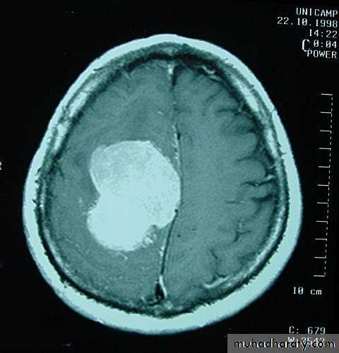
MeningiomaPost contrast CT
MeningiomaPre contrast MRI
MeningiomaPost contrast MRI
Meningioma Bone Involvement
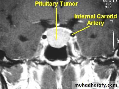
Meningioma Bone InvolvementPituitary Tumours
• Non-functioning pituitary adenomas• Functioning pituitary adenomas
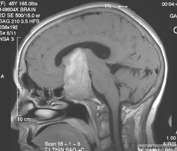
Non-functioning pituitary adenomas
• Accounts for about 30% of pituitary tumours.
• They are often seen in the fourth and fifth decades of life.
• Because they are non-functioning, they are not generally diagnosed until they are very large.
• Their presentation is by optic chiasm compression that cause visual field defect (bitemporal hemianopia).
• The usual treatment is microscopic trans-sphenoidal or trans-cranial excision.
Functioning pituitary adenomas
• Pituitary adenomas are classified according to the hormones they secrete. They include:• Prolactin secreting adenomas.
• Growth hormone secreting adenomas that produce acromegaly or gigantism.
• Glycoprotein secreting adenomas that produce excess amount of TSH, LH, or FSH.
• ACTH secreting adenomas that produce Cushing's disease.
• Some of these secrete more than one hormone, e.g. prolactin-growth hormone secreting adenomas.
Functioning pituitary adenomas
• 2. Adenomas may be further divided according to their sizes into Microadenomas that are less than 1 cm in diameter, and Macroadenomas that have a larger size.• 3. Functioning adenomas are diagnosed by:
• Clinical changes.
• Hormone assessment. And
• Radiology by using MRI and CT scan.
Functioning pituitary adenomas
4. Prolactinomas represent 40% of all pituitary adenomas. They cause amenorrhoea, galactorrhoea, and infertility in women, while in men they cause impotence or may be asymptomatic.
5. Bromocriptin (antiprolactin drug) has virtually replaced surgery as the treatment of choice for prolactinomas.
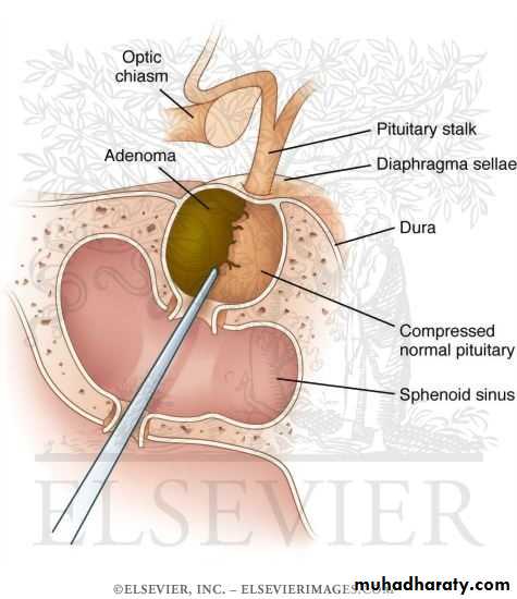
6. Surgical excision can preferably done by using the trns-sphenoidal route, whether sublabial or transnasal, although transcranial approach can also be used.
7. In case of an invasive tumour with incomplete excision, radiotherapy is required
Trans sphenoidal

DO NOT FORGET
• The most significant findings pointing to an intracranial tumour on physical examination are the presence of papilloedema and signs of focal damage to the nervous system.• If a cerebral tumour is suspected, LUMBAR PUNCTURE IS CONTRAINDICATED as it may precipitate a fatal coning of the brain stem through the foramen magnum.
THANK YOU