Premalignat disease of the cervix
Dr. Ahmed jasimAss.Prof.
MBChB-DOG-FICMS
COSULTANT OF GYN. & OBST.
Premalignat disease of the cervix
1.cervical disease2.Cervical intraepithelial neoplasia
(Cervical dysplasia)
cervical cancer is the common cancer affecting women after breast cancer.
Cervical cancer is a preventable disease because of:1. There is usually a phase of premalignancy intraepitheilial neoplasia and has relatively long natural history.
2. The cervix is a relatively accessible organ to examine.
3. The availability of a simple test for the presence of pre-malignancy.
4. Treatment for pre-invasive disease is highly effective.
Aetiology
Human papillomavirus (HPV) infection
HPV infection is extremely common and in majority of cases will not lead to development of cancer. Progression or regression depends on several factors that interfere with the host's ability to clear the virus such as in.
Transplant patient.
HIV-Positive women.
Smoking.
Screening for cervical intraepithelial neoplasia (CIN)
Medical screening methodDetect premalignant and malignant processes of cervix.
Prevent progression of abnormal cells to cancer.
This is NOT a diagnostic test!
Cervical cancer screening with cytology provides the opportunity for early effective intervention and has reduced morbidity and mortality
Papanicolaou
CervicoscopeyVisual inspection with acetic acid (VIA)
Visual inspection with acetic acid and magnification (VIAM): Gynescope or Aviscope
Colposcopy
Cervicography
Automated pap smears

Molecular (HPV/DNA) tests.
Co-testing using the combination of cytology plus HPV DNA testing is an appropriate screening test for women older than 30 years (applied in some places).Papanicolaou (Pap) smear test
What is a pap smearScreening test for Asymptomatic Women
To detect treatble pre-invasive squamous Abnormalities of the Cervix
Small number of women will develop invasive Cancer
Not diagnostic-rather screeing test to detect early changes on the cervix.
Exfolative cervical cytology was a technique to collect the cells that had been shed from the cervix.
It is a simple and painless test that may cause minor discomfort.
Cervical Smear aims to prevent cancer, not to detect cancer.
Cervical cancer screening should begin at age 21 years and not before age 21 because it may lead to unnecessary and harmful evaluation and treatment in women at very low risk of cancer.
Women who have been immunized against HPV-16 and HPV-18 should be screened by the same regimen as nonimmunized women.
frequency of cervical cytology screening
Cervical cytology screening is recommended every 2 years for women aged 21–29 years.*Women aged 30 years and older who have had three consecutive cervical cytology
test results that are negative for intraepithelial lesions and malignancy may be screened every 3 years.
frequency of cervical cytology screening
*women with any of the following risk factors may require more frequent cervical cytology screening:
• Women who are infected with human immunodeficiency virus (HIV)
• Women who are immunosuppressed (such as those who have received renal transplants).
• Women who were exposed to diethylstilbestrol in utero.
• Women previously treated for CIN 2, CIN 3, or cancer (continue to have annual screening for at least 20 years).
Pap Smear is not necessary in women in these categories:
Virginal patients.Total Hysterectomy for benign disease.
Recent result of pap smear.
Age over 65 and over 10 benign Pap Smears.
Preparation
To prepare for the Pap test, for two days before the test ,women should avoid:Vaginal Douching .
Using tampons.
sexual intercourse.
Using birth control foams, creams, or jellies or vaginal medications or creams.
the ideal time for a woman to have a Pap Smear is five days after her menstrual period has ended.
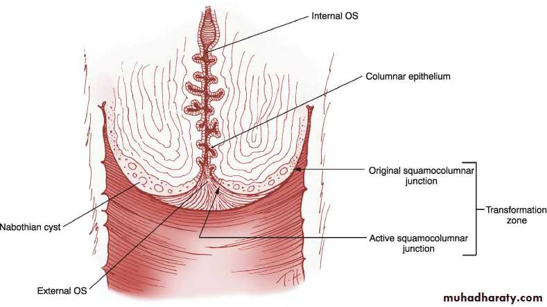
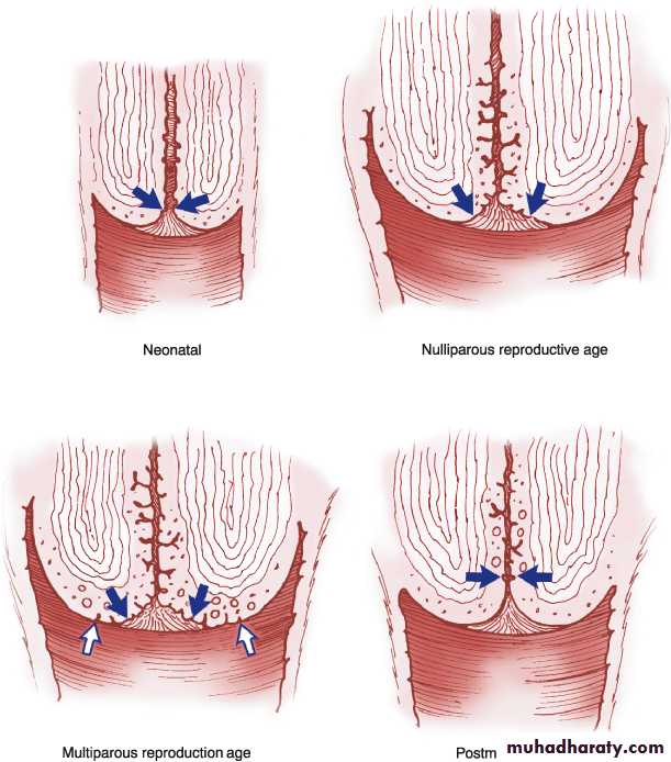
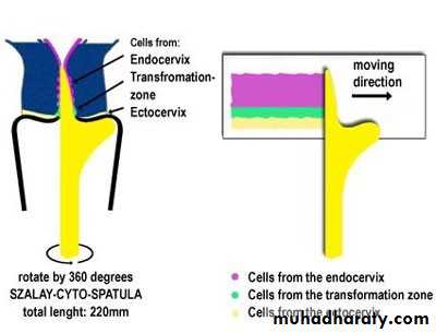
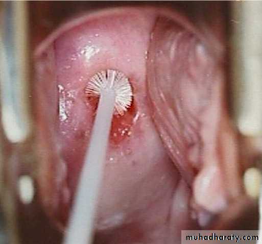
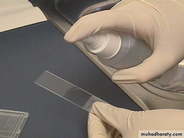
Exfoliated cells are collected from the transformation zone of the cervix by Use spatula of different size or brush.
There are two methods of preparing and processing cervical smear slides.
These methods are:
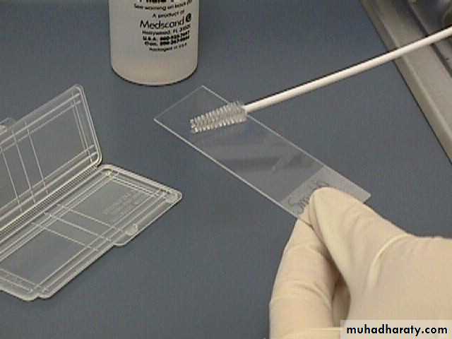
1. conventional cervical (Pap) smear test.
collecting the cells smears on a microscope slide and applies a fixative. The slide is sent to a laboratory for evaluation.


The Spatula with the optimal shape and size is chosen .

Broom type sampler

The 'tongue' of the spatula is introduced into the canal, whilst its 'shoulder' is positioned on the 3 o'clock position of the ectocervix at the beginning of the procedure.With gentle pressure the spatula is rotated in a clockwise direction.
2. liquid based cytology (LBC) test.
Cell transferred to a vial of liquid preservative that is processed in the laboratory to produce a slide for interpretation by light microscopy.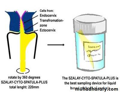
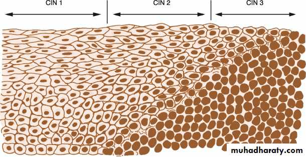
Classification of CIN
A. CIN classificationCIN 1 (mild dysplasia) involvement of the inner one-third of the epithelium.
CIN 2 (moderate dysplasia) involvement of inner one-half to two-third
CIN 3 (severe dysplasia/carcinoma in situ) full thickness involvement.
Figure 17.1 Diagram of cervical intraepithelial neoplasia compared with normal epithelium.
Or can be classified as:
*Low grade lesions (CIN1 and HPV-associated changes) in which there is a significant chance of regression and low progressive potential.*High grade lesions (CIN 2 and CIN 3) are likely to behave as cancer precursors.
A: Active metaplasia in the transformation zone. B: Maturing metaplasia in the transformation zone.
A diagnosis of CIN is based primarily on the presence of nuclear atypia and loss of normal squamous maturation (polarity).
Accurate grading of CIN lesions becomes important as we begin to understand the rates of regression, persistence and progression of the low-grade (CIN 1) and high-grade lesions (CIN 2 and 3), as their treatment and clinical follow-up are quite different.
Cervical pre-cancer has along natural history.
36% of women with CIN3 would develop invasive cancer if left untreated.
More than 40% of women with minor cytological abnormalities will revert to normal without treatment.
Clinical presentation
The disease is a symptomatic. The premalignant lesions cause no symptoms and are not recognizable with the naked eye.Results of the cervical smear test
The cytologist will classify the smear accordingly:Normal results:
Mean that no atypical, dysplastic, or cancer cells were detected, and the cervix is normal. It is seen in About 9 in 10 routine cervical screening tests.
(Note: a normal result means a very low chance of developing cancer of the cervix - not a 100% guarantee that it will not occur.)
Abnormal result:
Some changes in the cells are found in about 1 in 10 tests. There is a range of changes that may occur. In nearly all cases, these changes do not mean cancer.Inflammatory –excessive leucocytes, candida or trichomonas.Borderline.( Cellular appearance that cannot be described as normal).
Mild dysplasia
Moderate dysplasia.
Severe dysplasia.
Possible invasive carcinoma. Rarely, a cancer of the cervix is diagnosed by a cervical screening test.
Management of abnormal cervical smears
Inflammatory smears should be treated by antibiotics or antifungal agents accordingly. And the smear repeated 3-6 months later.Border line smear advice to Repeat smear in 6-12 months and refer for colposcopy if abnormalities persist.
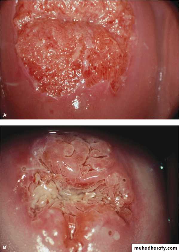
Ideally all women with abnormal cervical cytology(some mild ,moderate ,sever dysplasia) should have colposcopic assessment to exclude an invasive process and to identify the extent of abnormality.
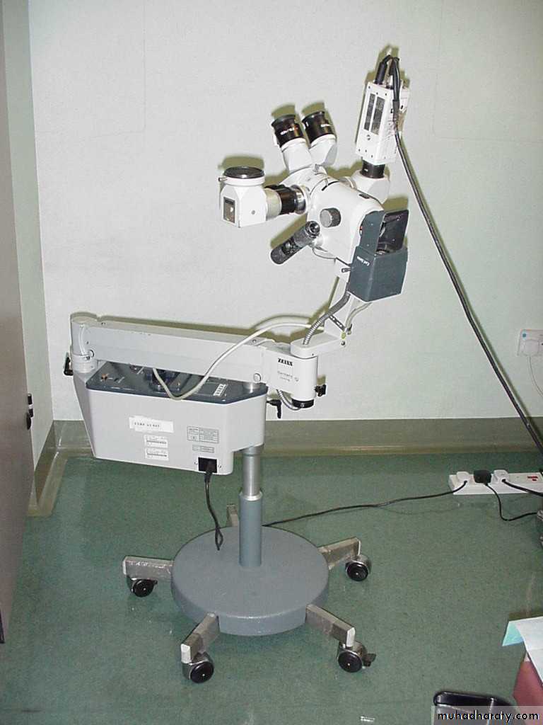
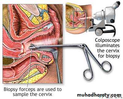
Colposcope
Minor or borderline abnormal changes are quite common. These often clear away on their own and most mild changes do not progress to anything serious. However, any change needs to be monitored as some may progress to become more serious in the future. A repeat test after 3-12 months is commonly advised, depending on the type and degree of change. Often the changes will have gone when the test is repeated. If the changes do not go, or the changes are more marked, then a referral to colposcopy is advised.
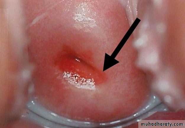
Any patient with a grossly abnormal cervix should have a punch biobsy regardless of the results of Papanicolaou smear.
Inflammation
CIN1
CIN2
CIN3
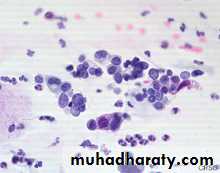

CIN3 ca in situ
Squamous cell carcinoma
CIN2
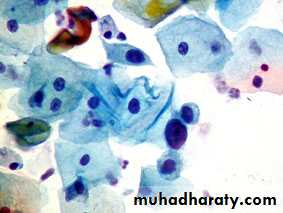
High Grade SIL
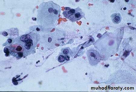
CIN1


Low grade SIL (L-SIL)
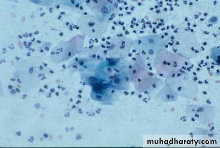
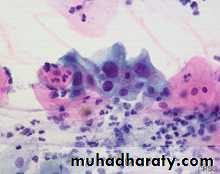
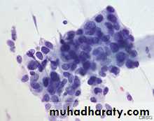
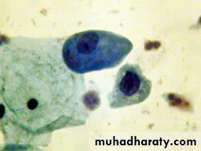
The cytologic features of normal squamous epithelial cells can be seen at the center top and bottom, with orange to pale blue plate-like squamous cells that have small pyknotic nuclei. The dysplastic cells in the center extending to upper right are smaller overall with darker, more irregular nuclei.


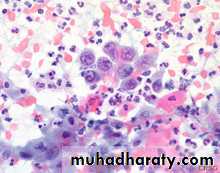
Gardnerella Vaginalis
Mixed bacterial flora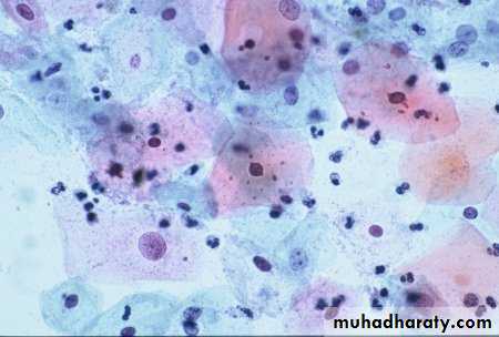
Candida
HERPS
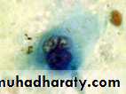
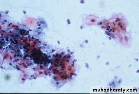
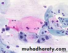
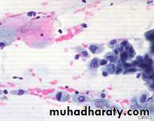
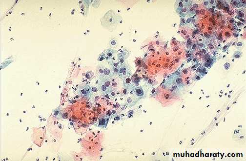
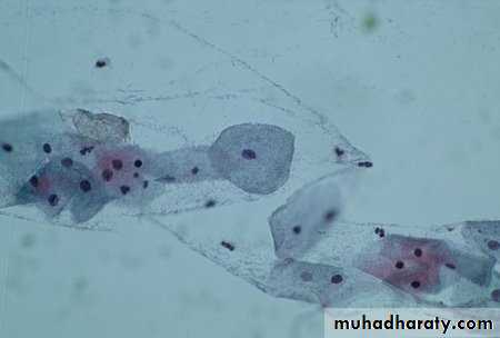
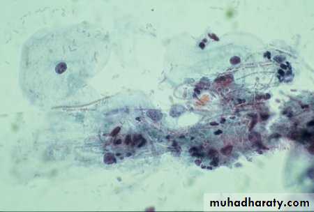
Abnormal Pap Smears
Treatment is based on the biopsy results.Colposcopy:
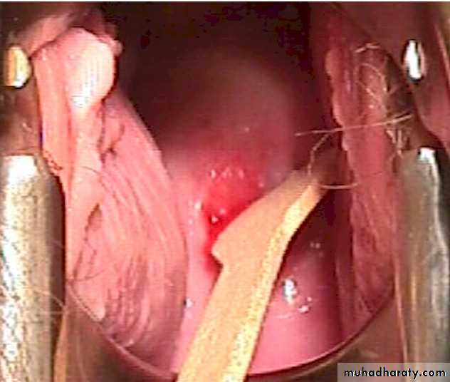
It is a binocular operating microscope withmagnification of 5-20 times. Indicated for further investigation of smear abnormalities.
It has been used to examine the cervix in detail to:
*Identify dysplastic abnormalities on the ectocervix.
*Detect changes in the cellular pattern and vascularity of the covering epithelium.
*Allow the accurate localization of the abnormal epithelium.
*Exclude an invasive process.

CIN has the potential to be an invasive malignancy but dose not have malignant properties.high grade lesions (CIN2 and CIN3) should be treated, but there is some debate about CIN1 they allow CIN 1 lesions to be treated or kept under close surveillance.
• The treatment for cervical dysplasia must be individualized for each woman, taking into
• account
• the grade of the dysplasia (CIN1, CIN2, or CIN3).
• the findings at colposcopy.
• the woman's age.
• reproductive status.
• and other factors.
Treatments for CIN include:
CIN has the potential to be an invasive malignancy but dose not have malignant properties. high grade lesions (CIN2 and CIN3) should be treated, but there is some debate about CIN1 as some allow CIN 1 lesions to be treated and others advice to be kept under close surveillance..
Treatment involves completely removing the abnormal epithelium.
This can be done by:1. Destroying the abnormal epithelium.( cryosurgery, laser vaporization)
2. Excisional techniques:(This allows better histopathological interpretation of the excised specimen).
These techniques include:
a. local excision
b. loop electrode excision procedure (LEEP).
c. cone biopsy.
d. trachelectomy (excision of cervix).
e. hysterectomy.
The success of treatment is usually defined as negative cytology 6 months following intervention.
Therapeutic vaccination aims to boost host's cell-mediated immunity but still experimental.
Follow up:
Follow up of patient treated for CIN is controversial between colposcopy or cytology. other tests such as a HPV DNA test may be advocated
LEEP (loop electrosurgical excision procedure(. After freezing the area with local anesthetic, an electrical wire loop is inserted into the vagina and all the abnormal tissue is removed. This procedure is also done in the physician's office.

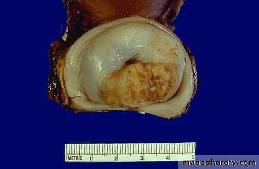
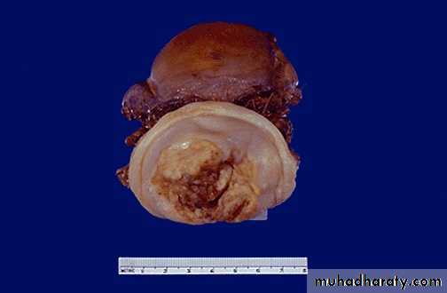
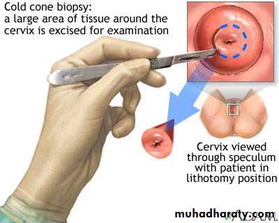
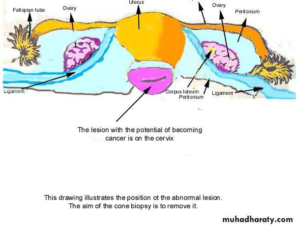
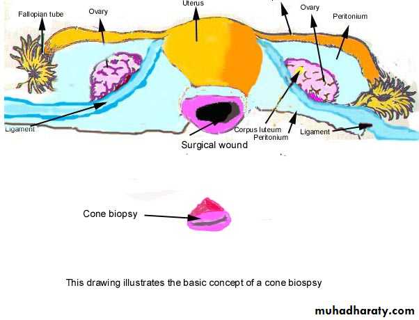
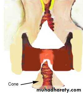
A cone biopsy
refers to removal of a cone-shaped piece of tissue. The tissue removed provides a more extensive sample for diagnosis than a simple biopsy. A cone biopsy is usually done in the operating room.
The cold cone biopsy is a surgical procedure requiring general anesthesia and is indicated by the presence of precancerous changes in the cervix.
What happens after treatment?
After treatment for dysplasia, patients are followed closely to make sure all the dysplasia is gone, and that new dysplasia does not occur. Typically, patients are followed with frequent Pap smears for two years after treatment, e.g. Pap smears every 3 to 4 months for the first year, and then every 6 months for the second year. If all the Pap smears come back negative, the patient is be cured, and is then followed with yearly Pap smears.A colposcopy-directed biopsy is a procedure in which the cervix is examined with a colposcope for abnormalities and a tissue sample is taken.
Cost of Pap smear screening
office visit and cytopathologycolposcopy plus the pathology
cancer therapy
hidden costs of cancer care
unquantifiable
cost of
loss of life
Further research is
needed to determine
what role
HPV testing should play
as part of
a cervical cancer
screening program.