عباره عن صور مختبر المايكرو من المحاضرات فقط جمعتها ووضعتها بهذا الملف
يحتوي على صور مختبرات فقط بعد نصف السنه
هنالك صور قليله مسحوبه من النت
مختبرات 13 و 14 أَخذت من العام الماضي لعم توفرها لدي
Done by me Hassan Almalah
Group – B -

Lab-12-
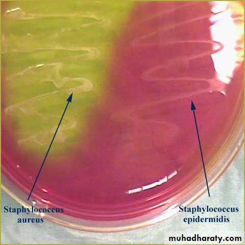
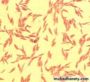
Staphylococci
Catalase tests
• (-) (+)
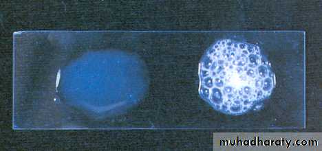
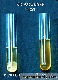
Coagulase test
Reaction to carbohydrate mannitol.
Dnase test


Novobiocin sensitivity test
Lab-13-
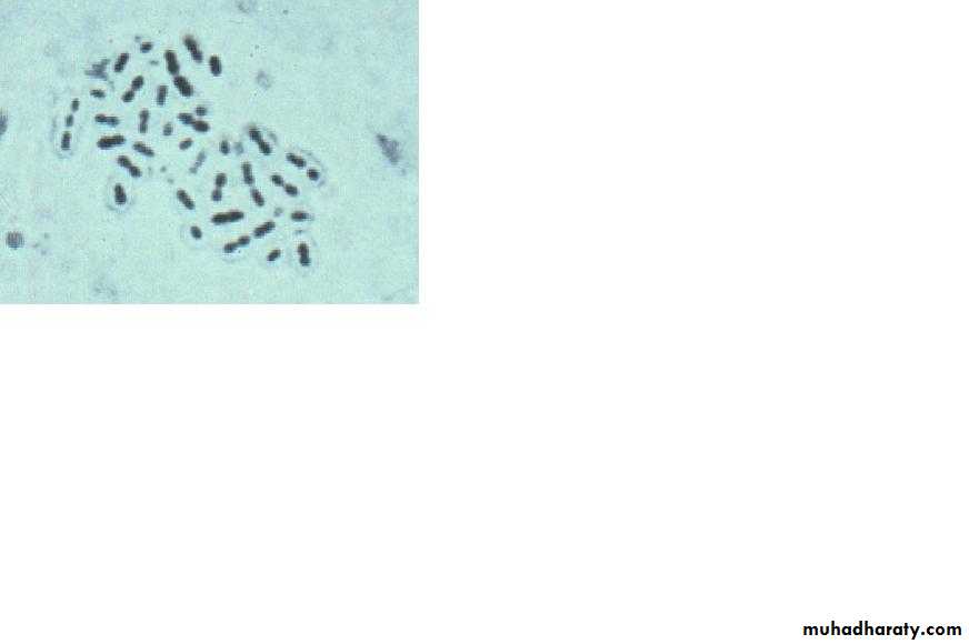
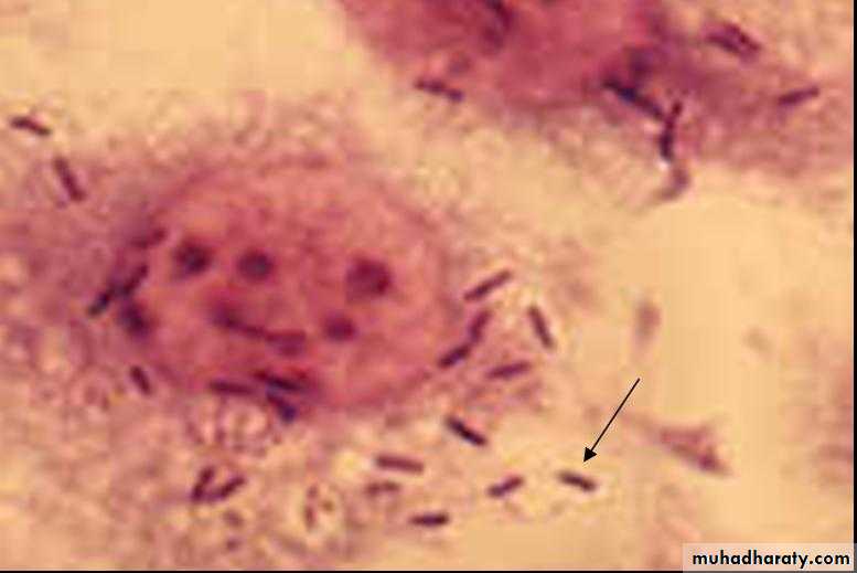
StreptococciStreptococci

a-chains b- pairs (S. Pneumonia)

β-hemolytic Sterptococci.α-hemolytic Sterptococci.Non-hemolytic streptococci
β-hemolytic Sterptococci.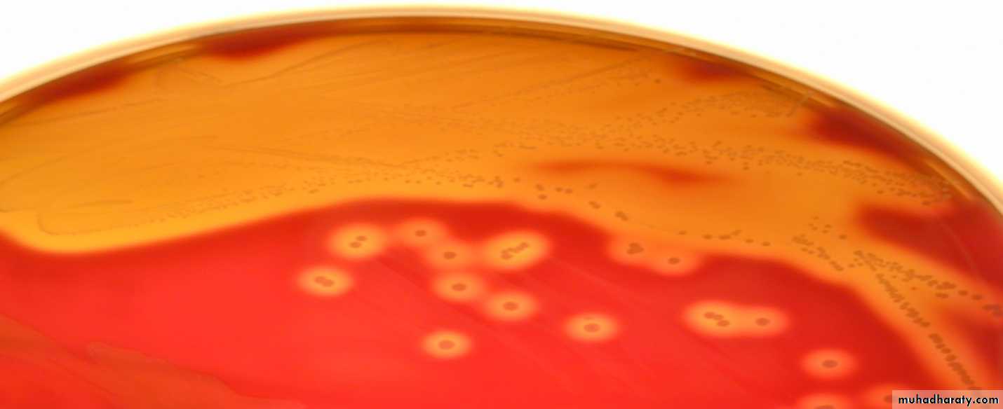
• It causes complete hemolysis to RBCs (caused by hemolysins) leading to formation of clear zone around the colonies
• Example: S. pyogenes (group A β-hemolytic Strept.), S.agalactiae
α-hemolytic Sterptococci.

• It causes:
• 1. Zone of greenish discolouration around the colonies.
• Example S.pneumonia ,s.viridans
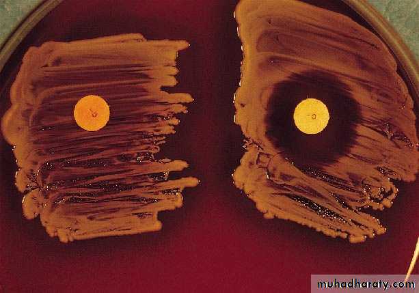
Bacitracin sensitivity
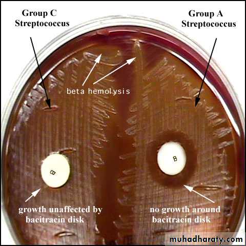
• Bacitracin will inhibit the growth of gp A Strep. pyogenes giving zone of inhibition around the disk
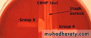
CAMP test

Optochin Susceptibility Test

Bile Solubility test

Lab-14-
Sterptococcus Pneumoniae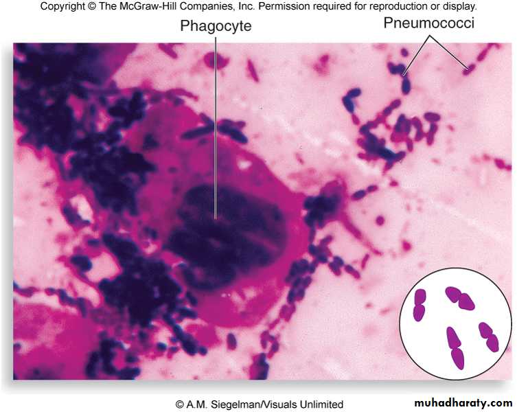
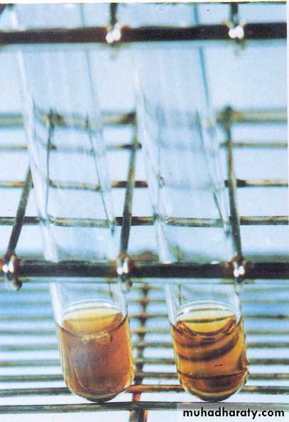
bile solubility test
• Result :• + ve clear
• - ve Turbidity
• Lyses of colonies means the microorganism become slouble in bile salt
Optochin disc test ( ethylhydro cupriene
• Result :• If the inhibition zone a greater than 18mm that means its colony of St. pneumoniae while other streptococcus hemolytic hare zone less than 18mm

Quelling reaction ( capsular swelling reaction ) ( Serological test )
• Result :• + ve capsule appear swollen
• - ve capsule quite invisible

Lab 15
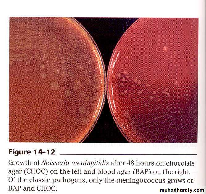
neisseriaLab 16
neisseria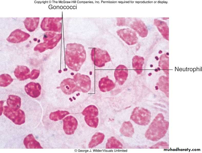
Associated with PMNs

• Intracellular ) )pathogenic Neisseria

• Neisseria (diplococci )appear in electron microscope
Gram stain of N.gonorrhoea
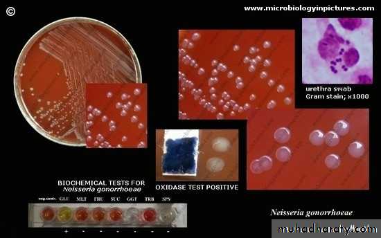

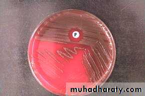
Growth of N. gonorrhea in the media (Thayer-Martin agar )

Gram stain of Neisseria meningitis
Lab-16
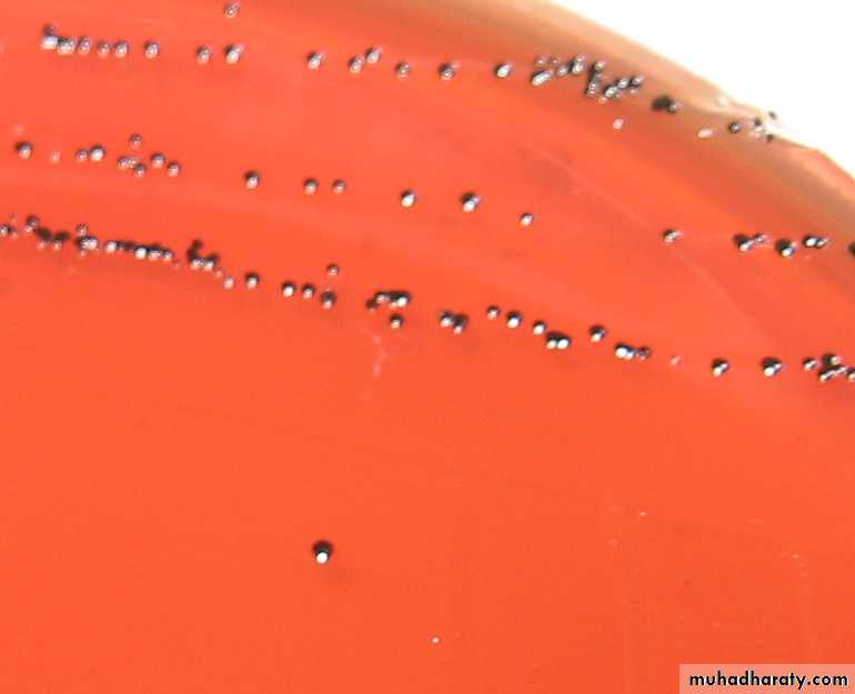
Corynebacterium sppCorynebacterium diphtheria
Loffler slope enriched media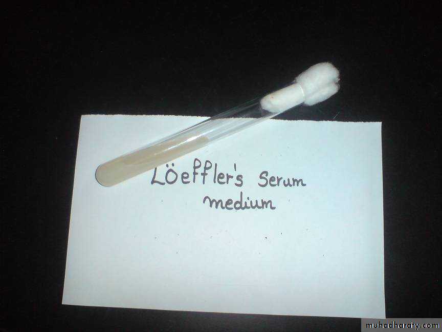
blood tellurite medium
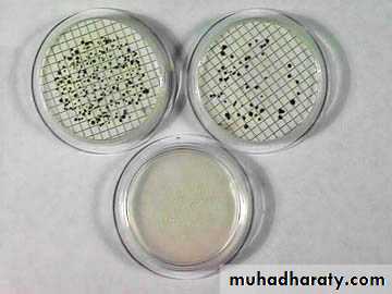
TOXIGENICITYIn vivo
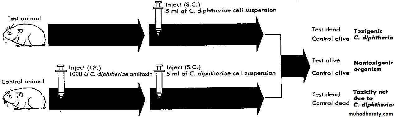
in vitro(Elek’s test)

+ve control
• UnknownLab 17
Lactobacilli
• Gram positive lactobacilli

• Lactobacilli under electron microscope
Tomato juice agar (pH 5)

• Tomato juice agar
Rogosa agar
• Rogosa agar

Counting of streptococcus mutans and lactobacillus species in the saliva
Snyder test
• Snyder test
Lab 18
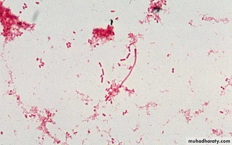
Enterobacteriaceae
Escherichia coli


Culture
1-Mac Conkey agar : Pinkcolonies
due to the lactose fermentation
2-Blood agar : Some strains
show beta haemolysis
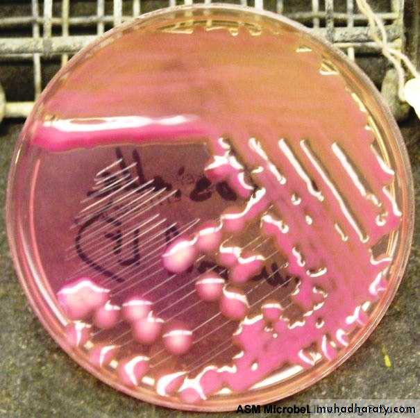
3-Eosin methylene blue
agar ( EMB ) : green to black
with metallic sheen.


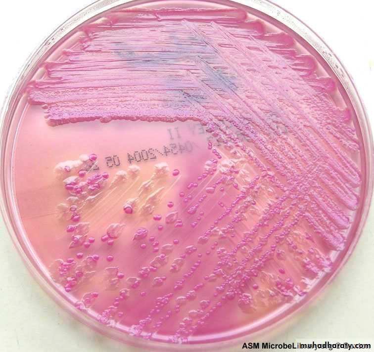
Klebisella Spp.

Klebisella Spp.

Culture :
Blood agar : mucoid colonies .MacConkey agar : large mucoid, pink colonies ( mucoid because accumulation of capsule ) .
Salmonella spp.

Culture :
Mac Conkey agar :Colonies colourless ( NLF )
Salmonella – Shigella agar : brown colonies
with black center ( SS agar ) .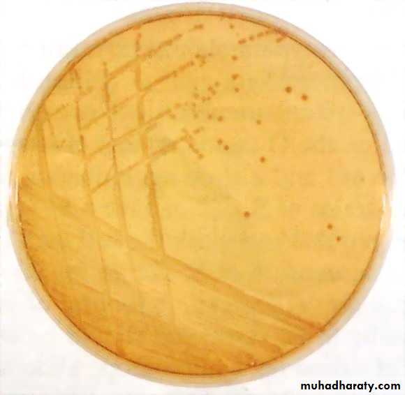

Shigella spp
Culture
MacConkey agar : Pale colonies except Sh. sonnei after 48hr .SS agar : Colourless
without black center .
5. Proteus spp.
swarmingpresent only in the absence
of bile salt , .


Culture
1-nutrient agars produce swarming, fishy odor2-urea agar.

Citrobacter :
Citrobacter :

Enterobacter

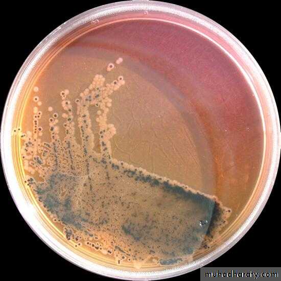
EMB PLATE: E. coli is seen on the left and E. aerogenes on the right

8.Serratia spp

Lab 19
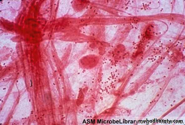
mycobacterium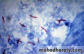

• Mycobacteria
Sputum testing
• Result:
• Acid fast bacteria appear as red• Non-acid fast bacteria appear as blue
Culture
Lowenstein-Jensen medium: special selective media

Lowenstein-Jensen medium: special selective media


(rough, tough, and buff).
Chest x-ray
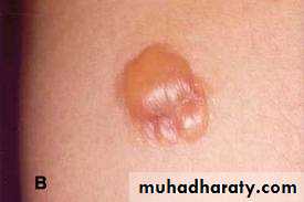
Tuberculin skin test (Mantoux skin test):


Tuberculin skin test (Mantoux skin test):

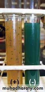
NIACIN TEST
+ve = yellowLab 20
Normal FloraGenus Streptococcus

Genus Stomatococcus

Genus Actinomyces

Genus Lactobacillus

Genus Propionibacterium

Genus Veillonella
Genus Haemophilus

Genus Fusobacterium
Genus Treponema

Throat Culture


With my love
