Tuberculosis of bone and joints
Tuberculosis is still a major cause of death and?disability in developing countries.
Tuberculosis again on the increase in last two decade, bones or joints are affected in about 5% of patients .
TUBERCULOSIS
Mycobacterium tuberculosis (human, less commonly bovine ), enter via the lung or gut or rarely through the skin.Tuberculosis may appear in any bone, synovial or bursal sheath.
Pathology of TB
1- Primary complexthe initial lesion in lung, pharynx or gut, with lymphatic spread to regional lymph node.
2- Secondary spread
If the resistance to the original infection is low, wide spread dissemination via the blood stream, giving rise to miliary TB ,or meningitis.Tertiary lesion
Mycobacterium tuberculosis gained a foot hold in bone or jointThere is predilection to vertebral bodies ( 50%), and the large synovial joints ( hip ,knee, less often shoulder or ankle).
These changes affected either the synovium (SYNOVITIS) or the bone (OSTITIS) is affected and then spread to the near by structure and whole joint affected ( ARTHRITIS).
Fibrous ankylosis of joint.
Spread to soft tissue lead to cold abscess.Followed with sinus and end with secondary pyogenic infection.
In endemic area skeletal TB is seen mainly in children.In non endemic area is seen in chronic debilitating disease (AIDS).
If the disease is arrested at an early stage, healing may be by resolution to normality.
If the cartilage is destroyed healing by fibrosis and incomplete ankylosis.

Clinical features of TB in large joints
History of previous infection.The patient usually a child ,or young adult
Pain .
Swelling.
Fever .
Lassitude .
Loss of weight.
Night cries
(the joint splinted by muscle spasm during the day, relaxes with sleep and damage tissues are stretched).Muscle wasting.
Movement in all direction limited.

In T B spine
BACKACHEKyphosis (gibbus).
Occasionally the presenting feature is weakness or loss of sensibility in the lower limb.
In neglected cases patient presented with paralysis (pott’s paraplegia).


Radiological features
early changes:Soft tissue swelling, Periarticular osteoporosis
Loclized osteoprosis (Bone ends ‘washed – out’ or localized decalcification ).
Narrowing and irregularity of the articular ends.
Late changes:
Erosions of the subarticular cartilage.
cystic changes appeared.
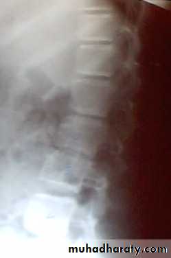


T B spondylitis may appear as localized bone erosion and collapse across an intervertebral disc space
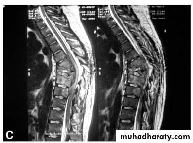
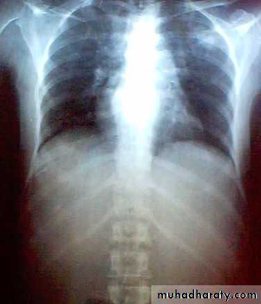
There may be soft tissue traces of paravertebral abscess.

investigations
E S R increased.Relative lymphocytosis.
Mantoux test positive.
Synovial fluid aspirate (high protein).
Polymerase chain reaction ( PCR ) is sensitive and specific .
Acid fast bacilli isolation and cultures is diagnostic.
Synovial biopsy is the more valuable .
Cardinal features of TBLong history.
Involvement only one joint.
Marked synovial thickening.
Marked muscle wasting.
Periarticular osteoporosis.
Differential diagnosis of tuberculosis.
1- transient synovitis.
2- Monoarticular rheumatoid arthritis.3- subacute arthritis ( reactive or brucellosis)
4- haemorrhagic arthritis.
5- pyogenic arthritis.
6- multiple myeloma and secondary metastases.
7- fungal infection and sarcodosis.
8- bone tumors.


Treatment of tuberculosis.
1- rest; splintage or skin traction, restricted activity continue for 6-12 months if diagnosis is early until joint resolved.2- Those with progressive joint destruction may need prolonged period of splintage to prevent ankylosis in bad position.
2- chemotherapy of TB
A- isoniazid , rifampicin, pyrazinamide, ethambutol for 2 months.B- isoniazid and rifampicin fore another 4 .
This is the recommendation of WHO, and BMRC.
C- some authors recommend to give chemotherapy for 18 months ( 4 HRZE, 14 HR)
3-SURGERY for TB
3- operative drainage or clearance of tuberculous foci may used.Arthrodesis or replacement arthroplasty may considers if articular surface is destroyed, years after healing, chemotherapy given 3 months before and after the operation.
arthrodesis

arthroplasty

TB tenosynovitis – compound ganglion.
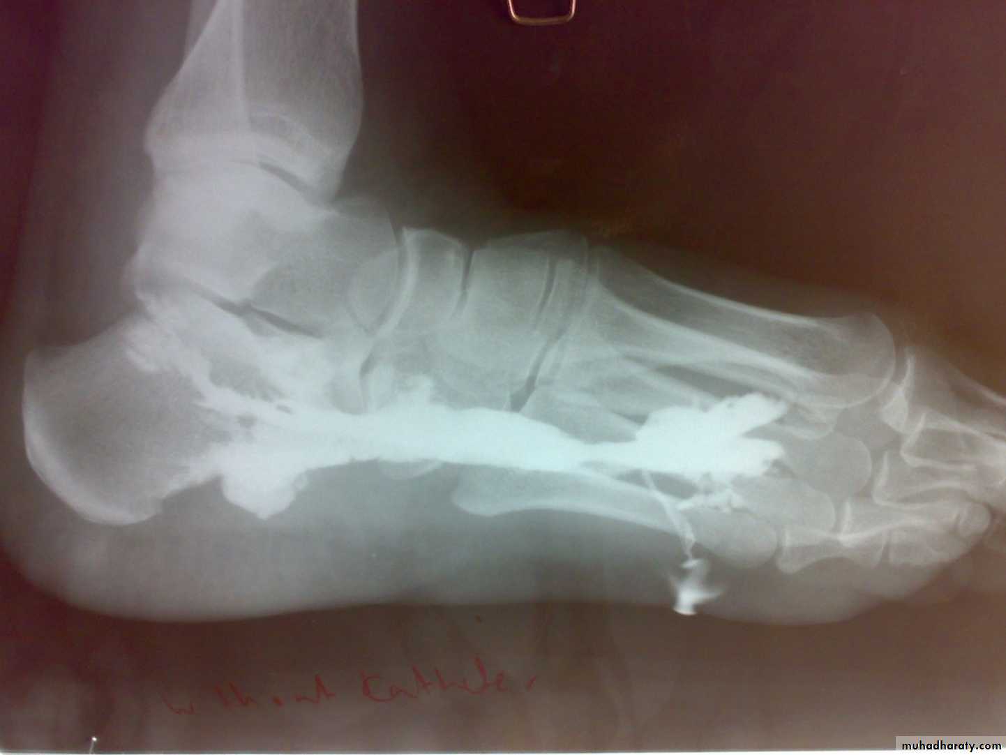
TB BURSITIS – trochrenteric bursitis.TB dactylitis – in children hand and feet digits , it should be differentiated from sickle cell anemia,
Brucellosis
It cause subacute or chronic arthritis.It has intermediate picture between pyogenic and TB.
Positive agglutination test is diagnostic.
Tissue culture positive if spine affected.
Treatment by rifampicin and 3rd generation cephalosporin.

Spirochaetal infections
SyphilisYaws
Lyme disease
Hydatid disease of bone
Hydatid disease affect bone in 1-2%.The cyst slowly enlarged in bone with little respect to cortical or epiphyeseal boundaries.
The bones most commonly affected are the vertebrae, pelvis, femur, scapula and ribs.
Patiens may complain of pain and swelling or pathological fractures.
It most differentiated from benign and malignant bone tumors.
Treatment by radical resection if possible with prolonged courses of albendazole.


